diagnosis and treatment of oral cavity metastases from the prostate gland. Hirshberg et al.2 stated that symptoms associ- ated with metastases include rapid swelling, pain and pares- thesia, which can be cardinal symptoms of jaw metastases.
The purpose of this study is to report a rare case of a me- tastasis from the prostate adenocarcinoma that was diagnosed with a biopsy. The Gleason system has been introduced to aid tumor evolution prediction and pathological staging. In addition, this case also discusses numb chin syndrome (NCS), which is an uncommon neurological manifestation caused by metastatic malignancy.
II. Case Report
An 80-year-old man was referred to our outpatient clinic by a dentist in December 2013, with complaints of numb- ness and swelling on the left site of the mandible. The patient stated that he had been suffering from this numbness for 7 months and swelling for 3 to 4 months.
A diffuse radiolucent lesion was observed in the left man- dibular angle region with a panoramic radiograph. Orophar-
I. Introduction
Prostate carcinoma is caused by development of cancer in the prostate, a gland in the male reproductive system.
Metastatic tumors from the primary region to the oral cavity are rare, and account for only 1% of all malignant oral neo- plasms. The proportional frequency of cases with oral and maxillofacial metastases that originate in the prostate to the total number of cases with oral and maxillofacial metastases that originate in all primary sites is about 6.2%1. Because of this low case frequency, it is important to determine the right
Dong-Hwan Lee
Division of Oral and Maxillofacial Surgery, Department of Dentistry, Inha University School of Medicine, 100 Inha-ro, Nam-gu, Incheon 22212, Korea TEL: +82-32-890-2470 FAX: +82-32-890-2475
E-mail: [email protected]
ORCID: http://orcid.org/0000-0002-1091-2543
This is an open-access article distributed under the terms of the Creative Commons Attribution Non-Commercial License (http://creativecommons.org/licenses/by-nc/4.0/), which permits unrestricted non-commercial use, distribution, and reproduction in any medium, provided the original work is properly cited.
CC
Prostate adenocarcinoma mandibular metastasis associated with numb chin syndrome: a case report
Il-Kyu Kim1, Dong-Hwan Lee1, Hyun-Young Cho1, Ji-Hoon Seo1, Seung-Hoon Park2, Joon-Mee Kim3
1Division of Oral and Maxillofacial Surgery, Department of Dentistry, Inha University School of Medicine,
2Department of Oral and Maxillofacial Surgery, Catholic Kwandong University International St. Mary’s Hospital,
3Department of Pathology, Inha University School of Medicine, Incheon, Korea
Abstract(J Korean Assoc Oral Maxillofac Surg 2016;42:301-306)
The purpose of this study is to report a rare case of mandibular adenocarcinoma that was diagnosed due to metastasis from the prostate. Numb chin syndrome (NCS), which was associated with this case, is also discussed. Computed tomography (CT) and an intraoral incisional biopsy of the left man- dibular area were performed. Urology consultation, hormone therapy, chemotherapy and follow-up radiographic images were administered. Histologi- cal examination of the incised specimen revealed moderately differentiated adenocarcinoma. The Gleason score was 8 (primary 4/secondary 4). Immu- nohistochemical features and radiographic results confirmed the diagnosis of metastasis from prostate adenocarcinoma, moderately differentiated. The patient’s prostate-specific antigen (PSA) level was very high. After hormone treatment, the patient’s PSA levels dropped gradually. Seventeen months later, in May 2015, the PSA level was elevated. The 18-month follow-up CT image indicated that the patient’s condition was aggravated. Docetaxel chemotherapy was started in June 2015 (18 months later), and the sixth cycle of the therapy is in progress. Oral metastases that originate from prostate adenocarcinoma are rare and can induce various periosteal reactions. Hormone therapy, chemotherapy and close follow-up could be additional, appro- priate treatment, and were applied in this case. Finally, NCS is a valuable indicator of metastatic disease in the mandible.
Key words: Prostate adenocarcinoma, Oral metastases, Hormone therapy, Chemotherapy, Numb chin syndrome
[paper submitted 2016. 4. 20 / revised 2016. 7. 20 / accepted 2016. 8. 17]
Copyright Ⓒ 2016 The Korean Association of Oral and Maxillofacial Surgeons. All rights reserved.
without pathologic cervical lymph nodes.(Fig. 1)
Biopsy was performed under local anesthesia. Histological examination (H&E staining) of the incised specimen revealed moderately differentiated adenocarcinoma. The Gleason score was 8 (primary 4/secondary 4) and it was predicted to be moderately aggressive. Immunohistochemical stain results ynx computed tomography (CT) showed a 6×7.7×7.2 cm3-
sized enhancing mass with a sunburst periosteal reaction in the ramus, angle and posterior body of the left mandible. It also showed invasion to the left medial pterygoid muscle and lateral pterygoid process, bulging into the left lateral mouth floor and shifting inferiorly to the left submandibular gland,
A
B C
Fig. 1. A 6×7.7×7.2 cm3 sized en- hancing mass with sunburst periosteal reaction in ramus, angle and posterior body of left mandible. A. Panorama (1st medical examination). B. Oropharynx computed tomography (CT) axial view (1st medical examination). C. Orophar- ynx CT coronal view (1st medical ex- amination). Arrows indicate enhancing mass with sunburst periosteal reaction.
Il-Kyu Kim et al: Prostate adenocarcinoma mandibular metastasis associated with numb chin syndrome: a case report. J Korean Assoc Oral Maxillofac Surg 2016
A B
Fig. 2. Immunohistochemical stain for prostate-specific antigen (PSA−positive; cytokeratin 7, cytokeratin 20, TTF-1, and S100−negative) reveals diffuse strong positive result. A. Submucosal tissue (H&E staining, ×100). B. Submucosal tissue (PSA immunohistochemical stain- ing, ×200).
Il-Kyu Kim et al: Prostate adenocarcinoma mandibular metastasis associated with numb chin syndrome: a case report. J Korean Assoc Oral Maxillofac Surg 2016
months follow-up). A follow-up positron emission tomogra- phy-CT after 10 months showed sclerotic change with mild fluorodeoxyglucose (FDG) uptake in the left mandible (max- imum standard uptake value, maxSUV=2.40; SUV=tissue radioactivity concentration [MBq/mL]/injected dose [MBq]×
BW [g]).(Fig. 4)
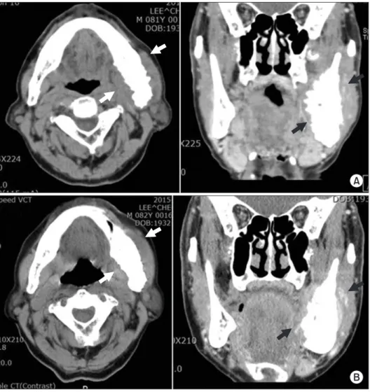
However, in May 2015 (17 months later), the PSA level was elevated to 315 ng/mL. The 18 month follow-up visit mandible CT showed an increased soft tissue mass that in- volved the left mandible and adjacent masticator space, de- creased extent of sclerotic changes and periosteal reaction.
(Fig. 5) The patient was evaluated as aggravated. Docetaxel chemotherapy (125 mg injection/1 cycle) was started in June 2015 (18 months later) and is currently in the sixth cycle.
Until now, the patient was moderately tolerant to hormonal and chemical treatment for several months and he has been in follow-up for 28 months.
III. Discussion
Prostate carcinoma is caused by cancer in the prostate, a gland in the male reproductive system. Jaw metastasis occurs less frequently, compared to the involvement of other bones of the body3 and represents 1% of all malignant mouth neo- plasms. Shen et al.1 showed that the proportional frequency of cases with oral and maxillofacial metastases that originate in the prostate to the total number of cases with oral and max- showed prostate-specific antigen (PSA−positive; cytokeratin
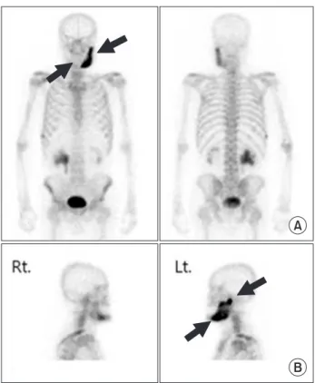
7, cytokeratin 20, TTF-1, and S100−negative).(Fig. 2) The patient was sent for medical consultation and an abdomen- pelvis CT, prostate magnetic resonance imaging, and bone scan were performed. The bone scan showed abnormal, increased bony uptake in the left mandible, right lateral 8th and 9th ribs, and right posterior 9th to11th ribs.(Fig. 3) These immunohistochemical features and radiographic results con- firmed the diagnosis of metastasis from the prostate adeno- carcinoma, moderately differentiated.
The PSA level was very high (>5,000 ng/mL; normal range, <4.4 ng/mL). The patient was started on leuprorelin acetate and given a 3.75 mg injection every 4 weeks, and a bicalutamide 50 mg tablet every day. After hormone treat- ment, the patient’s PSA level dropped to 505 ng/mL (after 1 month), 55 ng/mL (after 5 months), 28 ng/mL (after 9 months), and 47 ng/mL (after 14 months).
A mandible CT showed decreased extent of soft tissue mass, increased sclerotic changes, and a residual tumor that involved the left mandible and adjacent masticator space (5
Fig. 4. Sclerotic change with mild fluorodeoxyglucose (FDG) uptake in left mandible. Ten-month follow-up positron emission tomography-computed tomography. Arrows indicate sclerotic change in mandible.
Il-Kyu Kim et al: Prostate adenocarcinoma mandibular metastasis associated with numb chin syndrome: a case report. J Korean Assoc Oral Maxillofac Surg 2016
A
B
Fig. 3. Active bone lesion in left (Lt.) mandible and right (Rt.) lat- eral 8th and 9th ribs, right posterior 9th to 11th ribs. A. Whole body bone scan: abnormal increased bony uptake in right lateral 8th and 9th ribs, right posterior 9th to 11th ribs. B. Mandible bone scan: abnormal increased bony uptake in left mandible. Arrows indicate active bone lesion.
Il-Kyu Kim et al: Prostate adenocarcinoma mandibular metastasis associated with numb chin syndrome: a case report. J Korean Assoc Oral Maxillofac Surg 2016
venous plexus drainage is the predominant method for bony spread7. In the maxillofacial region, prostate carcinomas target the jawbone. In men, 11% of the jawbone metastases originated from the prostate gland compared with 1.5% in the soft tissues2. The mandible was found to be the most affected site for oral metastasis (35.6%). The molar and premolar region was the most frequently involved (53.0%), followed by the ascending ramus (31.6%), mandibular angle (17.8%), condyle (15.8%), and the mental region (6.1%)1. The poste- rior mandible is the most susceptible metastasis site because of its rich blood supply and hematosis7.
NCS is an uncommon neurological manifestation of an oral cavity metastatic tumor. Sensory discomfort could be the first sign of infiltration when the prostate cancer metastasizes to the mandible8-10. The first case of NCS was reported by Charles Bell in 1830 in his monograph ‘The nervous sys- tem of the human body’11. Clinical features of mandibular metastases present as toothache, rapid swelling, pain and paresthesia. These symptoms sometimes lead to extraction of illofacial metastases that originate in all primary sites is about
6.2%. Aksoy et al.3 determined that the most common sites of primary tumors in men were lungs (22.3%), prostate (12%), kidney (10.3%), bone (9.2%), and adrenals (9.2%). Because of this low frequency but common primary sites, it is impor- tant to accurately diagnosis and treat oral cavity metastases from the prostate gland4.
The Gleason system has been introduced for tumor evolu- tion prediction and to determine pathological staging. The Gleason score is the sum of the pattern-number of the pri- mary and secondary grades based upon microscopic appear- ances. A score of 2 to 4 (well differentiated) is mild, while a score of 7 to 10 is more aggressive and has a bad prognosis5. In this case, the primary tumor grade was 4 and the secondary tumor grade was 4, so the Gleason score was 4+4=8. Histo- logical features were moderately differentiated, and predicted to be moderately aggressive.
Prostate cancer usually metastasizes to bones, such as lum- bar vertebrae, thoracic vertebrae, and the pelvis6. Prevertebral
A
B
Fig. 5. Follow-up computed tomog- raphy (CT). A. Mandible CT axial and coronal view (5 months). B. Mandible CT axial and coronal view (18 months).
Arrows in Fig. 5. A indicate more de- creased soft tissue mass than Fig 1.
But arrows in Fig. 5. B show the ag- gravated soft tissue mass.
Il-Kyu Kim et al: Prostate adenocarcinoma mandibular metastasis associated with numb chin syndrome: a case report. J Korean Assoc Oral Maxillofac Surg 2016
was aggravated, and a sixth docetaxel chemotherapy session is in progress.
In summary, periosteal reactions identified in a panorama CT scan are helpful for diagnosing malignant tumors. Hor- mone therapy, such as chemotherapy in this case, can extend the patient’s lifespan in metastatic prostate cancer. NCS is also a useful indicator of metastatic prostate adenocarcinoma.
Conflict of Interest
No potential conflict of interest relevant to this article was reported.
ORCID
Il-Kyu Kim, http://orcid.org/0000-0003-3930-766X Dong-Hwan Lee, http://orcid.org/0000-0002-1091-2543 Hyun-Young Cho, http://orcid.org/0000-0003-3055-0591 Ji-Hoon Seo, http://orcid.org/0000-0003-3618-6380 Seung-Hoon Park, http://orcid.org/0000-0001-6847-0082 Joon-Mee Kim, http://orcid.org/0000-0003-1355-4187
References
1. Shen ML, Kang J, Wen YL, Ying WM, Yi J, Hua CG, et al. Meta- static tumors to the oral and maxillofacial region: a retrospective study of 19 cases in West China and review of the Chinese and English literature. J Oral Maxillofac Surg 2009;67:718-37.
2. Hirshberg A, Shnaiderman-Shapiro A, Kaplan I, Berger R. Meta- static tumours to the oral cavity: pathogenesis and analysis of 673 cases. Oral Oncol 2008;44:743-52.
3. Aksoy S, Orhan K, Kursun S, Kolsuz ME, Celikten B. Metastasis of prostate carcinoma in the mandible manifesting as numb chin syndrome. World J Surg Oncol 2014;12:401.
4. Daley T, Darling MR. Metastases to the mouth and jaws: a contem- porary Canadian experience. J Can Dent Assoc 2011;77:b67.
5. Menezes JD, Cappellari PF, Capelari MM, Gonçalves PZ, Toledo GL, Toledo Filho JL, et al. Mandibular metastasis of adenocarci- noma from prostate cancer: case report according to epidemiology and current therapeutical trends of the advanced prostate cancer. J Appl Oral Sci 2013;21:490-5.
6. Saijo H, Chikazu D, Mori Y, Hikiji H, Yonehara Y, Takato T. Me- tastasis of prostate cancer to the mandibular condyle. Asian J Oral Maxillofac Surg 2008;20:86-8.
7. Tchan MC, George M, Thomas M. Metastatic prostate cancer mimicking primary osteosarcoma of the jaw: an infrequent clinical case. South Med J 2008;101:657-9.
8. Soares EC, Costa FW, Rocha-Filho FD, Ferreira FV, Alves AP.
Metastatic prostate adenocarcinoma associated with numb chin syndrome. J Craniofac Surg 2011;22:2366-8.
9. Zaheer F, Hussain K, Rao J. Unusual presentation of 'numb chin syndrome' as the manifestation of metastatic adenocarcinoma of the lung. Int J Surg Case Rep 2013;4:1097-9.
10. Narendra H, Ray S. Numb chin syndrome as a manifestation of metastatic squamous cell carcinoma of esophagus. J Cancer Res Ther 2009;5:49-51.
11. Evans RW, Kirby S, Purdy RA. Numb chin syndrome. Headache
the affected tooth2. This patient had been suffering from this numbness for 7 months and swelling for 3 to 4 months with- out any detectable symptoms too. Accordingly, it is important to recognize NCS as an uncommon neurological manifesta- tion because of the possibility of metastatic malignancy.
Periosteal reactions can be classified as single layer, mul- tilayered, solid, speculated, perpendicular, sloping, com- plex, Codman triangle and sunburst12. The appearance of a
‘sunburst’ periosteal reaction is suggestive of rapid onset pathology7. Most tumors produce osteolytic-like lesions.
However, metastatic prostate carcinomas can secrete high amounts of the receptor activator of nuclear factor kappa-B ligand (RANKL) inhibitor osteoprotegerin, thereby weaken- ing osteoclastic reactions during metastasis. Based on these characteristics metastic prostate neoplasms were proposed to be osteoblastic or sclerotic2. The sunburst image is most com- monly associated with osteoblastic metastasis and approxi- mately 30% are secondary to prostatic carcinoma12. Sunburst periosteal reaction in the ramus, angle and posterior body of the left mandible with clinical features, in this case, strongly suggests a malignant bone forming tumor, such as an osteo- sarcoma.
There are several treatment approaches for prostate can- cer: radical prostatectomy, radiation therapy, chemotherapy, and endocrine therapy13. The first PSA level was very high (>5,000 ng/mL; normal range, <4.4 ng/mL). The patient was started on a leuprorelin acetate 3.75 mg injection every 4 weeks, and a daily bicalutamide 50 mg tablet. In response to these hormone treatments, the PSA level dropped to 505 ng/
mL (after 1 month), 55 ng/mL (after 5 months), 28 ng/mL (after 9 months), and 47 ng/mL (after 14 months). However, in May 2015 (17 months later) the PSA level was elevated to 315 ng/mL. Because of this PSA elevation, docetaxel chemo- therapy was administered.
The presence of oral metastases indicates a poor prognosis, with a mean interval of approximately 7 months between the occurrence of metastases and death2. Sometimes an ag- gressive case with high levels of PSA can rapidly progress to death within only 3 weeks after the detection of jawbone metastases8.
In this case, at the 18 month follow-up visit, a mandible CT image showed increased soft tissue mass that involved the left mandible and adjacent masticator space, decreased scle- rotic changes and a periosteal reaction.(Fig. 5) Evaluation of the patient indicated good disease control and good tolerance to treatment over several months; however, the PSA level elevation and CT image showed that the patient’s condition
2008;48:1520-4.
12. Wenaden AE, Szyszko TA, Saifuddin A. Imaging of periosteal re- actions associated with focal lesions of bone. Clin Radiol 2005;60:
439-56.
13. Reyes Court D, Encina S, Levy I. Prostatic adenocarcinoma with mandibular metastatic lesion: case report. Med Oral Patol Oral Cir Bucal 2007;12:E424-7.