INTRODUCTION
Since Brånemark introduced a protocol for dental implantation in 1977, implants have become increasingly successful in the restoration of the dentition of partially or completely edentulous patients [1-3]. In addition, root-form endosseous dental implants have proven to be excellent alternatives to conventional tooth replacement options [4]. Achieving good primary (biomechanical) stability is a prerequisite for successful implantation [5]. This de- pends on many factors, including the implant design and surface, bone quality, and implan- tation procedure [6,7]. During the last 20 years, implant designs and surfaces have been de- veloped to achieve faster and more stable osseointegration and higher success rates. Though the original Brånemark implant system had smooth endosseal surfaces, the currently mar- keted rough endosseal surfaces deliver higher success rates, especially in areas of cancellous bone quality [8,9]. Among the various rough surfaces, the sand-blasted, thermally acid-etched surface encourages more rapid implant osseointegration, thereby shortening the healing period, and it has the potential to become the gold standard soon.
Such an implant system, characterized by a sand-blasted, thermally acid-etched surface, machined collar, and internal hexagonal implant/abutment connection, was introduced in
This is an Open Access article distributed under the terms of the Creative Commons Attribution Non-Commercial License (http://creativecommons.org/licenses/by-nc/3.0/).
multicenter field study
Seung-Il Shin1,†, Jeong-Ho Yun2,†, Sung-Geun Kim3, Byoungkyou Park3, Yeek Herr1, Jong-Hyuk Chung1,*
1Department of Periodontology, Institute of Oral Biology, Kyung Hee University School of Dentistry, Seoul, Korea
2Division of Periodontology, Department of Dentistry, Inha University School of Medicine, Incheon, Korea
3Department of Periodontology, Kyung Hee University School of Dentistry, Seoul, Korea
Research Article
J Periodontal Implant Sci 2014;44:8-12 http://dx.doi.org/10.5051/jpis.2014.44.1.8
Purpose: The aim of this retrospective chart review was to evaluate the four-year survival rate of a titanium implant system.
Methods: A total of 352 sand-blasted, thermally acid-etched titanium implants were in- serted into 181 partially or completely edentulous patients. Their cumulative survival rate was evaluated retrospectively. Associated factors, such as the implant distribution and treatment type were included in the evaluation.
Results: The implants were equally distributed between the maxilla (52.3%) and the man- dible (47.7%). 48 implants (13.6%) were placed in the anterior region and 304 implants (86.4%) in the posterior region. The majority of the implants were inserted into bone of type II and III quality (89.8%) and volume (quantity B and C, 87.2%). Most of the implants (70.7%) were restored as single crowns; 28.7% supported a bridge construction and 0.6% a full denture. Only one implant failed, resulting in a four-year cumulative survival rate of 99.7%.
Conclusions: The implant system showed an excellent four-year survival rate. It proved to be a safe and predictable means for restoration of the dentition in partially or completely edentulous patients.
Keywords: Dental implant, Multicenter study, Survival rate.
Received: Dec. 11, 2013 Accepted: Jan. 4, 2014
*Correspondence:
Jong-Hyuk Chung
Department of Periodontology, Institute of Oral Biology, Kyung Hee University School of Dentistry, 26 Kyungheedae-ro, Dongdaemun-gu, Seoul 130- 701, Korea
E-mail: [email protected] Tel: +82-2-958-9382 Fax: +82-2-958-9387
†Seung-Il Shin and Jeong-Ho Yun contributed equally to this work.
2001. The implants are threaded, self-tapping, and have either cy- lindrical (constant diameter) or conical-cylindrical endosseous pro- files. They have a tapered and domed apical end that promotes smooth insertion into the implant bed. The state-of-the-art manu- facturing process results in an optimal implant surface and the self-tapping design contributes to achieving good primary stability.
Although entailing many advantages, these implants are newer than other brands and their use in clinical practice has not been documented for a prolonged time period; that is, the literature provides scant survival rate analysis. As these implants share many characteristics with currently used implant brands, a survival rate study of this implant line could provide the proof that its reliability and predictability is also comparable to competitor products. The goal of this study was thus to evaluate the medium-term survival rate of this implant system.
MATERIALS AND METHODS
A retrospective chart review was conducted with the aim of evaluating the survival rate of a newly introduced implant line.
Two university clinical centers have participated in the review.
Both clinics have collected ample experience with these implants.
Kyung Hee University Dental Hospital provided information from 140 patient charts comprising 262 implants, while Myong Ji Hos- pital contributed 41 patient charts with 90 implants. The protocol of this study, which used anonymized patient data, imposed no additional risk to the patients who were included and involved no procedures for which written informed consent was needed. It was approved by the Institutional Review Board (IRB) at the Dental Hospital of Kyung Hee University (KHD IRB-004-1), and the chart review was conducted according to the Declaration of Helsinki.
The 352 implants analyzed in this study were used for treatment of single or multiple tooth loss. The implants were manufactured
of pure titanium (grade 4). They had either a cylindrical (SPI ELE- MENT and SPI ONETIME, Thommen Medical AG, Waldenburg, Swit- zerland) or a conical-cylindrical design (SPI CONTACT, Thommen Medical AG). All of the implants are included in this report; in oth- er words, no patient was excluded from the analysis population.
All of the implants were inserted following standard surgical pro- tocols. Care was taken to ensure the ideal prosthodontic implant position and that the implant sites had the appropriate bone qual- ity and bone volume.
The placement of the implants was performed according to the manufacturer’s recommendations, after raising a muco-periosteal flap. The implants were allowed to heal either submerged or non- submerged. Permanent prosthetic rehabilitation was scheduled af- ter a healing time of 6 months.
The statistical analysis was performed using Cutler and Ederer’s life table method for survival analysis. The principal advantage of this method is that it enables the use of all survival information gathered up to the closing date of the study. Thus, the implants that entered observation 4, 3, 2, and even one year prior to the closing date also contributed useful information to the calculation of the 4-year survival [10].
RESULTS
Patient characteristics
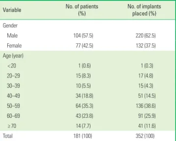
The age distribution of the patients in the study together with the corresponding implant breakdown is shown in Table 1. The av- erage patient age at the time of implant insertion was 55.2 years (19 to 87 years). A total of 352 implants was inserted in this pa- tient population between July 2006 and March 2009. 104 of the patients (57.5%) were male and 77 (42.5%) female. The largest group age group was 50–59 years, both in terms of the number of patients (64 patients, 36.8%) as well as the number of implants (136, 38.6%). In addition to systemic diseases, including hyperten- sion, diabetes, angina pectoris, asthma, hyperthyroidism, hyperlip- idemia, and tachycardia were seen in 61 patients (32.1%). 39 pa- tients (11.1%) were smokers, thus reflecting the general patient population in Korea.
Implant distribution
Table 2 shows the positions of the reported implants. 184 of the implants (52.3%) were placed in the maxilla. Among these, 37 (10.5%) were placed in the anterior and 147 (41.8%) in the poste- rior regions. One hundred sixty-eight implants (47.7%) were placed Table 1. Patient age and gender, and corresponding implant break-down.
Variable No. of patients
(%) No. of implants
placed (%) Gender
Male 104 (57.5) 220 (62.5)
Female 77 (42.5) 132 (37.5)
Age (year)
<20 1 (0.6) 1 (0.3)
20–29 15 (8.3) 17 (4.8)
30–39 10 (5.5) 15 (4.3)
40–49 34 (18.8) 51 (14.5)
50–59 64 (35.3) 136 (38.6)
60–69 43 (23.8) 91 (25.9)
≥70 14 (7.7) 41 (11.6)
Total 181 (100) 352 (100)
Table 2. Localization of inserted implants.
Anterior Posterior Total
Maxilla 37 (10.5) 147 (41.8) 184 (52.3)
Mandible 11 (3.1) 157 (44.6) 168 (47.7)
Total 48 (13.6) 304 (86.4) 352 (100)
in the mandible, with 11 (3.1%) in the anterior and 157 (44.6%) in the posterior region.
The distribution of the implant length and diameter is shown in Table 3. The length ranged from 8 to 12.5 mm. 21 of the implants (6.0%) were 8 mm long and 35 (9.9%) were 12.5 mm long; that is, the majority of the implants were either 9.5 mm (n=119, 33.8%) or 11 mm (n=177, 50.3%) long. The implant platform diameter ranged from 2.7 to 5 mm. Only a few implants (n=2, 0.6%) had a reduced diameter (platform, 3.5 mm); most of the inserted implants had a 4.5-mm platform diameter (n=218, 61.9%). The majority of the implants were inserted into bone with quality of types II and III (n=316, 89.7%) [11]. The bone quantity was classified as B and C (n=307, 87.2%) (Table 4).
Surgical protocols and prosthetics
The implants were placed using both nonsubmerged (1 stage) Table 3. Implant length and platform diameter.
Maxilla Mandible Total,
n (%) Anterior Posterior Anterior Posterior
Length (mm)
8 1 4 0 16 21 (6.0)
9.5 4 49 0 66 119 (33.8)
11 22 72 11 72 177 (50.3)
12.5 10 22 0 3 35 (9.9)
Total 36 147 11 157 352 (100)
Platform diameter (mm)
3.5 2 0 0 0 2 (0.6)
4.0 24 8 9 7 48 (13.6)
4.5 11 109 2 96 218 (61.9)
5.0 0 30 0 54 84 (23.9)
Total 37 147 11 157 352 (100)
Table 4. Alveolar bone quality and quantity.
Maxilla Mandible Total,
n (%) Anterior Posterior Anterior Posterior
Quality
Type I 0 0 2 1 3 (0.9)
Type II 10 6 5 115 136 (38.6)
Type III 25 117 3 35 180 (51.1)
Type VI 2 24 0 7 33 (9.4)
Total 37 147 10 158 352 (100)
Quantity
A 6 3 0 3 12 (3.4)
B 28 63 6 112 209 (59.4)
C 3 57 5 33 98 (27.8)
D 0 21 0 9 30 (8.5)
E 0 3 0 0 3 (0.9)
Total 37 147 11 157 352 (100)
Figure 1. Number of implants inserted by the 1 (nonsubmerged) and 2 stage (submerged) surgical protocols. Mx. Ant.: anterior maxilla, Mx. Post.: posterior maxilla, Mn. Ant.: anterior mandible, Mn. Post.: posterior mandible.
120 100
80 60 40
20 0
Mx. Ant.
Mx. Post.
Mn. Ant.
Mn. Post.
9 30
0 65
28 117
11 92
1 Stage 2 Stage
Figure 2. Implant restoration types.
Single crown (n=249, 70.7%) Bridge
(n=101, 28.7%)
Total prosthesis (n=2, 0.6%)
and submerged (2 stage) surgical protocols (Fig. 1). Fig. 2 summa- rizes the restoration types: single crowns (n=249, 70.7%), bridges (n=101, 28.7%), or full dentures (n=2, 0.6%). As shown in Table 5, 212 implants (60.2%) were placed in sites with bone grafting pro- cedures, such as crestal or lateral sinus floor elevation, guided bone regeneration, ridge expansion, etc.
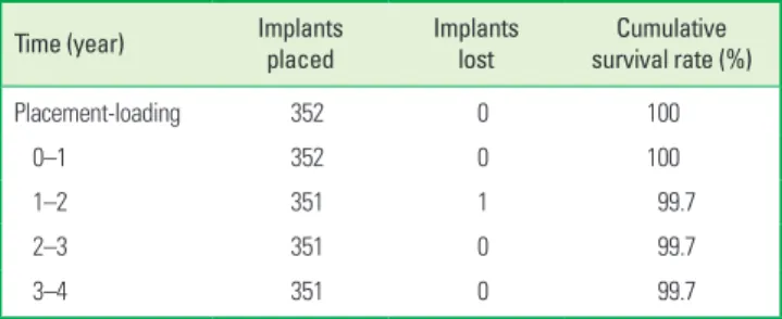
Implant survival
Implant survival was evaluated using the criteria of Albrektsson and Zarb [12]; specifically, implants still stable in situ were consid- ered to be survivors. During the follow-up period, only one of the 352 implants was lost. This implant (platform diameter, 4.5 mm;
length, 9.5 mm), was inserted into the mandible. It had to be re- moved due to osteomyelitis approximately 21 months after loading.
3 months following implant removal, the site healed uneventfully.
Using the above-mentioned criteria for implant survival, the life ta-
ble analysis resulted in a cumulative survival rate of 99.7% (Table 6).
DISCUSSION
Only a few of the currently marketed dental implant systems have documented mid- or long-term survival rates. When a new, substantially equivalent implant system is brought to market, cur- rent regulations do not require prospective clinical trials to deter- mine the predictability of the specific system. An efficient option to achieve this is thus a retrospective chart review study. One of the advantages of such a review is that its result is a true reflec- tion of general clinical practice with the system in question. The purpose of this study was to evaluate the medium-term (4-year) survival rate of 352 implants from a newly introduced titanium implant line. The results reported here were obtained in 181 pa- tients in two independent clinics. The 99.7% cumulative survival rate is the result of only one failure in the investigated timeframe.
This clearly indicates that the implants have still been present and stable in situ after the four year period studied. In addition, no se- vere complications, such as pain or mobility, were recorded in the 4-year period.
With other titanium implant lines, several prospective studies have been already published that document their survival rate.
These systems might differ somewhat in their endosseal surfaces and designs. A survival rate of 92.6% has been reported, for in- stance, for Brånemark implants, after 10 years of follow-up [3], while for another implant line with a titanium plasma-sprayed surface, a 96.7% 8-year cumulative survival rate was reported [13].
Recently, root-shaped implants with sand-blasted, thermally acid- etched surfaces showed a cumulative survival rate of 98.6% to 99.3% [14,15]. The results of the present retrospective multicenter chart review study comprise a total of 352 state-of-the art titani- um implants placed in 181 patients. The results showed quite a high four-year survival rate of 99.7%. Our findings, therefore, showed that the tested implants provide a safe and predictable clinical outcome that compares well with other implants for which the survival rates have been published earlier.
The single implant lost was due to osteomyelitis. At the time of implant placement in the right posterior mandible, the female pa- tient was 79 years old. After a 5-month healing period, prosthetic treatment was initiated. Some 21 months after the implant place- ment, the patient presented at the clinic with gingival swelling and pain in the implant area. Radiographic findings had confirmed os- teomyelitis and a sequestrectomy with removal of the implant and
the adjacent tooth was performed under general anesthesia. The patient recovered uneventfully.
It should be noted that a large number of implants, that is, more than half of the implants reviewed in this study, were placed in areas requiring additional interventions such as sinus floor elevation or guided bone regeneration due to poor bone quantity and/or quality at the implant site. Consequently, the majority of the cases can be classified as complex. Considering this background, the finding of only 1 failed implant among the 352 cases after 4 years is quite im- pressive. The implants that were used represent a very reliable alter- native when a substitution for natural teeth is required.
The tested implants feature a sand-blasted, thermally acid-etched endosseous surface. The roughness value, Sa, is 1.0–2.0 µm. This surface is therefore classified as moderately rough. The advantage of moderately rough surfaces is that they show stronger bone re- sponses than smoother or rougher surfaces, and contribute to achieving a favorable clinical performance [16]. Further advances in surface processing (conditioning with a week base) created a hydrophilic surface, which is known to accelerate the healing pro- cess by promoting the activity of osteoblasts, the bone-forming cells that play a key role in the integration of implants into bone [17]. The results from mechanical and histomorphometric analyses in animal studies have shown a significantly higher removal torque and percentage of bone-to-implant contact for rough surfaces when compared to machined surfaces [18,19]. A human histology study demonstrated that the rough surface developed a statisti- cally higher percentage of bone-to-implant contact than the ma- chined surface [20]. This implant system also provides different de- signs for the machined collar height. The availability of such an array of sizes enables the practitioner to select the fixture with an optimal design to ensure a successful treatment outcome. Depend- ing on the requirements of the particular implantation site, as well as the condition of the surgical site and additional surgical demands, the optimal implant can be effectively selected. The present retro- spective study focused on the 4 year cumulative survival rate. A lon- ger-term follow-up study and comparison with other implant lines (with different surface structures and geometry) would be feasible.
The cumulative survival rate of 352 titanium implants was de- termined to be 99.7% after four years. This calculation was based on a retrospective evaluation of implant survival. The availability of the assessment of other clinical parameters, mainly periodic ra- diographs, would have allowed the calculation of implant success Table 5. Bone grafting procedures at recipient sites.
CSE LSE GBR Bone
graft only CSE+
GBR LSE+
GBR Ridge
expansion Total
41 31 92 36 5 5 2 212 (60.2%)a)
CSE: crestal sinus floor elevation, LSE: lateral sinus floor elevation, GBR: guided bone regeneration.
a)Percentage of implants with augmentation.
Table 6. Life table analysis of cumulative implant survival rate.
Time (year) Implants
placed Implants
lost Cumulative
survival rate (%)
Placement-loading 352 0 100
0–1 352 0 100
1–2 351 1 99.7
2–3 351 0 99.7
3–4 351 0 99.7
to further support the outcome achieved with the tested implant line. Within the limitations of this retrospective chart review, we conclude that the newly introduced implant system is a safe and predictable option for restoring partially or completely edentulous patients and also in cases when additional bone grafting proce- dures are needed.
CONFLICT OF INTEREST
No potential conflict of interest relevant to this article was re- ported.
ACKNOWLEDGEMENTS
This study was funded by the Department of Periodontology, Kyung Hee University, Seoul, Korea.
ORCID
Seung-Il Shin http://orcid.org/0000-0001-8762-6169 Jeong-Ho Yun http://orcid.org/0000-0003-3929-4467 Sung-Geun Kim http://orcid.org/0000-0002-4698-5306 Byoungkyou Park http://orcid.org/0000-0003-1342-1413 Yeek Herr http://orcid.org/0000-0001-9243-7119 Jong-Hyuk Chung http://orcid.org/0000-0002-2678-1525
REFERENCES
1. Adell R, Lekholm U, Rockler B, Branemark PI. A 15-year study of osseointegrated implants in the treatment of the edentulous jaw. Int J Oral Surg 1981;10:387-416.
2. Henry PJ, Laney WR, Jemt T, Harris D, Krogh PH, Polizzi G, et al.
Osseointegrated implants for single-tooth replacement: a pro- spective 5-year multicenter study. Int J Oral Maxillofac Implants 1996;11:450-5.
3. Lekholm U, Gunne J, Henry P, Higuchi K, Linden U, Bergstrom C, et al. Survival of the Brånemark implant in partially edentulous jaws: a 10-year prospective multicenter study. Int J Oral Maxillo- fac Implants 1999;14:639-45.
4. Cochran D. Implant therapy I. Ann Periodontol 1996;1:707-91.
5. Martinez H, Davarpanah M, Missika P, Celletti R, Lazzara R. Opti- mal implant stabilization in low density bone. Clin Oral Implants Res 2001;12:423-32.
6. Meredith N. Assessment of implant stability as a prognostic de- terminant. Int J Prosthodont 1998;11:491-501.
7. Barewal RM, Oates TW, Meredith N, Cochran DL. Resonance fre- quency measurement of implant stability in vivo on implants with a sandblasted and acid-etched surface. Int J Oral Maxillofac
Implants 2003;18:641-51.
8. Buser D, Nydegger T, Oxland T, Cochran DL, Schenk RK, Hirt HP, et al. Interface shear strength of titanium implants with a sand- blasted and acid-etched surface: a biomechanical study in the maxilla of miniature pigs. J Biomed Mater Res 1999;45:75-83.
9. Cochran DL. A comparison of endosseous dental implant surfac- es. J Periodontol 1999;70:1523-39.
10. Cutler SJ, Ederer F. Maximum utilization of the life table method in analyzing survival. J Chronic Dis 1958;8:699-712.
11. Lekholm U, Zarb GA. Patient selection and preparation. In: Br- anemark PI, Zarb GA, Albrektsson T, editors. Tissue-integrated prostheses: osseointegration in clinical dentistry. Chicago: Quin- tessence; 1985. p.199-209.
12. Albrektsson T, Zarb GA. Current interpretations of the osseointe- grated response: clinical significance. Int J Prosthodont 1993;6:
95-105.
13. Buser D, Mericske-Stern R, Bernard JP, Behneke A, Behneke N, Hirt HP, et al. Long-term evaluation of non-submerged ITI implants.
Part 1: 8-year life table analysis of a prospective multi-center study with 2359 implants. Clin Oral Implants Res 1997;8:161-72.
14. Krennmair G, Seemann R, Schmidinger S, Ewers R, Piehslinger E.
Clinical outcome of root-shaped dental implants of various di- ameters: 5-year results. Int J Oral Maxillofac Implants 2010;25:
357-66.
15. Cochran D, Oates T, Morton D, Jones A, Buser D, Peters F. Clinical field trial examining an implant with a sand-blasted, acid-etched surface. J Periodontol 2007;78:974-82.
16. Albrektsson T, Wennerberg A. Oral implant surfaces: Part 1: review focusing on topographic and chemical properties of different surfaces and in vivo responses to them. Int J Prosthodont 2004;
17:536-43.
17. Lossdorfer S, Schwartz Z, Wang L, Lohmann CH, Turner JD, Wieland M, et al. Microrough implant surface topographies increase os- teogenesis by reducing osteoclast formation and activity. J Biomed Mater Res A 2004;70:361-9.
18. Buser D, Schenk RK, Steinemann S, Fiorellini JP, Fox CH, Stich H.
Influence of surface characteristics on bone integration of tita- nium implants: a histomorphometric study in miniature pigs. J Biomed Mater Res 1991;25:889-902.
19. Wennerberg A, Ektessabi A, Albrektsson T, Johansson C, Andersson B. A 1-year follow-up of implants of differing surface roughness placed in rabbit bone. Int J Oral Maxillofac Implants 1997;12:
486-94.
20. Lazzara RJ, Testori T, Trisi P, Porter SS, Weinstein RL. A human histologic analysis of osseotite and machined surfaces using im- plants with 2 opposing surfaces. Int J Periodontics Restorative Dent 1999;19:117-29.