Korean J Gastroenterol Vol. 64 No. 1, 59-61 http://dx.doi.org/10.4166/kjg.2014.64.1.59 pISSN 1598-9992 eISSN 2233-6869
IMAGE OF THE MONTH
Korean J Gastroenterol, Vol. 64 No. 1, July 2014 www.kjg.or.kr
소장내시경으로 진단된 메켈게실
정윤호, 고봉민
1순천향대학교 의과대학 천안병원, 부천병원1 내과학교실
Meckel’s Diverticulum Diagnosed by Enteroscopy
Yunho Jung and Bong Min Ko1
Department of Internal Medicine, Soonchunhyang University Cheonan Hospital, Cheonan, Soonchunhyang University Bucheon Hospital, Bucheon1, Korea
CC This is an open access article distributed under the terms of the Creative Commons Attribution Non-Commercial License (http://creativecommons.org/licenses/
by-nc/3.0) which permits unrestricted non-commercial use, distribution, and reproduction in any medium, provided the original work is properly cited.
교신저자: 고봉민, 420-767, 부천시 원미구 조마루로 170, 순천향대학교 부천병원 소화기내과
Correspondence to: Bong Min Ko, Division of Gastroenterology, Department of Medicine, Soonchunhyang University Bucheon Hospital, 170 Jomaru-ro, Wonmi-gu, Bucheon 420-767, Korea. Tel: +82-32-621-5087, Fax: +82-32-621-5080, E-mail: [email protected]
Financial support: None. Conflict of interest: None.
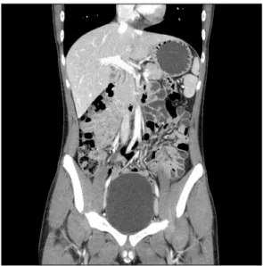
Fig. 1. Preoperative abdominal CT scan (coronal image) shows a tiny hepatic cyst in S5 and no abnormalities in the small bowel.
증례: 16세 된 남자 환자가 내원 당일 발생한 대량의 선홍 색 혈변을 주소로 내원하였다. 내원 3주전부터 건선을 진단받 고 스테로이드 제제를 복용한 경력이 있었으나 과거력과 가족 력에서 특이소견은 관찰되지 않았고, 흡연력 및 음주력도 없 었다. 내원 당시 대량의 혈변으로 인하여 전신 쇠약감을 호소 하였으나 복부 통증은 호소하지 않았다. 활력 징후는 혈압이 100/60 mmHg, 맥박수 75회/분, 호흡수 20회/분, 체온 36.9°C로 측정되었고 급성 병색소견을 보였다. 신체검사에서 창백한 결막이 관찰되었으나 복부는 부드러웠고 압통이나 반 발통은 관찰되지 않았다.
말초혈액검사에서 백혈구 18,350/mm3, 혈색소 4.8 g/dL, 혈 소판 405,000/mm3로 측정되었고, 생화학검사에서 AST/ALT 14/13 IU/L, T-bilirubin 0.47 mg/dL, PT (INR) 1.17로 측정 되어 혈액검사에서 심한 빈혈 소견을 보였고, 그 이외에 다른 이상 소견은 관찰되지 않았다. 내원 당일 시행한 복부 X선 촬영에서 소장 내 가스의 축적이 관찰되었으나 장폐쇄 소견은 보이지 않았고, 복부 전산화단층촬영에서 간낭종이 관찰되었 으나 소장의 종괴 및 폐쇄 소견은 관찰되지 않았다(Fig. 1).
내원 다음 날 시행한 상부위장관 내시경검사에서 출혈의 원인 으로 의심될 만한 병변은 관찰되지 않았고, 구불직장경 소견 에서 급성 출혈 소견은 보이지 않았으나 구불결장 상부까지 검붉은 혈액이 고여있었다. 대장 정결 후 시행한 전 대장내시 경에서 출혈의 원인이 될 만한 병변이 관찰되지 않아 소장
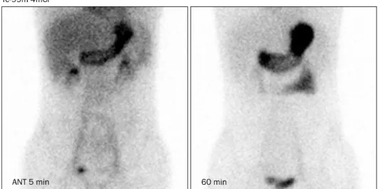
캡슐내시경검사를 시행하였다. 소장 캡슐내시경검사에서 말 단 공장 및 근위 회장으로 생각되는 위치에 게실을 의심할 수 있는 부위와 고여있는 검은 혈액을 관찰할 수 있어 메켈게 실을 감별진단하기 위해 99mTC pertechnetate 스캔을 시행하 였으나 비정상적인 국소 섭취 증가는 관찰되지 않았다(Fig.
2). 출혈의 병소를 확인하기 위해 시행한 이중풍선 소장 내시
60
정윤호, 고봉민. 소장내시경으로 진단된 메켈게실The Korean Journal of Gastroenterology
Fig. 4. Small bowel follow-through shows blind-ending diverticulum (arrows) with hemoclips.
Fig. 2. 99mTC pertechnetate scinti- graphy shows no areas of focal abnor- mal tracer accumulation within 60 minutes.
ANT, anterior.
Fig. 3. Endoscopic findings. (A) An orifice of the Meckel’s diverticulum (arrow) is visible in the proximal ileum. (B) Fresh blood and ulceration are evident within the diverticulum. (C) Hemostasis was successfully performed with hemoclips.
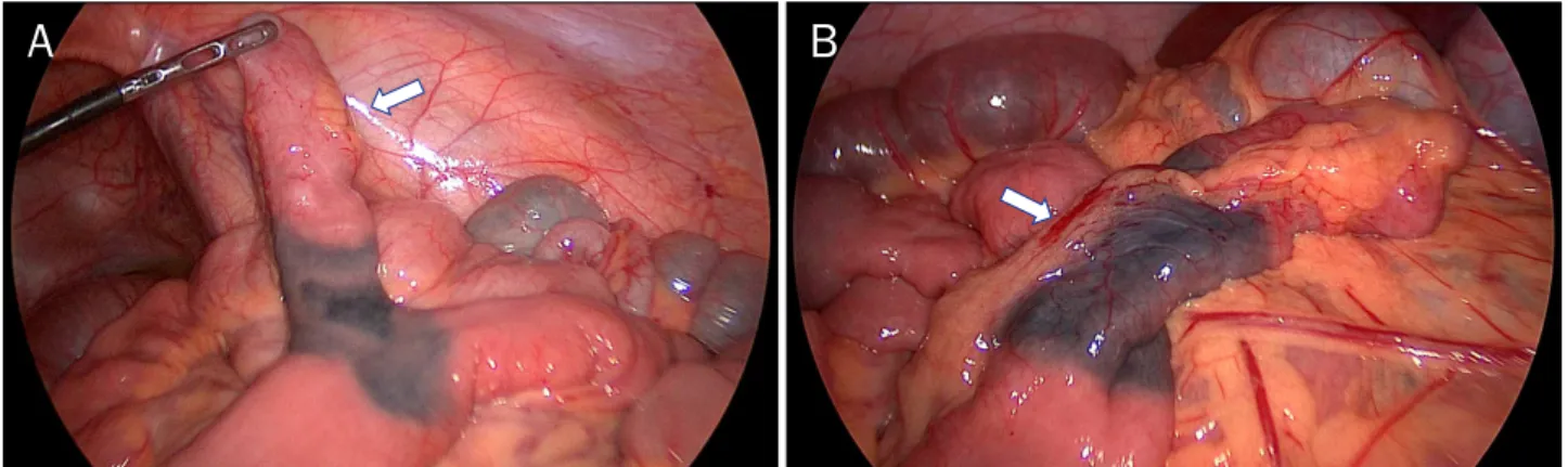
경검사에서 회맹판으로부터 약 130 cm 근위부 회장 부위에 서 메켈게실을 확인할 수 있었고, 게실의 끝부분에 도달하였 을 때에 궤양에 의한 출혈 소견이 관찰되어 내시경 클립지혈 술을 시행하였으나(Figs. 3, 4), 재출혈의 위험성 때문에 복강 경 수술을 시행하였다. 복강경 수술 소견에서 회맹판에서 약 130 cm 근위부 회장에서 5 cm 크기의 게실이 관찰되어 복강 경하 게실 절제술을 시행하였고(Fig. 5), 환자는 수술 후 합병 증 없이 퇴원하였으며 증상 재발없이 외래 추적관찰 중이다.
진단: 소장내시경으로 진단된 메켈게실
메켈게실은 난황관이 완전히 막히지 않아 발생하는 위장관 의 가장 흔한 선천성 기형으로 1809년에 이를 자세히 기술한 독일의 해부학자 Johann Friedrich Meckel의 이름에서 유래 되었다.1 전 인구의 약 1-2%에서 발견되고 남자가 여자보다 2배 더 흔하며 대부분의 환자에서는 증상이 없지만 약 4%의 환자에서 증상 및 합병증이 발생되는 것으로 보고되고 있 다.2,3
메켈게실은 장막, 근층, 점막하 조직, 점막 등 장의 모든 층을 포함하고 그 자체가 혈액을 공급받는 진성 게실이다. 크
Jung Y and Ko BM. Meckel’s Diverticulum Diagnosed by Enteroscopy
61
Vol. 64 No. 1, July 2014
Fig. 5. Laparoscopic operative findings. (A) About 5 cm sized diverticulum which had been marked with tattoo (arrow) is visible. (B) A feeding artery (arrow) of the diverticulum is evident.
기는 다양하게 보고되고 있는데 평균 길이는 5 cm이고 주로 회맹부에서 상방 100 cm 이내의 장간막 부착부의 반대쪽에 위치하며 나이가 젊을수록 회맹부에 가깝게 위치한다. 상장간 막동맥에서 분지되는 우난황동맥(right vitelline arteries)이 주된 혈액공급원으로, 흔하지 않지만 회결장동맥에서 분지되 기도 한다.2,4 난황관을 이루는 세포는 다능성을 띠므로 게실 내에서 이소성 조직을 흔히 찾아볼 수 있어 조직학적으로 50-70%의 메켈게실은 소장의 점막으로만 덮여 있으나, 30-50%에서는 이소성 위점막 혹은 이소성 췌장조직이 발견 되고 드물게는 대장 점막이 발견되기도 한다.5
메켈게실 환자를 분석한 대규모 연구에서 장폐쇄, 장중첩 증, 염증 또는 게실염, 출혈, 천공 등의 증상 및 합병증이 나타 날 수 있고 주로 10세 이하의 소아 시기에 40% 이상이 발생 하며, 무통성의 출혈이 흔하여 혈변 및 흑색변으로 나타나고 출혈의 대부분 위산을 분비하는 게실 내의 이소성 위점막에 인접한 회장 점막의 소화성 궤양에 의해서 유발되는 것으로 알려져 있다.2,3 하지만 성인에서 흔한 합병증은 장폐쇄로 22-50%에서 발생된다고 보고되고 있다.6
99mTC-pertechnetate를 이용한 메켈 스캔은 위점막이 pertechnetate를 흡수하고 분비한다는 점을 이용하여 이소성 위점막을 가진 메켈게실을 진단하기 위한 비침습적 일차 진단 법으로 사용되고 있는데 소아의 경우는 80-90%의 진단율을 나타내지만 성인의 경우 위양성과 위음성이 높아 진단율이 46% 정도로 보고되고 있다.7 메켈게실은 일반적인 대장 바리 움 조영이나 혈관 조영술로 거의 진단이 되지 않는 것으로 알려져있고, 복부 전산화단층촬영에서도 메켈게실과 소장을 구분하는 것이 거의 불가능하기 때문에 진단적 가치가 적은 것으로 알려져 있으나 최근 해상도의 비약적인 발전으로 진단 이 이루어지는 경우가 종종 보고되고 있다.8 최근에는 메켈게 실의 진단에 있어서 캡슐내시경과 소장내시경의 중요성이 보
고되고 있는데, 특히 메켈게실이 강력히 의심되는 경우에는 이중풍선 소장내시경을 시행하는 것이 안전하고 효과적인 검 사법이라 보고하고 있다.9 증상이 나타난 메켈게실의 치료는 수술 절제이며 단순한 게실 절제나 소장절제술 후 단단문합을 시행할 수 있다.10
REFERENCES
1. Opitz JM, Schultka R, Göbbel L. Meckel on developmental pathology. Am J Med Genet A 2006;140:115-128.
2. Ymaguchi M, Takeuchi S, Awazu S. Meckel's diverticulum.
Investigation of 600 patients in Japanese literature. Am J Surg 1978;136:247-249.
3. Park JJ, Wolff BG, Tollefson MK, Walsh EE, Larson DR. Meckel di- verticulum: the Mayo Clinic experience with 1476 patients (1950-2002). Ann Surg 2005;241:529-533.
4. Rossi P, Gourtsoyiannis N, Bezzi M, et al. Meckel’s diverticulum:
imaging diagnosis. AJR Am J Roentgenol 1996;166:567-573.
5. Artigas V, Calabuig R, Badia F, Rius X, Allende L, Jover J. Meckel's diverticulum: value of ectopic tissue. Am J Surg 1986;151:631- 634.
6. Dumper J, Mackenzie S, Mitchell P, Sutherland F, Quan ML, Mew D. Complications of Meckel's diverticula in adults. Can J Surg 2006;49:353-357.
7. Schwartz MJ, Lewis JH. Meckel's diverticulum: pitfalls in scinti- graphic detection in the adult. Am J Gastroenterol 1984;79:
611-618.
8. Bennett GL, Birnbaum BA, Balthazar EJ. CT of Meckel's divertic- ulitis in 11 patients. AJR Am J Roentgenol 2004;182:625-629.
9. He Q, Zhang YL, Xiao B, Jiang B, Bai Y, Zhi FC. Double-balloon en- teroscopy for diagnosis of Meckel's diverticulum: comparison with operative findings and capsule endoscopy. Surgery 2013;
153:549-554.
10. Peoples JB, Lichtenberger EJ, Dunn MM. Incidental Meckel's di- verticulectomy in adults. Surgery 1995;118:649-652.
![외국인 PCR검사 가능 기관 [Referral Laboratories : 11sites]](data:image/gif;base64,R0lGODlhAQABAIAAAP///wAAACH5BAEAAAAALAAAAAABAAEAAAICRAEAOw==)