대한소화기학회지 2006;47:77-81
서 론
총담관의 양성종양은 대부분 유두종, 선종 등의 상피 종 양이고, 평활근종, 지방종, 카르시노이드, 혈관평활근종, 섬 유종 등의 비상피 종양은 매우 드물다.1,2 담관의 평활근종 은 평활근 세포 또는 고유근층 내의 전구체에서 기원하는 양성 종양으로, 현재까지 4예가 보고되었으며,3-6 국내에서 는 아직 보고된 바 없다. 담관의 평활근종은 임상 소견이나 방사선 소견이 악성 종양과 뚜렷한 감별점이 없고, 수술 전에 조직검사가 어려워 임상에서 주의가 필요한 질환이다.
저자들은 반복적인 간기능 이상과 담관염 증상을 보인 환 자에서, 수술로 진단한 총담관의 평활근종 1예를 경험하여
문헌고찰과 함께 보고한다.
증 례
39세 여자가 내원 하루 전부터 심해진 전신 무력감과 피로, 열감을 주소로 응급실로 내원하였다. 환자는 5년 전에 진행 위암으로 본원에서 광범위 아전 위절제술과 Billoth II 재건술, 항암화학치료를 받았다. 이후 외래 추 적 중, 내원 5개월 전에 혈액 검사에서 AST 698 IU/L, ALT 477 IU/L, 총 빌리루빈 1.3 mg/dL, 알칼리 포스파타 제 1,962 IU/L, γ-GT 538 IU/L로 간기능 이상이 발생하 였다가 저절로 호전되었다. 또한 내원 1개월 전에도 간 ꠏꠏꠏꠏꠏꠏꠏꠏꠏꠏꠏꠏꠏꠏꠏꠏꠏꠏꠏꠏꠏꠏꠏꠏꠏꠏꠏꠏꠏꠏꠏꠏꠏꠏ
Correspondence to: Jae-Woon Choi, M.D.
Department of General Surgery, Chungbuk National University College of Medicine and Medical Research Institute, 62 Gae- sin-dong, Heungdeok-gu, Cheongju 361-711, Korea
Tel: +82-43-269-6358, Fax: +82-43-273-3252 E-mail: [email protected]
총담관 평활근종 1예
충북대학교 의과대학 의학연구소 내과학교실, 외과학교실*, 병리학교실†
구자충․이미연
․전원중
․서정철
․채희복
․박선미
․윤세진
․김석형
†․최재운*
A Case of Leiomyoma in the Common Bile Duct
Ja Chung Goo, M.D., Mi Yeoun Yi, M.D., Won Joong Jeon, M.D., Jeong Chul Seo, M.D., Hee Bock Chae, M.D., Seon Mee Park, M.D., Sei Jin Youn, M.D.,
Seok Hyoung Kim, M.D.†, and Jae Woon Choi, M.D.*
Departments of Internal Medicine, General Surgery*, and Pathology†, Chungbuk National University College of Medicine and Medical Research Institute, Cheongju, Chungbuk, Korea
Leiomyomas, originating in the bile duct, are very rare, and only few cases have been reported in the literature.
We experienced a case of leiomyoma of the distal common bile duct, mimicking bile duct cancer. A 39-year-old woman presented with intermittent jaundice and general weakness for three months. Clinical profiles showed obst- ructive jaundice, and the abdominal computed tomography and cholangiography revealed diffuse bile duct dilata- tion with distal common bile duct stricture. A pylorus-preserving pancreaticoduodenectomy was performed and the pathologic specimen disclosed leiomyoma of the common bile duct accompanying severe fibrosis. This is the first case of leiomyoma in the bile duct reported in Korea. (Korean J Gastroenterol 2006;47:77-81)
ꠏꠏꠏꠏꠏꠏꠏꠏꠏꠏꠏꠏꠏꠏꠏꠏꠏꠏꠏꠏꠏꠏꠏꠏꠏꠏꠏꠏꠏꠏꠏꠏꠏꠏꠏꠏꠏꠏꠏꠏꠏꠏꠏꠏꠏꠏꠏꠏꠏꠏꠏꠏꠏꠏꠏꠏꠏꠏꠏꠏꠏꠏꠏꠏꠏꠏꠏꠏꠏꠏꠏꠏꠏꠏꠏꠏꠏꠏꠏꠏꠏꠏꠏꠏꠏꠏꠏꠏꠏꠏꠏꠏꠏꠏꠏꠏꠏꠏꠏꠏꠏꠏꠏꠏꠏꠏꠏꠏꠏꠏꠏꠏꠏ
Key Words: Leiomyoma; Common bile duct
ꠏꠏꠏꠏꠏꠏꠏꠏꠏꠏꠏꠏꠏꠏꠏꠏꠏꠏꠏꠏꠏꠏꠏꠏꠏꠏꠏꠏꠏꠏꠏꠏꠏꠏ 접수: 2005년 5월 4일, 승인: 2005년 10월 10일
연락처: 최재운, 361-711, 충북 청주시 흥덕구 개신동 62번지 충북대학교 의과대학 외과학교실
Tel: 043-269-6358, Fax: 043-273-3252 E-mail: [email protected]
* 이 논문은 2004년도 충북대학교 학술연구지원사업의 연구 비 지원에 의하여 연구되었음.
78 대한소화기학회지: 제47권 제1호, 2006
기능 이상이 생겼다가 호전되었으며, 당시 시행한 복부 초음파검사는 정상이었다. 간헐적으로 전신 피로감과 열 감 등이 발생하였고 그때마다 감기약 등의 약물을 복용 한 병력이 있어서, 독성 간염이 발생하였다가 호전된 것 으로 판단하고 외래 추적 관찰 중이었다. 내원 3일 전 또 다시 증상이 발생하여 감기약 등을 복용했으나 효과가 없었고, 내원 1일 전 증상이 더욱 심해져서 내원하였다.
가족력은 특이사항이 없었으며, 음주나 흡연은 하지 않 았고, 내원 5개월 전부터 간헐적으로 약국에서 구입한 감기약을 복용한 병력이 있었다. 내원 당시 혈압 130/90 mmHg, 맥박 90회/분, 호흡수 20회/분, 체온 36.5oC였다.
복부 신체검사에서 장음은 정상이었고, 압통, 반발통, 촉 지되는 종괴는 없었으며, 이전의 위암 수술 자국이 있었 다. 검사실 소견은 혈색소 9.0 g/dL, 헤마토크릿 27.6%로 저색소 저용적 빈혈이 있었고, 백혈구 19,100/mm3, 혈소
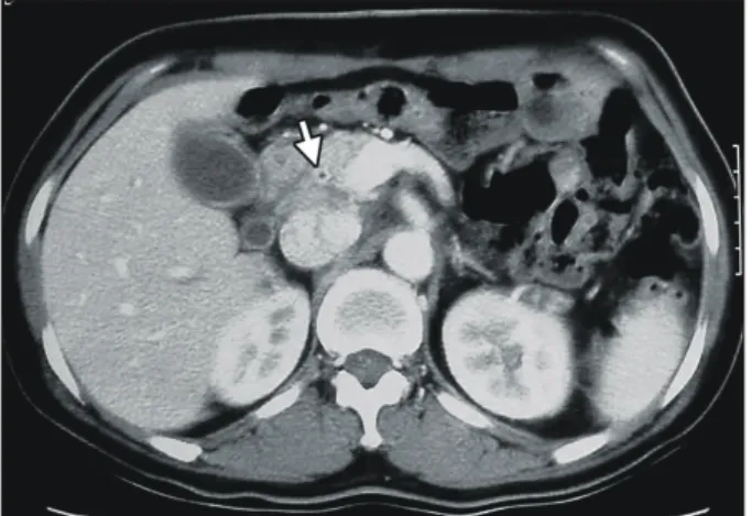
판 241,000/mm3, CRP 7.0 mg/L였다. 간기능 검사는 AST 709 IU/L, ALT 574 IU/L, 총 빌리루빈 2.3 mg/dL로 이전 의 검사에 비해 악화된 소견이었다(Fig. 1). 내원 2일째 체온이 38oC로 상승하고 상복부 통증을 호소하였으며 복 부 진찰에서 상복부 압통이 있었다. 급성 담관염으로 추 정하여 항생제를 정맥 주입하였고, 방사선 검사를 하였 다. 복부 전산화단층촬영에서 간내 담관과 총담관이 10 mm 정도로 확장되었고 원위부 담관에서 갑자기 좁아져 있었으며 담관벽이 조영 증강되었다(Fig. 2). 내시경역행 담관조영에서 유두부 모양은 정상이었고, 담관은 약 2.5 cm에 걸친 총담관 원위부의 협착이 있었으며, 내부에 종 괴나 담석은 관찰되지 않았다(Fig. 3). 담관암을 의심하여 솔 세포진 검사 후 내시경 담즙배액관을 삽입하였다. 솔 세포진 검사는 염증 소견 외 악성 세포는 관찰되지 않았 으며, CEA와 CA19-9은 모두 정상이었다. 담즙배액 이후 복부 통증과 담관염 증상은 모두 호전되었고, 내원 13일 째 담관암 의심하에 수술을 시행하였다. 수술 소견은 원 위부 담관 주위가 돌처럼 단단하였고 총담관은 1.5 cm 정도로 확장되어 있었다. 확장된 담관의 바로 아래에 1.5 cm 크기의 둥근 모양의 종괴가 발견되어 동결 절편 생검 후 양성 종양이라는 결과를 듣고, 유문부 보존 췌-십이지 장절제술을 시행하였다. 병리조직은 동결 절편 생검 조 직에 국한된 1.5×1.0 cm 크기의 평활근종이 관찰되었고, 수술 후 절제된 조직에서는 종양은 관찰되지 않았으며, 담관 벽 전체에 심한 섬유화를 동반한 염증 소견이 관찰
Fig. 2. Abdominal CT scan. It shows the contrast enhanced luminal wall with stenosis of the distal bile duct (arrow).
Fig. 1. The profile of liver function tests. It shows fluctuation of liver function tests for 3 months. ENBD, endoscopic nasobiliary drainage; ALP, alkaline phosphatase.
Fig. 3. ERCP finding. A cholangiography demonstrates a 2.5 cm length smooth narrowing of the distal common bile duct (arrows).
구자충 외 8인. 총담관 평활근종 1예 79
되었다(Fig. 4). 조직 소견에서 종괴는 끝이 무딘, 시가 모 양의 균일한 핵을 가진 평활근으로 구성되어 있으면서, 세포 분열, 괴사와 주위 조직으로의 침범 소견이 없었다.
면역조직화학검사에서 smooth muscle actin 양성, CD117
과 CD34에 음성이어서 평활근종으로 진단하였다(Fig. 5).
수술 이후 임상증상과 검사 소견 모두 호전되었고, 10개월 이 지난 현재 외래 추적 관찰 중에 있다.
Fig. 4. Resected specimen and frozen biopsy. (A) Gross speci- men shows distal bile duct ste- nosis with periductal fibrosis (ar- row) and frozen biopsy site (ar- row head). (B) Frozen specimen shows a 1.5×1.2 cm sized solid mass (H&E stain, ×1).
A
B
Fig. 5. Microscopic findings. (A) The tumor is consisted of spindle cells with blunt-ended, cigar shaped, uniform nuclei with absence of mitosis, necrosis and invasion (H&E stain, ×200). (B-D) Immunohistochemical study shows that the tumor cells are positive for smooth muscle actin (B, ×200) and negative for CD117 (C, ×400) and CD34 (D, ×400), indicating that the tumor cells are originated from smooth muscle.
A B
C D
80 The Korean Journal of Gastroenterology: Vol. 47, No. 1, 2006
고 찰
간외 총담관에서 발생하는 종양은 대부분 악성 종양이며, 양성 종양은 전체 종양의 6%, 수술한 종양의 0.1% 정도로 매우 드물게 발생한다.2 양성 종양의 대부분은 유두종, 선종 등의 상피 종양으로 약 90% 정도를 차지하고, 평활근종, 지 방종, 카르시노이드, 혈관평활근종, 섬유종 등의 비상피 종 양은 매우 드물다.2 20,000건의 담관 외과 수술 결과 양성 종양은 오직 4예(0.02%)뿐이었고,1 총담관의 양성종양 88예 를 분석한 보고에선 유두종과 선종이 80예였다.2
간외 담관의 평활근종은 현재까지 모두 4예가 보고되어 있으며, 국내에는 아직 보고된 적이 없는 매우 드문 종양이 다. 간외 담관에 평활근이 존재하느냐에 대해서는 아직까지 논란이 있다. 총담관의 병리 조직을 분석한 보고7에 의하면, 담관은 표피층, 섬유탄성(fibroelastic)층, 외막층으로 구성되 어 있고, 100예 중에서 12예에서만 섬유탄성층에서 콜라젠 이나 엘라스틴 섬유 등과 얽혀 있는 평활근이 발견되었다.
담관의 평활근은 담관의 근위부와 원위부에 고루 분포하며 나이와 무관하였다. 약 12%에서만 평활근이 발견되는 담관 의 구조 특징이,7 담관에서 평활근종이 드문 이유 중의 하나 이다.
이번 증례를 포함한 5예의 담관 평활근종의 임상 양상을 살펴보면, 포합형 빌리루빈혈증과 알칼리 포스파타제와 γ- GT가 상승하고 방사선 검사에서 담관 확장 소견이 관찰되
는 폐쇄 황달 소견을 보였다. 그 외 체중 감소, 소화불량, 발 열, 복부 통증 등의 증상은 일부에서 관찰되었다. 평활근종 의 발생 부위는 총간담관 부위에 생긴 1예5를 제외한 4예는 원위부 담관이었다. 평활근종의 크기는 0.4-4.0 cm 정도의 타원형으로 조직 소견은 일반적인 평활근종의 특징을 보였 다.8 복부 초음파, 복부 전산화단층촬영과 담관조영술 등의 방사선 소견은 담관 확장과 부분적인 담관 협착이 주요 소 견으로, 일반적인 유두부 주위 악성 종양과 감별하기가 어 려워서 수술 전에 평활근종으로 진단한 증례는 없었고 악성 담관암, 췌장두부암, Klatskin 종양 등으로 진단되었다. 보고 자들은 수술 도중에 세침 흡인술을 한다면 진단에 도움이 될 것으로 추천하였다.6 치료는 모두 외과 절제술을 시행하 였으며, 수술 후 재발하지 않는 양호한 예후를 보였다(Table 1).
이번 증례는 5년 전에 진행 위암으로 아전 위절제술을 시 행 받은 병력이 있어서 위암의 재발 가능성을 고려하였으나 진단 검사 후에는 담관암을 의심하였다. 그러나 수술 전에 시행한 솔 세포진 검사가 음성이어서 확진할 수가 없었다.
수술 후 조직 소견에서 1.5 cm 크기의 평활근종 이외에 심 한 섬유화를 보였다는 점은 다른 증례에서는 볼 수 없는 특 이 소견이었다. 이러한 섬유화는 평활근종으로 유발된 반복 적인 담즙 정체로 인해 장기간에 걸쳐 담관염의 악화와 호 전이 반복되면서 형성되었을 것으로 생각한다. 또한 평활근 종으로 유발된 담즙 정체는 간헐적으로 간기능 악화를 일으
Table 1. Five Cases of Leiomyoma in the Extrahepatic Bile Duct Author Sex
Age
Clinical symptoms
Symptom
duration Site Abdominal image Operation Tumor
size Archambault3 F
31
Nausea, vomiting, itching, jaundice
1 year, intermittent
dCBD Nonvisualization of the gallbladder
Excision &
choledochojejunostomy
2.0 cm
Kune4 M
49
Jaundice 3 months dCBD Pancreatico-
duodenectomy
4.0 cm
Mandeville5 F 55
Epigastric pain, nausea, vomiting
4 days CHD Dilated IHD Excision &
hepaticojejunostomy
3.0 cm
Yamaoka6 M
49
Jaundice dCBD Dilated IHD & CBD Pancreatico- duodenectomy
0.4 cm
Present case F 39
Jaundice, fatigue
5 months, intermittent
dCBD Dilated IHD & CBD Pancreatico- duodenectomy
1.5 cm
IHD, intrahepatic duct; dCBD, distal common bile duct; CHD, common hepatic duct.
Goo JC, et al. A Case of Leiomyoma in the Common Bile Duct 81
켰을 것이다. 이번 증례와 유사하게 간헐적인 황달 소견을 보인 경우가 1예3에서 보고되었으나, 나머지 증례들은 진행 황달 소견을 보여서, 간헐적인 간기능 이상을 양성 담관 종 양의 일반적인 특징으로 받아들이기는 어렵다.
총담관에 생긴 평활근종은 담관 확장과 담즙정체성 황달 소견을 보이므로 담관암과 감별 진단이 매우 어렵다. 그러 나 담관암과 달리 양성 질환으로 수술로 완치될 수 있는 질 환이므로 적극적인 진단과 치료가 필요하며, 특히 반복적인 간기능 이상을 보이며 담관 확장 소견이 동반될 경우엔 드 문 감별 질환의 하나로 고려해야 한다.
참고문헌
1. Marshall JM. Tumors of the extrahepatic bile ducts. Surg Gynecol Obstet 1932;54:6-12.
2. Burhans R, Myers RT. Benign neoplasms of the extrahepatic
biliary ducts. Am Surg 1971;37:161-166.
3. Archambault H. Archambault R. Leiomyoma of the common bile duct. AMA Arch Surg 1952;64:531-534.
4. Kune GA, Polgar V. Leiomyoma of the common bile duct causing obstructive jaundice. Med J Aust 1976;1:698-699.
5. Mandeville GA, Stawski WS. Obstructing leiomyoma of the common hepatic duct bifurcation simulating a Klatskin tumor.
Am Surg 1991;57:676-678.
6. Yamaoka K, Tozuka S, Ikeda T, et al. Leiomyoma of the common bile duct. Am J Gastroenterol 1993;88:469-470.
7. Mahour HG, Wakin KG, Soule EH, et al. Structure of the common bile duct in man: presence or absence of smooth muscle. Am Surg 1967;166:91-94.
8. Sohn TA, Lillemoe KD. Tumors of the gallbladder, bile ducts, and ampulla. In: Feldman M, Friedman LS, Sleisenger MH, eds. Sleisenger & Fordtran's gastointestinal and liver disease.
Volume 1. 7th ed. Philadelphia: Sanders, 2002;1153-1166.