대한발사선의학호|지 1998; 38: 1013-1019
소아의 바장 둔상시 경도관 동맥색전술의 효과1
박시균 · 김영주 · 권택상 · 김종진 · 고성민 · 성기준
목 적 : 복부 둔상후 소아의 비장 손상시 비수술적인 방법의 경도관 동맥색전술 (Tran
scathter arterial embolization ; 'l' AE)을 시행함으로써 그 효과와 장점에 대하여 알아보고 자한다.
대상 및 방법 : 복부 둔상후 시행한 복부 CT에 의하여 비장 출혈 및 비실질내 동맥 파열이 확진된 비장 손상 환아 총 9명을 대상으로 경도관 동액색전술을 시행하였다. 대상 환자 총 9
명중남자가 5명이고여자가 4명이였으며,환자의 연령 분포는 2-14세였고평균연령은 8.0 세였다.
경도관 동맥색전술은 수상 초기에 저혈압, 혈역학적인 불안정 (Hb/Hct 의 감소), 및 맥박 수의 증가등을 보일 때 수액 요법이나 수혈후에도 혈역학적으로 교정이 잘 되지 않는 환자 를 대상으로 하여 시행하였다. 색전술은 총 9예중 2예는 코일, 3예는 첼폼, 그리고 4예는 코 일과 젤폼을 각각 어용하여 혈관조영술상 조영제의 혈관외 유출이 관찰된 혈관에 직접 시행 하였으며, 색전술을 시행한 혈관의 위치는 총 9예중 비장 동맥의 주 체간이 7예이고, 비장 동맥의 분지가 2예였다.
경도관 동액색전술을 시행한 후 CT나 방사성 동위원소 주사법으로 추적 검사하여 비장 의 모양,혈종및 혈복강의 감소여부,그리고방사능섭취정도를분석하였다.
결 과 : 경도관 동맥색전술을 시행하기전에 Mirvis등이 발표한 복부 둔상시 비장의 손상 정도를 파악하여 단계별로 수술적 또는 비수술적 방법의 치료여부를 결정하는데 유용하다 는 CT grading system을 이용하여 분석한 결과 총 9예 중 각각 grade 1 과 2는 O예, grade 3
이 2예,그리고 grade 4가 7예 였다.
혈관조영술상 8예에서 조영제의 혈관외 유출이 관찰되었고, 1 예에서는 조영제의 전반적 인 삼출성 (oozing) 양상을 보였다. 경도관 동맥 색 전술을 시 행한 후에 조영제의 혈관외 유출 은총 9예 모두에서 관찰되지 않았다.
1 개월후 추적 검사상 전 예에서 혈종 빛 혈복강의 감소와 방사능 섭취의 증가를 보였으 며, 계속된 출혈이나 합병증없이 비장 기능이 소생되는 것을 알 수 있었으며, 6개월 이후의 추적 검사에도 재출혈은 없었다.
결 론 : 경도관 동맥색전술은 소아에서 비장절제술 후에 발생할 수 있는 패혈증 (sepsis) 이나 문맥 혈전증등의 합병증을 막고, 성공적인 조기 지혈과 비장 기능의 소생이 가능한 비 수술적인방법이다.
비장은 복부 둔상으로 인하여 손상받는 복부 실질 장기중 가 장많은약 25%를차지하며(1),특히 소아에서 비장손상에 의 해 비장적출술을 시행한 후에는 합병증으로 패혈증의 위험이 생기므로 (2-4) , 그 위험을 피하고 비장 소생을 위한 보존적 치 료가 많이 보고되고 있다 (5).
소아 비장 손상 환자에게 개복술대신 관찰과 침상 안정으로
1 연세대학교 원주의과대학 원주기독병원 진단방사선과학교실 이 논문은 1998년 2월 24 일 접수하여 1998년 4월 13일에 채택되였음
성공적으로 비수술적 치료를 한 보고가 있으며 (6), Sclafani 등 (7 -9) 은 경도관 동맥색전술 (Transcatheter arterial embol ization : 이하 TAE로 약함)이 비장 둔상 환자의 치료에 효과 척이라고 하였으나, 국내 및 국외의 문헌을 고찰하여 본 결과 소아에서 비장 둔상의 경우에 TAE를 시행한 보고는 전무한 실 정이다.
한편 복부 둔상의 평가 (evaluation) 에는 CT가 좋은 진단 방 법으로 알려져 있지만 혈관 손상에 대한 정보는 부정확하므로
- 1013
혈관조영술을 통한 세멸한 검사가 필요하다 (7, 8, 10 , 11).
본 연구의 목적은 소아에서 복부 둔상에 의한 비장 손상시 TAE를 시행함으로써 그 효과와 장점에 대하여 알아 보고자한 다.
대상및방법
1994년 8월부터 1998년 2월까지 복부 둔상에 의한 비장 손상 이 확진된 소아 9명을 대상으로 후향적 연구 (retrospecti ve study) 를 하였다. 총 9명의 환자중 비장 손상의 원인으로 6명
A
/-1셜
c
E
은 교통사고, 3명은 추락에 의한 손상으로 나타났다. 대상 환자 중 5명이 남자이고 4명이 여자였으며, 연령 분포는 2세에서 14
세였고 평균 연령은 8.0세였다.
전 예에서 초기에 CT를 시행하였고 활력 정후및 Hb/Hct 치를 평가하였고, 비장 손상의 CT grading system(Table 1) 은 Mirvis등의 분류법을 이용하였다(1 0). CT grading sys- tem은 비장의 피막하 혈종, 실질의 파열, 그리고 3개이상의 비 장 절편의 존재 유무에 의하여 평가한다. 총 9명의 환자중 grade 1과 2는 O예, grade 3이 2예, 그리고 grade 4가 7예였다.
혈관조영술은 전 예에서 standard seldinger technique을
fκ
j B
D
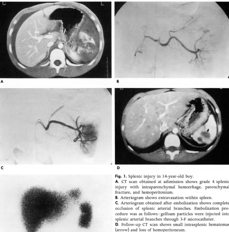
Fig. 1. Splenic injury in 14-year-old boy.
A. CT scan obtained at adimission shows grade 4 splenic injury with intraparer때ymal hemorrhage, parenchymal fracture, and hemoperitonium.
B. Arteriogram shows extravasation within spleen.
C. Arteriogram obtained after embolization shows complete occlusion of splenic arterial branches. Embolization pro- cedure was as follows : gelfoam particles were injected into splenic arterial branches through 3-F microcatheter.
D. Follow-up CT scan shows small intrasplenic hematomas (arrow) and loss of hemoperitoneum.
E. Follow-up 99mTc-sulfur colloid scintigram (anterior view) obtained shows preservation of splenic function
1014
대한방사선의학회지 1998; 38: 1013-1019
이용하였으며, 대퇴동맥을 통한 혈관조영은 5F Imager Tor- 사용하여 색전숨을 시행하였다.
que Cobra Catheter(Boston scientific, California, USA)를 혈관조영술상 조영제의 혈관외 유출 (extravasation) 은 각각 사용했고, 비장 동맥의 분지는 3F FasTRACKER(Target 상극 (upper pole), 중간극 (middle pole), 그리고 하부극(lower therapeutics / Boston scientific, California, USA) 를 사용하
여 초선택적 조영 을 하였다
Table 1. CT Grading System of Splenic Injury 조영제는 62% iopamidol(Ultravist 300: Korea Schering,Seoul, Korea) 을 사용하여 초당 5-20ml로 주업한 후 복부대 동맥 조영상과 복강동맥 조영상을 얻은 후 비장동맥 조영상을 초당 4-5ml로 iopamidol을 주사하여 얻었다. 영상은 Optim us DVI system(Philips, Einthoven, Netherland) 으로 DSA (6 frames / sec)를 얻었다.
Grade Findings on CT scan
색전 물질은 코일 (Target therapeutics complex helical fibered platinum coil- 18-2-20 or - 18-3- 30:Boston scientific, California, USA) 이나 젤폼 절편(lxlxlmrn) 을
A
c
Fig. 2. Splenic injury in 4-year-old girl
B
D
Capsular avulsion, superficial laceration(s), or subcapsular hematoma < 1 cm
2 parenchymal laceration(s) 1 - 3 cm deep, central or s있ubc떠apsu띠lla값r he잉matom베s야))3cm
3 Laceration(s) ) 3 cm deep, central/subcapsular hematoma(s) ) 3 cm
4 Fragmentation of three or more sections, devascularized spleen(nonenhancement)
A. CT scan ob바ta없ined a따t ad띠ml염ss잉ion shows grade 4 splenic lllJury with lacerations, parenchymal fragmentations, and massive hemoperitoneum.
B. Splenic arteriogram shows extravasation of contrast medium within spleen and fracture of mid-pole.
C. Arteriogram obtained after embolization shows occlusion of splenic artery. Embolization procedure was as follows First, gelfoam particles were injected into splenic arterial branches through 3-F microcatheter. Next, coil was placed in main trunk of splenic artery through 5-F standard catheter
D. Follow-up CT scan shows small intrasplenic hematoma and loss of hemoperitoneum
- 1015
pole) 으로 세분하여 관찰하였고, 색전 부위는 비장 동맥 분지부 의 초선택과 주 체간(또는 근위부)으로 나누어 시행되었다.
모든 환자는 TAE시행후 진찰, 활력 정후의 감시, 그리고 Hb/Hct치를 집중 치료실에서 평가하였다.
추적 관찰은 TAE를 시행한 후 1개월 빛 6개월 후에 CT에 의하여 혈종 및 혈복강의 감소 여부를 평가하였으며 99mTc_
sulfur colloid scintigraphy에 의하여 비 세망내피기능 (splen
ic reticuloendothelial function)을 방사능 섭취의 증가 유무 로평가하였다.
결 과
초기 CT상 전 예에서 혈복강이 동반되어 있었으며, 다른 복 강내 실질 장기의 동반 손상은 관찰되지 않았으며, Mirvis 등 (1 이 이 발표한 CT grading system상 grade 3이 2예 , grade 4 가 7예 였고, grade 1과 2는 없였다.
혈관조영술상 8예에서 조영제의 혈관외 유출 (extrava
sation) 이 관찰되었는데, 각각 -"J--극에서 4예, 중간극에서 5예, 그리고 하부극에서 3예가 관찰되었다. 그리고 나머지 1예에서 는전반적인 삼출성( oozing) 양상으로관찰되었다.
색전술에 이용한 색전물은 3예에서는 젤폼만을(Fig. 1), 2예
、
A
c
에서는 코일만을 각각 사용하였고 (Fig. 3), 4예에서는 젤폼과 코얼을 함께 사용하였으며 (Fig. 2), 색전술을 시행한 위치는 주 체간이 (Fig. 3) 7예였고, 주 체간 및 분지에 함께 시행한 경우 가 2예였q(Table 2).
추적 관찰에서 전 예에서 혈역학적으로 안정되었으며, 추적 CT소견상 혈종 및 혈복강의 감소 (Fig. 1)를 보였으며 99mTc_
sulfur colloid scintigraphy상 비 세망내피 기능이 소생 (Fig.
1) 된 것을 관찰할 수 있었으며, 색전술을 시행한 후에 l예에서 단기간의 혈소판 증가증이 있었으나 추적 관찰에서 1주일이내 에 해소되었고,나머지 8예에서는어떤 합병증도관찰되지 않았 다.
고 찰
비장은 항원이나 피막화된 세균 (encapsulated bacteria) 의 제거와 손상된 적혈구를 제거하는 여과 기능을 하며 면역학적 으로 손상된 혈소판과 과립구의 여과와 제거에도 관여하고,
opsonins과 tuftsin같은 색소와 지질 대사, 탐식작용 그리고 적 혈구의 성숙에 관계되는 특별한 항체의 생성에도 관여하며 순 환 혈소판의 약 30%를 저장하는 저장고(resorvoir) 로서도 작 용한다.
’
B
Fig. 3. Splenic injury in 12-year-old girl.
A. Splenic arteriogram shows multiple sites of disruption of splenic branch and extravasation(arrow)
B. Late-phase arteriogram shows wedge-shaped defect of spleen and persistent extravasation extending beyond the spleen(arrow).
c. Splenic arteriogram obtained after embolization with two coils shows complete occlusion of splenic artery.
- 1016 -
대한방사선의학회지 1998; 38: 1013-1019
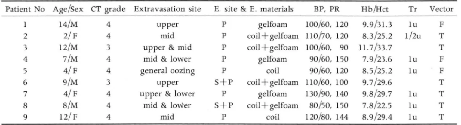
Table 2. Clinical Data and Results of Embolization
Vector F T T F F T T T T 1u 1/2u
Tr
1u 1u
1u 1u 1u Hb 떠ct 9.9/31.3 8.3/25.2 11.7/33.7 7.9/23.6 8.5/25.2 9.7/29.6 9.8/29.7 7.8/22.5 8.9/29.4 BP, PR
100/60, 120 110/70, 120 100/60, 90 90/60, 150 90/60, 120 110/60, 100 130/90, 140 80/50, 150 120/80, 144 gelfoam
coil
+
gelfoam coil+
gelfoamgelfoam coil coil
+
gelfoamgelfoam coil
+
gelfoamcoil E. site & E. materials
P P P P P
S 十 P
P S+P
P Extravasation site
upper mid upper & mid mid & lower general oozing
upper upper & lower
mid & lower mid CT grade
4 4 3 44 4 3 4 4 4 Age/Sex
14/M 2/F 12/M 7/M 4/F 9/M 4/F 8/M 12/ F Patient No
1i
1ι 「j
A*
ζJ
rb
7l
。。
QJ
No : number, E. : embolization, Tr: transfusion of pack celL T: traffic accident, F: falling down injury S : superselection of branch, P: proximal area of splenic artery
방법으로 비장 혈관내에 pitressin의 주입, 첼폼 절편 색전술,
그리고 코일 폐쇄를 보고하였다. Sclafani 등 (8) 은 혈역학적으 로 안정되고 혈관조영술상 조영제의 혈관외 유출이 관찰되었던 비둔상 환자에서 비수술적 방법인 코일 색전술에 의한 비장 소 생율이 84%라고 보고하였고 이것은 수술에 의한 비장 동맥 결 찰술과 유사한 결과라고 하였다.
Hagiwara 등(17) 은 혈역학적으로 안정되고 혈관조영술상 조영제의 혈관외 유출이 있고 동맥 파열이 확진된 경우에 TAE 를 시행하였는데 비장 소생율은 93% 였다.
본 연구에서는 CT scan상 비장 손상이 확진되고 혈역학적인 불안정 (hemodynamic instabili ty) 이 있으며 혈관조영술상 조 영제의 혈관외 유출이 관찰되었던 환아 총 9명 모두에서 TAE 를 시행하였다. 비장 손상에 대한 CT grading system은 Mirvis 등(10) 이 보고한 방법을 이용하였으며 총 9예 중 grade 3이 2예, grade 4가 7예였고 grade 1과 2는 없었다.
TAE를 시행한 후 전 예에서 조영제의 혈관외 유출이 관찰되 지 않았으며 빠르게 혈역학적인 안정성을 회복동}는 것을 관찰 할 수 있었다. 이러한 높은 성공률 (9/9, 100%) 은 보다 세밀한 혈관조영술 검사와 적절한 TAE의 이용으로 인한 것이라 생각 하며(1 7) , 비장적출술을 시행하는 것보다 좋은 결괴흘 얻을 수 있다고생각한다.
본 연구에서는 총 9예중 7예에서 비장 동맥의 주 체간에 성 공적인 색전술이 시행되었다 (Fig. 3). 이와같이 비장 통맥의 주 체간에 직접 색전술을 시행하는 것은 주변의 좌위동맥 (left gastric artery), 좌위대망막동맥 (left gastroepiploic artery), 그리고 후훼장동맥 (dorsal pancreatic artery) 등에 의한 풍부 한 측부 순환으로 인하여 비장 경색 (splenic infarction) 이 없 이 비장 기능이 보존되기 때문이다 (8, 11). 나머지 2예에서는 비장 동맥의 주 체간 및 분지에 미세도관으로 초선택하여 젤폼 절편이나 코일로 성공적인 색전술이 시행되였다.
CT scan이 나 99mTc-sulfur colloid scintigraphy에 의 한 추 적 검사상 전 예에서 초기 CT scan에서 보였던 혈종 및 혈복강 이 감소되었고 비 세망내피기능(splenic reticuloendothelial function) 이 잘 보존되였다. 추적 기간중에 l예에서만 단기간 의 혈소판 증가증이 있었으나 1주일내에 해소되였고, 나머지 8 1Ol7
비장에는 여러 가지 인대가 부착되어 있고 실질이 혈액 원소 (blood elements) 와 세망내피 세포와의 최대한의 계면 (maxi
mal interface)을 허용핸 최소한의 해연 양상으로 구성되어 있어서 둔상에 매우 민감하여 복부 둔상시 가장 흔히 손상받는 장기이다.
소아에서 복부둔상으로인한비장손상환자에게 비장적출술 을 시행한 경우에 다양한 합병증이 생길 수 있는데 그 중에 비 장적출후 패혈증(overwhelming postsplenectomy sepsis) 이 가장 심하고 위험한 합병증으로 알려져 있으며 이것은 피막화 된 세균에 의한 전격적 감염 (fulminant infection) 과 손상된 연역 체계 (impaired immune system) 로 인해 유발된다 (2 4) 그리고 혈소판 수의 증가로 인하여 문액, 장간막 정맥과 비 장 정맥의 혈전증이 생길 수 있으며 (4, 12). 적혈구의 변이성 (deformabi li t y )의 감소와 동반되어 전혈의 점도 (whole blood viscosi ty) 가 증가하여 허혈성 심질환을 유발 할 수 있다(13). 그 외에 흔한 재출혈이나 복강내 비증 (intraabdominal splen- osis) 도 (4, 14) 나타날 수 있다.
비둔상 환자의 비장 손상의 평가에는 CT가 좋은 진단 방법 으로 알려져 있으며 (7, 8, 10, 15, 18), 몇몇 연구자들은 개복술 을 필요로 하는 환자의 선택에 유용한 CT소견을 보고하기도 했다(1 0, 16). 그러나 조영증강 CT scan은 비장 손상의 유무 뿐만 아니라 손상의 형태, 위치 및 정도, 그리고 복강내 출혈 여 부도 알게 해 주지만 CT 소견만으로는 비장 손상에 대한 결과 를 예측하기 어렵고 혈역학적 인 평가에 유용한 충분한 혈관 분 포 상태 ( vasculari t y)를 제공해주지는 못해서 (7, 8, 10, 11, 18) 실제적으로는 개복 여부를 결정하는데 큰 도움을 주지는 못한 다.
비장 손상 환자에게 시행하는 보존적 치료의 이론적 근거는 비장적출후 발생될 수 있는 광벙위한 패혈증으로 인한 사망의 위험성을감소시키고자하는데 있다(7).
혈역학적으로 안정된 소아 비둔상의 경우에는 TAE의 이용 없이 보존적 인 비수술적 치료에 대한 많은 보고가 있었 3며 그 런 경우 성공률은 95-100% 였다 (6).
Sclaf ani ( 9) 가 처음으로 비장 손상으로 인한 출혈을 조절하 기 위 하여 경 도관 지 혈 ( tr ansca theter hemos tasis) 의 3가지
예에서는 합병증이 관찰되지 않았다.
그러므로 TAE가 비장 손상에 의한 비장의 계속된 출혈을 안 전하게 막을 수 있으며 비장 소생 (splenic salvage) 에 좋은 비 수술적 방법이라고 생각한다. 하지만 본 연구는 대상군이 9예로 서 소규모이며 추적 기간이 짧고, 추적 검사시 측부 순환 여부 의 확인을 위한 혈관조영술이 시행되지 못한 제한점은 있다.
결론적으로 소아에서 복부 둔상에 의한 비장 손상이 확진된 경우에 TAE를 시행함으로써 비장절제술후 발생할 수 있는 패 혈증, 비증, 재출혈, 그리고 혈전증과 같은 합병증도 없이 성공 적인 조기 지혈 및 혈역학적 불안정의 회복과 비장 기능의 소생 이 가능한 비수술적인 방법으로 생각된다.
"'~ 며 • --;l 프므 ;
1. Trunkey DD: The spleen. In Blaisdell FW, Trunkey DD (eds):
Abdominal Trauma. New York: Thieme-Stratton, 1982: 185-192 2. Green JB, Shackford SR, Sise MJ, et al. Late septic complica- tions in adults following splenectomy for trauma ‘ a prospecti ve analysis of 144 patients. J Trauma 1986; 26: 999-1004
3. Balfanz JR, Nesbit ME, JR., Jarvis C, et al. Overwhelming sep- sis following splenectomy for trauma. J Pediatr 1976; 88: 458-460
4. Ellison EC, Fabri PJ. Complications of splemectiomy: etiology, prevention and management. Surg Clin North Am 1983; 63 1313-1330
5. Ein SH, Shandling B, Simpson JS, et al. Nonoperative manage- ment of traumatized spleen in children: How and Why. J Pediatr Surg 1978; 8: 117-119
6. Pearl RH, Wesson DE, Spence LJ, et al. Splenic injury: a 5-year update; improved results and changing criteria for conservative management. J Pediatr Surg 1987; 743: 310-313
7. Sclafani SJA, Weisberg A, Scalea TM, Phillips TF, Duncan AO‘
Blunt splenic injuries: nonsurgical treatment with CT, arteriography, and transcatheter arterial embolization of splenic artery‘ Radiolo
‘
gy 1991; 181: 189-1968. Sclafani SJA, Shaftan GW, Scalea TM, Patterson LA, Kohl L, et al. Nonoperative salvage of CT-diagnosed splenic injuries:
utilization of angiography for triage and embolization for hemostasis. J Trauma 1995; 39: 818-827
9. Sclafani SJA: The use of angiographic hemostasis in salvage of the injured spleen. Radiology 1981; 141 : 645
10. Mirvis SE, Whitley NO, Gens DR. blunt splenic trauma in adults : CT -based classification and correlation with prognosis and treatment. Radiology 1989; 171: 33-39
11. Yoshioka H, Huroda C, Hori S, et al. Splenic embolization for hypersplenism using steel coils. AJR 1985; 144’ 1269
12. Petit Philippe, Bret PM, Atri M, Hreno Andrew, et al. Splenic vein thrombosis after splenectomy ‘ frequency and role of imaging. Radiology 1994; 190: 65-68
13. Robertson DAF, Simpson FG, Losowsky MS. Blood viscosity after splenectomy. Br Med J 1981 ; 283 ’ 573-575
14. Pearson HA, Johnston D, Smith KA, Touloukian RJ. Return of splenic function after splenectomy for trauma. N Engl J Med 1978; 298: 1389-1392
15. Jeffrey RB, Laing FC, Federle MP, Goodman PC. Computed tomography of splenic trauma. Radiology 1981; 141 : 729-732 16. Ulmas SL, Cronan JJ. Splenic trauma: can CT grading systems
enable prediction of successful nonsurgical treatment? Radi- ology 1991; 178 ‘ 481-487
17. Hagiwara A, Yukioka T, Ohta S, Nitatori T, Matsuda H, Shimazaki S. Nonsurgical management of patients with blunt splenic injury: efficacy of transcatheter arterial embolization AJR 1996;167:159-166
18. Taylor GA, Fallat ME, Potter BM, Eichelberger MR. The role of computed tomography in blunt abdominal trauma in children.
J Trauma 1988; 28: 1660-1664
1018 -
대한밤시선의학회지 1998;38: 1013-1019
J Korean Radiol Soc 1998; 38: 1013-1019
The Efficacy of Transcatheter Arterial Embolization (TAE) in Children with Blunt Splenic Injuryl
Si Kyun Park, M.D., Young Ju Kim, M.D., Taek Sang Kwon, M.D.
Jong Jin Kim, M.D., Sung Min Ko, M.D., Ki Joon Sung, M.D.
1 Department of Radiology, Yonsei University Wonju College of Medicine
Purpose: To evaluate the efficacy of transcatheter arterial embolization (TAE) in children with blunt splenic injury.
Materials and Methods: The results of transcatheter splenic arterial embolization in nine children who suffered splenic injury after blunt abdominal trauma were retrospectively studied. This injury was demonstrated by CT, and the findings were evaluated according to the classification of Mirvis et al.; two patients were grade 3 and seven were grade 4. All were carefully observed in intensive care before embolization.
TAE was performed if a patient satisfied the following criteria: (1) transfusion and/or fluid re- placement required to maintain hemodynamic stability; or (2) rapid Hb/Hct decrease; or (3) both.
Splenic function was subsequently estimated according to the results of 99rnTc-sulfur colloid scintigraphy and/or CT scanning
Results:TAE was suscessful in all nine children. Two were embolized with a coil only, three with gelfoam, and four with gelfoam and a coil. Seven were embolized in the main trunk of the splenic artery and others in both the main trunk and its branches. Splenic function was preserved in all nine children, during follow-up, none suffered rebleeding.
Conclusion : TAE of the splenic artery can be a safe and effective nonsurgical approach to the management of blunt splenic injury in children, and can preserve splenic function.
Index words: Arteries, therapeutic blockade Spleen, angiography
Spleen, trauma Children, injuries
Address reprint requests to: Young Ju Kim, M.D., Depaπment of Radiology, Yonsei University Wonju College of Medicine,
U 162, Ilsan-Dong, Wonju, Kangwon-Do, 220-701, Korea.
Tel. 82-371-741-1474 Fax. 82-371-732-8281
- 1019
」 1998년도 제 54 자 학술대회 사전등록 신정서
@ 연락처 @
성 명:
Jι ...L. ~.
- , •
^
ικ...
회원구분:정회원口 전공의口 비회원口
@ 학술대회 등록 쌍
전 화:
우편번호:
평생회원口 년 차口 전공과목:
1998년도 제 54 차 학술대회에 사전등록을 하시겠습니까?
예 口 아니오口
예口를 선택하신 분은 아래에 해당하는 금액을 온라인 구좌로 업금하십시오 . . 사전등록 : 전문의 70,000원, 전공의 30,000원, 비회원 70,000원(’98.9.15. 마감)
· 현장등록 : 전문의 80,000원, 전공의 40,000원, 비회원 80,000원(’98. 9. 15. 이후)
@ 만 65세 이상 원로회원은 등록비가 변제되오나 신청서는 보내셔야 등록이 됩니다.
밟 평생회원이 아닌 분은 년회비 30,000원을 추가로 납업하여야 합니다.
@ 범주별 연수과정 (Categorical Course) 등록 @
1998년도 제 54 차 학술대회 범주별 연수과정 (Categorical Course) 에 사전등록을 하시겠습니까?
1.
위장관의 방사선 진단 및 중재적 시술 예 口 아니오 口 ll. 간담도계의방사선진단및중재적시술 예口 아니오口예口를 선택하선 분은 과목당 교재비 (5,000원씩)를 학술대회 등록비와 함께 온라인 구좌로 입금하십시오.
· 사전등록 : 전문의 5,000원, 전공의 5,000원, 비회원 5,000원(’98.9.15. 마감) . 현장등록 : 전문의 7,000원, 전공의 7,000원, 비회원 7,000원(’98. 9.15. 이후)
쌍 온라인 송금 @
해당금액을 아래 구좌로 송금하신 후 학회 Home Page에 사전등록 내용을 업력하시거나 본신청서를우편또는 Fax로송부하여 주시기 바랍니다.
온라인 번호:평화은행, 구좌번호: 025-25-0005-373, 예금주: 대한방사선의학회
Home Page : http://radiol.medikorea.net E-mail: [email protected]
학회 주소 : 서울시 서초구 양재동 121 옹 (!) 137 -130 Tel (82-2) 578-8003 Fax (82-2) 529-7113
솜금자성명:
~그 。 1.
~ t::l ë등~ •
- 1020 -