․교신저자 : 나창수, 동신대학교 한의과대학 경혈학교실, Tel.
061-330-3522, E-mail: [email protected]
․본 연구는 한국한의학연구원의 2007년 침구경락연구거점 기반구 축 사업 기초연구의 위탁연구에 의하여 수행되었음.
․투고 : 2007/09/02 심사 : 2007/09/02 채택 : 2007/09/10
直刺, 迎隨 및 捻轉手技法에 따라 시행한 經渠·復溜 鍼刺가 中大腦動脈 閉塞에 의하여 誘發된 局所 腦虛血 白鼠 hippocampus의 抗細胞自滅死 및 神經保護에 미치는 影響
윤대환1⋅변정윤1⋅최찬헌1⋅백진웅1⋅정지연2⋅정연진2⋅나창수1
1동신대학교 한의과대학,2전남대학교 치의학전문대학원, 치의학연구소
Effects of acupuncture at the LU8·KI7 on Anti-apoptotic cell death and neuroprotection in Rat hippocampus following focal brain ischemic injury
induced by Intraluminal Filament insertion in Rats
Dae-Hwan Youn
1, Jeng-Yun Byun
1, Chan-Hun Choi
1, Jin-Ung Baek
1, Ji-Yeon Jeong
2, Yeon-Jin Jung
2, Chang-Su Na
11
College of Oriental Medicine, Dongshin University
2
Dental Science Research Institute, School of Dentistry Chonnam National University Abstract
Objectives : Aims of this study is to investigate the effects of LU8·KI7 in rat induced by experimental focal ischemia.
Materials and methods : The focal ischemia was induced by intraluminal filament insertion into middle cerebral artery. The groups divided into 6groups, control; no therapy group after ischemia-induced, AT1;
acupuncture therapy group at LU8·KI7 after ischemia-induced, AT2; acupuncture therapy at LU8·KI7 bilaterally and the needle was twirled and rotated forward with the thumb of the right hand 9times, AT3;
acupuncture therapy at LU8·KI7 bilaterally and the needle was twirled and rotated forward with the forefinger of the right hand 9times, AT4; acupuncture therapy at LU8·KI7 bilaterally and the needle was inserted to the direction following the flowing route of the meridian(digital direction), AT5; acupuncture therapy at LU8·KI7 bilaterally, the needle was inserted to the direction following the flowing route of the meridian(digital direction) and the needle was twirled and rotated forward with the thumb of the right hand 9times. Acupuncture therapy was carried out 7times during 2weeks after focal ischemia-induced. The anti-apoptotic and neuroprotective effects of acupuncture are observed by Bax, Bcl-2, mGluR5, cytochrome c, Cresyl violet and ChAT-stain.
Results : The intensity of Bax was decreased in AC1, AC4, AC5 group, was increased in AC2, AC3 group. The intensity of Bcl-2 was increased in AC2, AC3, AC4, AC5 group. The intensity of mGluR5 was decreased in AC1 group, was increased in AC4, AC5 group. The intensity of Cytochrome c was increased in AC1, AC2 group, was decreased in AC4, AC5 group. The density of neurons stained by Cresyl violet was increased in all group without control group. The density of ChAT was increased in AC2, AC5 group.
Conclusions : Our study suggests that AC5 group show anti-apoptotic and neuroprotective effects on cholinergic neuron in focal cerebral ischemia of the stroke in rats
Key words : Acupuncture, Apoptosis, mGluR5, Cresyl violet, ChAT
Ⅰ. 緖 論
뇌혈관의 병변으로 인한 중추신경계의 손상
이 유발되어 진행되는 것을 효과적으로 조기 에 방어할 수 있는 치료법이 밝혀진다면 현재 다발되는 중풍환자에 큰 도움이 될 것이다.
중풍을 연구하기 위한 모델로는 전뇌 허 혈을 유발하는 방법1-3)과 국소 뇌허혈을 유 발하는 방법4)이 다용되고 있으며, 본 연구 에서 활용한 국소 뇌허혈 유발법은 총경동 맥에서 내경동맥을 통하여 이어지는 중뇌동 맥을intraluminal filament를 삽입하여 그 혈 류를 차단하여 hippocampus 부분의 신경세 포를 손상시키는 것으로, 前腦에만 국한하기 때문에 後腦에서의 혈류가 영향을 받지 않 아 호흡과 체순환에 영향을 주지 않음으로 써 뇌허혈 중 白鼠의 사망을 최소화 할 수 있는 장점이 있다. 중대뇌동맥의 혈류 차단 으로 인한 hippocampus 부분의 신경세포 손 상은 5~7일이 경과되면 apoptosis와 비슷한 세포 손상으로 인지 및 학습장애가 일어나 게 된다5,6). 그 기전에 대해서는 아직 명확 히 밝혀지지 않았으나, 허혈 후 주어진 자극 에 대한 예민성 증가로 인한 신경세포의 과 활동, 칼슘을 매개로 하는 글루탐산 흥분독 성, 미토콘드리아 손상과 단백질 합성의 장 애, 유리산소기에 의한 손상, 에너지 대사와 뇌의 혈액순환 이상 등이 관여하는 것으로 여겨지고 있다7). Hippocampus는 medial septal nucleus에 위치한 세포체로부터 투사 되는 콜린성 신경전달경로로8) 학습수행 및 기억력에 관여하는데, hippocampus의 손상 은 방사형 미로와 같은 공간 기억과제를 학 습하는 능력에 장애를 보이게 된다9,10).
한의학에서 中風은 卒然昏倒, 不省人事, 口眼喎斜, 半身不隨, 言語障碍 등을 주 증상 으로, 僵仆, 大闕, 薄厥, 偏枯, 偏風, 身偏不
用, 痺風 등의 病症으로 인식되어 왔으며11), 老年에 빈발하는 뇌허혈로 인한 뇌경색은 虛證에 해당되는데, 이 경우 鍼治療法 중 五 行鍼法에서는 足少陰 腎經을 補하는 穴位들 이 응용되고 있다12).
침자치료를 중풍 모델에 적용한 연구로는 장13)등은 太衝(LR3)에 대한 艾葉 藥鍼이 白 鼠의 일과성 전뇌 허혈손상에 대하여 예방 및 치료효과를, 김14)등은 對金飮子 약침이 알코 올 독성 흰쥐의 해마에서 신경세포생성과 NOS 발현에 미치는 영향을, 한15) 등은 혈해 에 대한 당귀약침이 허혈성 국소뇌손상에 미 치는 영향을, 윤16)은 太衝과 光明에 대한 자 침이 국소 뇌허혈손상에 대한 신경보호효과 를, 임17)등은 다종의 태충침자요법이 해마손 상에 미치는 영향을, 박18)등은 태충에 대한 영수 및 염전보사수기법이 국소 뇌허헐손상에 미치는 영향 등을 보고하였으나, 뇌허혈 모델 을 腎虛로 보아 五行鍼法 중 ‘虛則補其母’의 穴位인 經渠·復溜에 대한 鍼刺 및 捻轉手技에 관한 연구보고는 아직 접하지 못하였다.
이에 본 연구에서는 中大腦動脈 閉塞에 의 하여 실험적으로 유발된 국소 뇌허혈 백서에 經渠·復溜 혈위에 直刺, 迎隨 및 捻轉 補瀉 手技法을 시행하여 hippocampus 부위의 Bax, Bcl-2, mGluR5, Cytochrome c, cresyl violet 염색법에 의한 신경세포 손상방어 효과 및 ChAT 발현 등抗細胞自滅死 및 神經保護 효 과를 관찰한 바 다음과 같은 지견을 얻었다.
Ⅱ. 材料 및 方法
1. 동물
실험동물은 삼육동물센터로부터 구입한 260~300g, 8주령의 수컷 Sprague-Dawley계 白鼠로, 일주일간 실험실 환경(온도 22±3℃, 습도 50±10%)에 적응시킨 후 사용하였다.
각 cage당 3~4마리씩 넣었으며, 물과 사료 (pellet, Samyang, Korea)를 자유로이 섭취 하도록 하였다.
2. 군 분리
실험군들은 좌측중대뇌동맥을 폐색시켜 뇌허혈 誘發 후 3일째부터 대조군(Control, n=7)은 침자를 시행하지 않는 군으로, 經渠
·復溜 直刺群(AC1, n=7)은 經渠·復溜에 鍼
刺時 直刺한 군으로, 經渠·復溜 拇指向前 捻 轉 手技群(AC2, n=7)은 經渠·復溜에 鍼刺 한 후 拇指向前(시계방향)으로 9회 捻轉 手 技한 군으로, 經渠·復溜 食指向前 捻轉 手技 群(AC3, n=7)은 經渠·復溜에 鍼刺한 후 食 指向前(반시계방향)으로 9회 捻轉 手技한 군으로, 經渠·復溜 迎隨 補法 手技群(AC4, n=7)은 經渠·復溜에 經絡의 흐름에 循하는 손가락 및 발가락 방향으로 鍼刺한 군으로, 經渠·復溜 迎隨 補法․指向前 捻轉 手技群 (AC5, n=7)은 經渠·復溜에 經絡의 흐름에 循하는 손가락 및 발가락 방향으로 鍼刺한 후 拇指向前(시계방향)으로 9회 捻轉 手技 한 군 등으로 분리하였다(Table 1).
군분류 내용 뇌허혈 유발 유발 후 처치 내용
대조군 (Control) 좌측중대뇌동맥을 폐색시켜
뇌허혈을 유발시킴 유발후 3일째부터 14일간 침자를 시행하지 않음 經渠·復溜 直刺群
(Acu-1) 좌측중대뇌동맥을 폐색시켜
뇌허혈을 유발시킴 유발후 3일째부터 14일간 2일에 1회씩 經渠·復溜에 直 刺로 鍼刺함
經渠·復溜 拇指向前
捻轉 手技群 (Acu-2) 좌측중대뇌동맥을 폐색시켜
뇌허혈을 유발시킴 유발후 3일째부터 14일간 2일에 1회씩 經渠·復溜에 鍼 刺한 후 拇指向前(시계방향)으로 9회 捻轉 手技함 經渠·復溜 食指向前
捻轉 手技群 (Acu-3) 좌측중대뇌동맥을 폐색시켜
뇌허혈을 유발시킴 유발후 3일째부터 14일간 2일에 1회씩 經渠·復溜에 鍼 刺한 후 食指向前(반시계방향)으로 9회 捻轉 手技함 經渠·復溜 迎隨 補法
手技群 (Acu-4) 좌측중대뇌동맥을 폐색시켜
뇌허혈을 유발시킴 유발후 3일째부터 14일간 2일에 1회씩 經渠·復溜에 經 絡의 흐름에 循하는 손가락 및 발가락 방향으로 鍼刺함 經渠·復溜 迎隨
補法․指向前 捻轉 手技群 (Acu-4)
좌측중대뇌동맥을 폐색시켜 뇌허혈을 유발시킴
유발후 3일째부터 14일간 2일에 1회씩 經渠·復溜에 經 絡의 흐름에 循하는 손가락 및 발가락 방향으로 鍼刺 한 후 拇指向前(시계방향)으로 9회 捻轉 手技함 Table 1. Distribution of Groups
3. 취혈 및 침자
經渠(LU8)는 Radiocarpal joint의 Carpal groove위의 Radius의 Styloid process 아래에서, 復溜(KI7)는 Flexor hallucis longus muscle과 Calcaneal tendon 사이에서 취하였으며, 모두
인체에 상응하는 부위로 취하였다. 각각 1회씩 白鼠의 양측의 穴位에 자침하였으며, 鍼은 毫鍼 (No.3-0.5, haenglim, korea)을 이용하였다.
정상군과 대조군을 제외한 각 군들은 Ischemia 誘發 3일 후부터 각 군들은 1回/2 日(오전 10시) 치료를 시작하여, 14일간 총
7회에 걸쳐 30초 동안 留鍼 및 시계방향 및 반시계방향으로 9회의 捻轉手技를 시행하였 으며, 15일째에 白鼠를 희생시켜 腦를 신속 히 적출하거나 관류 고정하였다. 鍼刺 深度 는 피하를 통과하여 捻轉 및 迎隨 手技法 시행이 가능할 수 있는 정도의 깊이로 하였 다. 침자 手技法은 전 실험기간 동안 숙련된 전문가 1인이 계속하여 시술하였다.
4. Occlusion에 의한 허혈성 국소 뇌손 상 유발
局所 腦虛血은 Longa 등4)의 방법에 따라 좌측중대뇌동맥을 폐색시켜 만들었다. 白鼠 를 80% O2와 20% CO2가 혼합된 가스에 5% isoflurane (Choongwae, Korea)을 이용 하여 흡입마취 유도를 한 후 2% isoflurane 으로 계속 유지시켰다. 白鼠의 직장에 체온 측정 probe를 삽입하고 가온 등과 가온 메 트리스를 이용하여 마취유지 기간동안 체온 을 38℃로 유지하였다. 좌측 중대뇌동맥을 폐색하기 위하여 경부 정중선을 따라 피부 를 절개하고 흉골혀근과 흉골저작근 사이에 좌측 총경동맥을 노출한 후 좌측 내경동맥 의 원위부를 최대한 확보한 후 미세혈관 Clip (WPI, USA)을 이용하여 결찰하였고, 좌측 내경동맥과 intraluminal filament 사이 의 출혈을 방지하기 위하여 6-0 silk suture 실을 이용하여 좌측 총경동맥 근위부 및 좌 측 외경동맥 분지부를 결찰하였다. 좌측 총 경동맥의 좌측 외경동맥과 좌측내경동맥 분 지부에서 1㎝정도 되는 좌측 내경동맥의 원 위부에 미세혈관 가위(W.P.I., USA)를 사용 하여 구멍을 내었다. 미세혈관 클립을 제거한
후 좌측 내경동맥내로 20mm의 치과 인상제 (Durelon, Germany)가 발라진 intraluminal filament(직경 0.28mm rounded tip)를 좌측 내경동맥상의 구멍에 intraluminal filament 가 faint resistance의 느낌이 느껴질 때 까 지(약 17mm) 부드럽게 삽입하여 그 끝이 좌측 중대뇌동맥까지 도달하도록 하였으며, 이로써 좌측 중대뇌동맥부위에 국소 허혈을 유발하였다. 출혈을 방지하기 위하여 좌측 내 경동맥상의 intraluminal filament 삽입부위를 가볍게 묶은 후 절개된 피부를 봉합하였다.
국소뇌허혈 유발에 대한 검증은 실험 수행이 완료된 후 白鼠를 희생시켜 삽입된 intraluminal filament가 중대뇌동맥과 전대뇌동맥의 기시 부 사이에 위치된 것이 확인된 개체만을 실 험 대상으로 삼아 관찰을 진행하였다.
5. Total RNA 분리 및 Reverse Transcription Polymerase Chain Reaction (RT-PCR) 1) Total RNA 분리
Total RNA의 분리는 좌측 hippocampus 부위의 조직(3g)을 500㎕ TRIZOL reagent (Gibco-BRL, USA)로 균질화하고 균질액에 대해 1 ㎖의 chloroform(Sigma, USA)을 가 하여 15초 동안 흔들어 잘 혼합한 후, 실온 상태에서 15분 방치하고 난 다음 세포 유잔 물을 제거하기 위하여 4℃, 14,000rpm에서 15분 동안 원심분리하였다. 원심분리로 얻어 진 상층액과 1㎖의 isopropanol(sigma, USA) 을 첨가하여 실온상태에서 5분 동안 방치한 후 RNA pellet을 얻기 위하여 4℃, 14,000 rpm 에서 8분간 원심분리하고, 원심분리로 생긴
pellet에 냉장 보관된 70% ethanol과 함께 DEPC를 넣고 4℃, 7,500rpm에서 5분간 원 심분리 후 pellet만 남기고 모두 제거하고, 남은 ethanol은 실온에서 10분간 방치시켜 건조시킨 다음 DEPC-treated water에 녹여 spectrophotometer(Eppendorf, Germany)에서 OD260 값을 읽어 RNA의 순도 및 농도를 정 량하였다. 이들 뇌조직의 total RNA는 사용시 까지 -70℃에서 보관하였다.
2) RT-PCR
분리된 total RNA 2㎍과 2.5㎕ Oligo (dT), DEPC-treated water를 RT Premix (Bioneer, Korea)에 넣어 Mastercycler gradient (Eppendorf, Germany)를 이용하여 50㎕ cDNA 를 합성하여 PCR 증폭을 위한 template로 사 용하였다. Reverse transcription temperature cycle은 42°C에서 1시간 동안 cDNA synthesis, 94℃에서 5분 동안 denature 그리고 4°C에 서 5분 동안 cooling시키는 단계를 거쳤다.
이때 housekeeping 유전자인 glyceraldehyde -3-phosphate dehydrogenase(GAPDH)(sense primer: 5'-TGCATCCTGCACCACCAACT- 3', antisense primer: 5'-CGCCTGCTTCAC CACCTTC-3')를 internal control로 사용하 였다. 白鼠의 특이 primer는 PCR-premix kit (Bioneer, Korea)를 사용하였다. Bax Polymerase chain reaction은 cDNA 2㎕, Bax sense primer 1㎕, Bax antisense primer 1㎕, DEPC -treated water를 PCR Premix(Bioneer, Korea) 에 넣는다. PCR temperature cycle은 cDNA 의 증폭을 위하여 95°C에서 300초 동안 pre- denaturation, 94°C에서 40초 동안 melting,
54°C에서 40초 동안 annealing, 72°C에서 90 초 동안 extension하는 과정을 30회 반복 수 행하고 마지막 cycle에서 72°C에서 600초 동 안 extension 단계를 거쳐 Bax 유전자증폭 을 Primer(sense primer: 5'-GTTTCAAGG ATCGAGCAG-3', antisense primer: 5'-CA TCTTCTTCCAGATGGTGA-3')룰 이용하여 Mastercycler gradient(Eppendorf, Germany) 에서 시행하였다.
Bcl-2 Polymerase chain reaction은 cDNA 2㎕, Bcl-2 sense primer 1㎕, Bcl-2 antisense primer 1㎕, DEPC-treated water를 PCR Premix(Bioneer, Korea)에 넣는다. PCR temperature cycle은 cDNA의 증폭을 위하여 95°C에서 300초 동안 pre- denaturation, 94°C에서 40초 동안 melting, 55°C에서 40초 동안 annealing, 72°C에서 90초 동안 extension 하는 과정을 30회 반복 수행하고 마지막 cycle에서 72°C에서 600초 동안 extension 단 계를 거쳐 Bcl-2 유전자증폭을 Primer (sense primer: 5'-ACTTTGCACAGATGTCCAG T-3', antisense primer: 5'-CGGTTCAGGTACTC AGTCAT-3')룰 이용하여 Mastercycler gradient (Eppendorf, Germany)에서 시행하였다.
mGluR5 Polymerase chain reaction은 cDNA 2㎕, mGluR5 sense primer 1㎕, mGluR5 antisense primer 1㎕, DEPC- treated water를 PCR Premix(Bioneer, Korea) 에 넣는다. PCR temperature cycle은 cDNA 의 증폭을 위하여 95°C에서 300초 동안 pre-denaturation, 94°C에서 40초 동안 melting, 55°C에서 40초 동안 annealing, 72°C에서 90 초 동안 extension하는 과정을 30회 반복 수 행하고 마지막 cycle에서 72°C에서 600초 동
안 extension 단계를 거쳐 mGluR5 유전자증 폭을 Primer(sense primer: 5'-TCCAATCTGC TCCTCCTACC-3', antisense primer: 5'-CA ACGATGAAGAAC TCTGCG-3')룰 이용하여 Mastercycler gradient (Eppendorf, Germany) 에서 시행하였다.
이렇게 증폭된 DNA를 ethidium bromide (EtBr, 10㎎/㎖)를 포함한 1.5% agarose gel 상에서 0.5x TBE buffer (80mM Tris-HCL, 80mM boric acid, 2mM EDTA, pH8.3)로 50 V에서 전기 영동시킨 후 transmitter scanning videodensitometer (Bioneer, Korea) 에 넣고 감광시킨다. Intensity는 image master VDS(1D ver.2.1, Pharmacia biotech, USA) 로 정량하였다.
6. Western blotting 1) Protein preparation
적출된 뇌의 좌측 hippocampus를 신속히 액체 질소에 급속 냉동시키고 분석할 때까 지 -70℃에서 보관하여, Cytochrome C 단백 발현을 Western blot방식을 이용하여 관찰 했다. 白鼠의 hippocampus에 NP40 lysis buffer (pH8.0 50mM Tris HCl 150mM NaCl, 5mM EDTA, 0.2mM PMSF (Phenylmethyl-sulfonylfluoride), 1% NP-40, 1mM Benzamidine, 1㎍/㎖ Trypsin inhibitor) 와 1x Protease Cocktail inhibitor (Roche) 혼 합액 500 ㎕를 넣어 homogenization한다. 이 sample을 20분간 ice상태에 놓아둔 후 12,000rpm에서 20분간(4℃상태) 원심분리한 후 상층액을 분리한다. 이 sample은 bicinchonic
acid assay kit(Bio Rad, CA)를 사용해 정 량한다. 96 well plate에 BCA 용액(A:B=
20:1) 100㎕을 넣고 protein 5㎕를 넣은 후 37℃에서 20분간 방치시킨 후, 570㎚에서 ELISA reader (Biorad, USA)를 사용해 측 정하였다.
2) Electrophoresis
12% polyacrylamide resolving gel은 30분 간 굳히고, 4% polyacrylamide staking gel은 resolving gel 위에 붓고 comb을 꽂아 30분 간 굳힌다. 각 sample은 4x SDS sample buffer(pH6.8 100mM Tris, 4% SDS, 20%
glycerol, 0.2% bromophenol blue, 200mM DTT, 10% 2-mercaptoethanol) 5㎕에 정량 된 단백질 sample을 넣어 100℃에서 5분간 끓인다. Gel은 Gel running tank에 장착하고 pH8.3의 running buffer(0.025M Tris, 0.192M glycine, 10% SDS)를 붓는다. 80V (200㎃)로 전기 영동한다.
3) Electrotransfer
전기영동이 끝난 gel은 transfer buffer (25mM Tris, 192mM glycine, 20%
methanol)에 filter paper, gel, nitrocellulose membrane 순서로 얹고 20mA에서 20시간 transfer한다. Membrane은 5% nonfat milk 와 TBST (0.1% Tween20 in pH7.4 Tris- based saline buffer)에 1시간 동안 blocking하 고 TBST로 3회 washing한다. Cytochrome C antibody(BD, USA)를 TBST(1:2000 dilution) 에 희석시켜 4℃에서 overnight한다. TBST
로 4회 washing후 horseradish peroxidase- labeled anti-mouse IgG(Cell signalling, USA) 를 TBST(1:2000 dilution)에 희석시켜 1시 간 배양하고 TBST로 4회 washing한다.
Membrane은 TBST를 제거한 후 enhanced chemiluminescence plus(Amersham, UK)를 넣은 후 3분 동안 반응시킨다. 발현된 Cytochrome C의 band는 ECL film(Amersham, UK)을 develper(vivid, 1:5)와 Fixer(vivid, 1:5)에서 현상하였다. 발현된 Cytochrome C 의 intensity는 image master VDS(1D ver.2.1, Pharmacia biotech, USA)로 정량하였다.
7. Immunohistochemistry 1) Cresyl violet 염색
모든 실험이 끝난 직후 白鼠를 pentobarbital sodium (100mg/kg, i.p. Hanlim, Korea)로 마취시킨 후, 0.9% saline 200ml에 이어 phosphate buffer로 준비한 4% formalin 용 액(fixative) 800ml로 심장을 통해 관류하였 다. 처음 고정액 200ml는 2분간 빠른 유속으 로, 그리고 나머지 800ml는 25분간에 걸쳐 천천히 관류하였다. 고정이 끝난 쥐는 뇌를 꺼내 같은 고정액으로 2시간 경과 후 고정시 키고, 20% sucrose가 함유된 phosphate buffered saline(PBS)에 넣어 4℃에서 하루 동안 보관하였다. 다음날 뇌를 급속 냉동한 후 뇌 조직을 hippocampus 부위를 30μm의 두께로 잘랐다. PBS로 조직을 몇 차례 씻고 xylene(5min), 100% alcohol(2min), 95%
alcohol(1min), 70% alcohol(1min), D.W.
(2min)순으로 옮겨 담아 탈지, 탈수를 시킨
다음 cresyl violet buffer(5min)로 염색을 하였 다. 염색이 끝난 조직은 광학현미경(Olympus, Japan)을 사용하여 40배로 확대하여 관찰하였 고, 신경세포의 밀도를 Scion image program (Scion Corp. MD, USA)을 이용하여 측정 하였다.
2) Choline acetyltransferase(ChAT)
뇌 조직을 PBS에 3회 정도 세척한 후 ChAT 유전자 발현 연구에 가장 널리 사용 되고 있는 primary sheep ChAT anti-body (1:1,500, polyclonal, Cambridge Research Bio chemicals, USA)를 사용하였다. 1차 항 체는 PBS에 0.3% Triton X-100을 첨가한 PBST에서 2% 토끼 혈청과 0.1% sodium acid(Sigma, USA)로 2000배 희석하여 준비 하였다. 뇌 조직은 1차 항혈청에 4℃에서 72 시간 동안 지속적으로 흔들어 주면서 배양 하였다. 그 후 3번 이상 조직을 PBST로 씻 은 다음 2시간 동안 실온에서 2% 토끼 혈 청을 함유하는 PBST에서 200배 희석한 biotinylated anti-sheep serum (Vector Laboratories, USA)에 반응시켰다. PBST로 3번 씻은 다음, 뇌 조직은 실온에서 2시간 동안 Vectastain Elite ABC reagent(Vector Laboratories, USA)에 위치시켰다. PBS로 몇 번 헹군 다음 조직을 nickel chloride로 강 화시키고 착색제로서 diaminobenzidine(DAB) 을 사용하여 발현시켰다. 통제군 조직에는 1 차 항체를 생략하거나 nonimmuno sheep serum으로 대체하였는데, 이 경우 특정 표 지가 나타나지 않았다.
모든 처리를 거친 뇌 조직을 gelatine-
coated slide에 고정하고 공기를 제거하면서 커 버글라스를 덮은 후 광학현미경으로 40배로 확 대하여 hippocampus에서 ChAT-immunoreactive 신경 세포의 밀도를 Scion image program (Scion, USA)을 이용하여 측정하였다.
8. 통계처리
모든 측정값은 Excel statistic program (Microsoft, USA)을 이용하여 평균치와 표준오 차(mean±standard error)로 표시하였고, 각 실험 군 간의 통계학적 분석은 Window용 SPSS (SPSS, USA)를 사용하여 비모수적 방법으로 Mann-Whitney U test를 시행하였다. 각 실험군 은 대조군에 비하여 α=0.05 수준(P<0.05)과 α
=0.01 수준(P<0.01)에서 유의성을 검정하였다.
Ⅲ. 實驗 成績 1. Bax 발현 변화
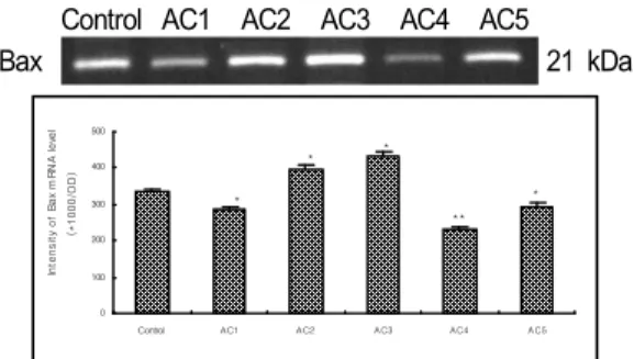
뇌허혈을 유발시킨 백서에게 大經渠·復溜 혈위에 수기법에 따라 침자를 시행하여 hippocampal CA1 부위의 Bax 발현정도를 관 찰한 결과, control군은 335.5±7.11(×1000 OD), AC1군은 80.9±3.88(×1000 OD), AC2군은 399.1±8.93(×1000 OD), AC3군은 435.7±9.13 (×1000 OD), AC4군은 230.2±11.24(×1000 OD), AC5군은 294.8±9.55(×1000 OD)로, control 군에 비해 AC1군(P<0.05), AC4군(P<0.01) 과 AC5군(P<0.05)은 유의하게 감소하였으 나, AC2군(P<0.05)과 AC3군(P<0.01)은 유 의하게 증가하였다(Fig. 1).
Control AC1 AC2 AC3 AC4 AC5
Bax 21 kDa
0 100 200 300 400 500
Control AC1 AC2 AC3 AC4 AC5
*
*
**
*
*
Intensity of Bax mRNA level (*1000/OD)
Fig. 1. Effect of acupuncture by needle manipulation at the LU8·KI7 on the intensity of Bax mRNA in the hippocampal CA1. Results are shown as mean±S.E.. control; no therapy group after ischemia- induced. AT1; acupuncture therapy group at LU8·KI7 after ischemia-induced. AT2; acupuncture therapy at LU8·KI7 bilaterally and the needle was twirled and rotated forward with the thumb of the right hand 9times. AT3; acupuncture therapy at LU8·KI7 bilaterally and the needle was twirled and rotated forward with the forefinger of the right hand 9times. AT4;
acupuncture therapy at LU8·KI7 bilaterally and the needle was inserted to the direction following the flowing route of the meridian. AT5; acupuncture therapy at LU8·KI7 bilaterally, the needle was inserted to the direction following the flowing route of the meridian and the needle was twirled and rotated forward with the thumb of the right hand 9times. *, P<0.05, **, P<0.01 as compared with the control group.
2. Bcl-2 발현 변화
뇌허혈을 유발시킨 백서에게 大經渠·復溜 혈위에 수기법에 따라 침자를 시행하여 hippocampal CA1 부위의 Bcl-2 발현정도를 관 찰한 결과, control군은 340.3±7.35(×1000 OD), AC1군은 400.4±44.03(×1000 OD), AC2군은 488.4±38.55(×1000 OD), AC3군은 571.9±76.91 (×1000 OD), AC4군은 660.8±11.71(×1000 OD), AC5군은 582.5±26.37(×1000 OD)로, control군 에 비해 AC2군(P<0.05), AC3군 (P<0.05), AC4군(P<0.01)과 AC5군(P<0.01)은 유의하 게 증가하였다(Fig. 2).
Control AC1 AC2 AC3 AC4 AC5
Bcl-2 28 kDa
0.0 200.0 400.0 600.0 800.0 1000.0
Control AC1 AC2 AC3 AC4 AC5
*
* **
**
Intensity of Bcl-2 mRNA level (*1000/OD)
Fig. 2. Effect of acupuncture by needle manipulation at the LU8·KI7 on the intensity of Bcl-2 mRNA in the hippocampal CA1. Results are shown as mean±S.E.. control ;no therapy group after ischemia- induced. AT1; acupuncture therapy group at LU8·KI7 after ischemia-induced. AT2; acupuncture therapy at LU8·KI7 bilaterally and the needle was twirled and rotated forward with the thumb of the right hand 9times. AT3; acupuncture therapy at LU8·KI7 bilaterally and the needle was twirled and rotated forward with the forefinger of the right hand 9times. AT4;
acupuncture therapy at LU8·KI7 bilaterally and the needle was inserted to the direction following the flowing route of the meridian. AT5; acupuncture therapy at LU8·KI7 bilaterally, the needle was inserted to the direction following the flowing route of the meridian and the needle was twirled and rotated forward with the thumb of the right hand 9times. *, P<0.05, **, P<0.01 as compared with the control group.
3. mGluR5 발현 변화
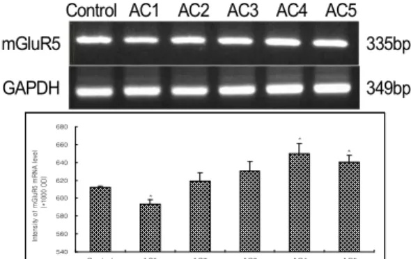
뇌허혈을 유발시킨 백서에게 大經渠·復溜 혈위에 수기법에 따라 침자를 시행하여 hippocampal CA1 부위의 mGluR5 발현정도 를 관찰한 결과, control군은 612.3±1.45 (×1000 OD), AC1군은 593.2±5.06(×1000 OD), AC2 군은 619.2±9.07(×1000 OD), AC3군은 630.4±10.86 (×1000 OD), AC4군은 649.7±11.71(×1000 OD), AC5군은 640.7±7.45 (×1000 OD)로, control군에 비해 AC1군은 유의하게 감소하였고, AC4군과 AC5군은 유의하게 증가하였다(P<0.05)(Fig. 3).
Control AC1 AC2 AC3 AC4 AC5
mGluR5 335bp
GAPDH 349bp
540 560 580 600 620 640 660 680
Control AC1 AC2 AC3 AC4 AC5
Intensity of mGluR5 mRNA level (*1000 OD)
*
*
*
Fig. 3. Effect of acupuncture by needle manipulation at the LU8·KI7 on the intensity of mGluR5 mRNA in the hippocampal CA1. Results are shown as mean±S.E.. control;no therapy group after ischemia- induced. AT1; acupuncture therapy group at LU8·KI7 after ischemia-induced. AT2; acupuncture therapy at LU8·KI7 bilaterally and the needle was twirled and rotated forward with the thumb of the right hand 9times. AT3; acupuncture therapy at LU8·KI7 bilaterally and the needle was twirled and rotated forward with the forefinger of the right hand 9times. AT4;
acupuncture therapy at LU8·KI7 bilaterally and the needle was inserted to the direction following the flowing route of the meridian. AT5; acupuncture therapy at LU8·KI7 bilaterally, the needle was inserted to the direction following the flowing route of the meridian and the needle was twirled and rotated forward with the thumb of the right hand 9times. *, P<0.05, as compared with the control group.
4. Cytochrome c 발현 변화
뇌허혈을 유발시킨 백서에게 大經渠·復溜 혈위에 수기법에 따라 침자를 시행하여 hippocampal CA1 부위의 Cytochrome c 발현 정도를 관찰한 결과, control군은 128.8±2.91 (×1000 OD), AC1군은 158.1±7.50(×1000 OD), AC2군은 149.9±7.75(×1000 OD), AC3군은 122.8±5.38(×1000 OD), AC4군은 114.0 ±5.05 (×1000 OD), AC5군은 111.6±6.44 (×1000 OD) 로, control군에 비해 AC1군과 AC2군은 유 의하게 증가하였고, AC4군과 AC5군은 유의 하게 감소하였다(P<0.05)(Fig. 4).
Control AC1 AC2 AC3 AC4 AC5
Cytochrome c
14
kDa
0 20 40 60 80 100 120 140 160 180
Control AC1 AC2 AC3 AC4 AC5
Intensity of Cytochrome c level(×1000 OD)
* *
* *
Fig. 4. Effect of acupuncture by needle manipulation at the LU8·KI7 on the intensity of Cytochrome c in the hippocampal CA1. Results are shown as mean±S.E.. control ;no therapy group after ischemia- induced. AT1; acupuncture therapy group at LU8·KI7 after ischemia-induced. AT2; acupuncture therapy at LU8·KI7 bilaterally and the needle was twirled and rotated forward with the thumb of the right hand 9times. AT3; acupuncture therapy at LU8·KI7 bilaterally and the needle was twirled and rotated forward with the forefinger of the right hand 9times. AT4; acupuncture therapy at LU8·KI7 bilaterally and the needle was inserted to the direction following the flowing route of the meridian. AT5; acupuncture therapy at LU8·KI7 bilaterally, the needle was inserted to the direction following the flowing route of the meridian and the needle was twirled and rotated forward with the thumb of the right hand 9times. *, P<0.05, as compared with the control group.
5. Cresyl violet을 이용한 신경세포 손상 방어효과
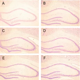
뇌허혈을 유발시킨 백서에게 大經渠·復溜 혈위에 수기법에 따라 침자를 시행하여 hippocampal CA1 부위를 cresyl violet 염색 법으로 염색하여 신경세포 손상방어 효과를 관찰한 결과, control은 14.4±0.76(density), AC1 군은 21.4±1.76(density), AC2군은 23.6±2.93 (density), AC3군은 24.6±1.89(density), AC4 군은 24.2±1.04(density), AC5군은 23.0±0.58 (density)로, control군에 비해 모든 실험군에서 유 의하게 증가된 밀도를 보였다(P<0.05)(Fig. 5, 6).
0 5 10 15 20 25 30
Control AC1 AC2 AC3 AC4 AC5
Density
*
* *
*
*
Fig. 5. Effect of acupuncture by needle manipulation at the LU8·KI7 on the density of cresyl violet- stained neural cell sections in the hippocampal CA1.
Results are shown as mean±S.E.. control;no therapy group after ischemia- induced. AT1; acupuncture therapy group at LU8·KI7 after ischemia-induced. AT2;
acupuncture therapy at LU8·KI7 bilaterally and the needle was twirled and rotated forward with the thumb of the right hand 9times. AT3; acupuncture therapy at LU8·KI7 bilaterally and the needle was twirled and rotated forward with the forefinger of the right hand 9times. AT4; acupuncture therapy at LU8·KI7 bilaterally and the needle was inserted to the direction following the flowing route of the meridian. AT5; acupuncture therapy at LU8·KI7 bilaterally, the needle was inserted to the direction following the flowing route of the meridian and the needle was twirled and rotated forward with the thumb of the right hand 9times. *, P<0.05, as compared with the control group.
A B
C D
E F
Fig. 6 Representive microphotographs of coronal
sections in the hippocampal CA1. A;control group,
B;AC1 group, C;AC2 group, D;AC3 group, E;AC4
group, F;AT5 group. Cresyl violet-stain. ×40.
6. Choline acetyltransferase(ChAT)발현
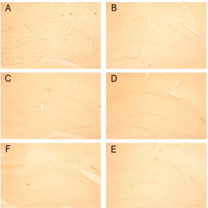
뇌허혈을 유발시킨 백서에게 大經渠·復溜 혈위 에 수기법에 따라 침자를 시행하여 hippocampal CA1 부위의 ChAT 발현정도를 관찰한 결 과, control은 11.6±1.04(density), AC1군은 12.8±1.80(density), AC2군은 18.8±1.53(density), AC3군은 13.8±0.76(density), AC4군은 16.2±2.75 (density), AC5군은 15.8±0.50(density)로, control군에 비해 AC2군과 AC5군에서 유의하 게 증가된 밀도를 보였다(P<0.05)(Fig. 7, 8).
0 5 10 15 20 25
Control AC1 AC2 AC3 AC4 AC5
Density
*
*
Fig. 7. Effect of acupuncture by needle manipulation at the LU8·KI7 on the density of Choline acetyltransferase (ChAT)-stained sections in the hippocampal CA1. Results are shown as mean±S.E..
control;no therapy group after ischemia-induced. AT1;
acupuncture therapy group at LU8·KI7 after ischemia induced. AT2; acupuncture therapy at LU8·KI7 bilaterally and the needle was twirled and rotated forward with the thumb of the right hand 9times. AT3; acupuncture therapy at LU8·KI7 bilaterally and the needle was twirled and rotated forward with the forefinger of the right hand 9times. AT4;acupuncture therapy at LU8·KI7 bilaterally and the needle was inserted to the direction following the flowing route of the meridian. AT5;
acupuncture therapy at LU8·KI7 bilaterally, the needle was inserted to the direction following the flowing route of the meridian and the needle was twirled and rotated forward with the thumb of the right hand 9times. *, P
<0.05, as compared with the control group.
A B
C D
F E
Fig. 8. Representive microphotographs of coronal sections in the hippocampal CA1. A;control group, B;AC1 group, C;AC2 group, D;AC3 group, E;AC4 group, F;AT5 group. ChAT-stain. ×40.
Ⅳ. 考 察
中風은 《內經》에 風이 인체를 손상시키 면 혹 熱中風하고, 혹 寒中風하고, 혹 癘風 하고, 혹 半身不遂한다고 하였으며, 歷代 醫 家中 河間은 心火暴甚하고 腎水虛衰하여 制 止不能하면 陰虛陽實의 熱氣怫鬱로 心神昏 冒, 筋骨不用하며, 卒倒無所知하게 된다고 하였다19).
中風에 대한 다양한 鍼 應用法중에는 四 肢肘膝以下에 있는 井滎輸經合의 다섯 特定 穴인 五輸穴을 應用응용하여 難經의 “瀉南 補北”과 “虛則補其母 實則瀉其子”설의 기본 원리에 따라 장부 허실을 조정하는 五行鍼 法이 임상에서는 多用된다20,21). 五行鍼法에 서는 그 辨證에 따라 補瀉 穴位를 선정하여 치료를 시행하고 있는데, 中風의 證狀中에 言語蹇澁, 半身不遂, 全身關節 虎咬 등은 腎
虛로 變證하여 腎을 補할 수 있는 穴位들이 응용되고 있다22).
본 연구에서 응용된 혈위는 經渠와 復溜 로, 腎虛로 인한 中風證狀에는 《難經․六 十九難》23)의 “虛則補其母”에 해당하는 金 에 해당하는 手太陰肺經의 金性穴이 經渠와 本經인 足少陰腎經의 金性穴인 復溜를 응용 하였다.
五行鍼法 응용함에 있어 장부의 허실을 조절하기 위해서는 혈위 자극방법인 補瀉法 의 개념이 중요시 되고 있는데24,25), 補瀉法 은 虛할 때 自經의 母穴과 母經의 母穴을 補하고, 實할 때는 自經의 自性穴과 自經의 自性穴을 瀉하여 병사를 정상적인 생리상태 로 회복시키는 방법으로26,27), 이에는 鍼向, 手技法, 刺入順序 및 方法등에 따라 여러 가 지 종류가 있지만 흔히 迎隨補瀉, 捻轉補瀉, 九六補瀉가 임상에서 함께 응용되고 있다.
본 연구에서는 五行鍼法의 穴位 활용에
대하여 실험적으로 탐색하기 위하여
intraluminal filament 삽입술로 중대뇌동맥 을 폐색시켜 뇌허혈을 유발한 모델에 대하 여4), 中風 疾患중 腎虛證에 응용되는 五行 鍼法을 혈위인 經渠·復溜의 鍼刺 및 迎隨捻 轉補瀉手技法을 시행한 후 hippocampus의 항세포자멸사 및 뇌신경세포의 손상보호효 과를 관찰하였다. 세포고사기전에 작용하는 인자들 중 본 연구에서 관찰한 지표는 Bax, Bcl-2, mGluR5, Cytochrome c를 선택하였 다.
Bax는 세포자멸사를 촉진하는 인자이고 Bcl-2는 세포자멸사를 억제하는 인자이다28). Bcl-2의 세포자멸사 억제 효과는 Bax의 영
향을 받는다고 알려져 있다29). 본 연구에서 Bax의 발현을 관찰해 본 결과 control군에 비해 control군에 비해 經渠·復溜 直刺群인 AC1군, 經渠·復溜 迎隨 補法 手技群인 AC4 군과 經渠·復溜 迎隨 補法․指向前 捻轉 手 技群인 AC5군이 유의하게 감소하였으나, 經 渠·復溜 拇指向前 捻轉 手技群인 AC2군과 經渠·復溜 食指向前 捻轉 手技群인 AC3군 이 유의하게 증가하였다. 이를 통해 Bax 발 현에 유의한 감소를 나타낸 經渠·復溜 直刺 群인 AC1군, 經渠·復溜 迎隨 補法 手技群인 AC4군과 經渠·復溜 迎隨 補法․指向前 捻 轉 手技群인 AC5군이 뇌세포사멸을 완화하 는 작용에 유의성이 있는 것으로 사료되며, 유의하게 증가한 經渠·復溜 拇指向前 捻轉 手技群인 AC2군과 經渠·復溜 食指向前 捻 轉 手技群인 AC3군은 다른 인자들과의 비 교 검토가 필요하다고 생각된다. Bcl-2의 발 현을 관찰한 결과 control군에 비해 經渠·復 溜 直刺群인 AC1군을 제외한 모든 군에서 유의하게 증가하였다. 이는 直刺후 留鍼의 자극보다는 적극적인 迎隨補瀉手技가 세포 사멸인자를 감소시키고 세포생존인자를 촉 진하는 쪽으로 작용하여 뇌손상을 감소시키 는 것을 의미한다고 볼 수 있다.
뇌허혈로 인한 신경세포의 손상에 흥분성 신경전달물질의 역할이 중요함이 잘 알려져 있으며 이러한 흥분성 신경전달물질은 허혈 기간 동안에 증가하기 시작하여 재관류 직 후에 상당한 증가하는데, 특히 glutamate의 역할은 흥분성 신경독성발현에 중요한 역할을 하고 있는데30) mGluR5(Metabotropic glutamate receptors)는 뇌에 있어서 주요한 신경독성
물질이며 허혈이나 저혈당 및 산소결핍 후 에 손상을 일으키게 하는 glutamate의 수용 체 아형 단위로, glutamate의 조정반응을 매 개하게 한다31). mGluR5는 신경세포 흥분효 과와 억제효과 등의 이중성이 연구 보고되 고 있는데, 현재 glutamatergic terminal에서 의 시냅스 전 억제작용을 함으로써 신경보 호 작용이 있다고 보고되고 있다32). 본 연구 에서 hippocampal CA1 부위의 mGluR5 발 현 변화를 관찰한 결과 control군에 비해 經 渠·復溜 直刺群인 AC1군이 유의하게 감소 하였고, 經渠·復溜 迎隨 補法 手技群인 AC4 군과 經渠·復溜 迎隨 補法․指向前 捻轉 手 技群인 AC5군이 유의하게 증가하였다. 이를 통해 迎隨 補法 및 拇指向前의 捻轉補法의 시행이 mGluR5의 활성을 통하여 뇌신경독 성작용을 완화하는 작용을 발휘하는 것으로 사료되며, 유의하게 감소한 經渠·復溜 直刺 群인 AC1군은 Bax발현의 감소와는 상반된 결과이나 Cytochrome c 발현과는 일치된 양 상을 보여 이에 대한 추가적인 연구가 요구 된다.
Cytochrome c는 세포자멸사를 유발하는 인자로 Bax와 Bcl-2와 밀접한 관계를 맺고 있으며 이33) 등의 실험에서 cytochrome c의 세포내 미세주입으로 세포자멸사가 매우 빠 르게 진행됨이 관찰되어져 세포자멸사를 관 찰하는데 주요한 지표가 된다. 본 연구에서 는, control군에 비해 經渠·復溜 直刺群인 AC1군과 經渠·復溜 拇指向前 捻轉 手技群 인 AC2군이 유의하게 증가하였고, 經渠·復 溜 迎隨 補法 手技群인 AC4군과 經渠·復溜 迎隨 補法․指向前 捻轉 手技群인 AC5군이
유의하게 감소하였다. 이를 통해 보면 迎隨 補法이 보다 효과적으로 抗細胞自滅死 작용 이 발휘된 것으로 볼 수 있는데, 이러한 결 과는 앞의 mGluR5의 발현 증가와 일관된 결과라고 볼 수 있다.
Hippocampal CA1 부위를 cresyl violet 염 색법에 의한 신경세포 손상방어 효과를 관 찰한 결과, control군에 비해 모든 실험군에 서 유의하게 증가된 밀도를 보였다. 이는 허 혈성 손상에 방어기전이 침자 및 영수염전 보사수기의 효과에 의하여 작동되어, 이를 회복하려고 하는 작용을 활성화 된 것으로 사료되며, 이는 항세포고사기전 등과는 다르 게 진행됨을 추정할 수 있었다.
Cholinergic system은 신경전달물질인 acetylcholine(Ach), Ach의 합성효소인 ChAT, Ach을 분해하는 분해효소인 AchE 및 Ach 전달물질의 수용체가 포함된다. 그 중 cholinergic synapse에 존재하는 AchE는 신경 전달 물질인 Ach를 choline과 acetic acid로 가 수분해 시키는데34),이 효소는 post-sysnaptic membrane의 수용체에 결합하는 Ach를 가 수분해시켜 수용체의 정상적인 기능을 유지 시키며, Ach의 생합성에 필요한 choline을 공급함으로써, 신경계가 원활하게 작용하는 데 매우 중요한 역할을 하게 된다. 특히 hippocampus는 기억장애에 관계되는 구심성 및 원심성 신경섬유가 가장 많이 연결되는 부위이다. Hippocampus와 신피질로 투사하 는 기저전 뇌콜린계 신경단위의 상실 즉, medial septum, diagonal band, nucleus basalis of Meynert에 있는 choline계 신경단 위의 상실로 인해 투사 부위인 대뇌피질 및
hippocampus 부위에서 pre-synaptic choline 계 지표가 극도로 감소하게 되며35), basal forebrain cholinergic system내의 ChAT활성 도 감소는 Ach 감소를 초래하여 기억력 및 여러 학습능력 저하와 집중력의 이상을 유 발하게 된다36,37).
Hippocampal CA1 부위의 ChAT 발현정 도를 관찰한 결과, control군에 비해 AC2군 과 AC5군이 유의하게 증가된 밀도를 보였 다. 이는 뇌허혈에 대한 조절 작용 중 ChAT 발현에 經渠·復溜 拇指向前 捻轉 手 技群과 經渠·復溜 迎隨 補法․指向前 捻轉 手技群이 유효함을 보인 것으로 보아 모지 향전의 염전보법이 주요하게 작용함을 시 사한다고 사료된다.
이상에서 본 바와 같이 실험적으로 유발 된 국소 뇌허혈에 대하여 腎虛에 활용되는
‘虛則補其母’의 혈위인 經渠·復溜에 直刺, 迎 隨 및 捻轉補瀉手技法을 시행해 본 결과 迎 隨 補法과 拇指向前 捻轉補法을 함께 응용 한 침자법에서 항세포사멸사 및 신경세포 손상방어 효과가 발현됨을 관찰할 수 있었 으며, 迎隨 및 捻轉補瀉 手技法을 시행하지 않은 直刺로 침자한 경우에 mGluR5 발현의 감소와 Cytochrome c 발현의 증가를 보임에 도 불구하고 일정한 신경세포 손상방어의 효과를 보인 점 등에 대하여서는 향후 심도 있는 후속 연구가 필요하리라고 사료된다.
Ⅴ. 結 論
中大腦動脈 閉塞에 의하여 실험적으로 유 발된 국소 뇌허혈 백서에 經渠·復溜 혈위에
直刺, 迎隨 및 捻轉 補瀉 手技法을 시행하여 hippocampus 부위의 Bax, Bcl-2, mGluR5, Cytochrome c, cresyl violet 염색법에 의한 신경세포 손상방어 효과 및 ChAT 발현 등 에 미치는 영향을 관찰한 바 다음과 같은 결론을 얻었다.
1. Hippocampal CA1 부위의 Bax 발현에서, control군에 비해 AC1군, AC4군과 AC5 군이 유의하게 감소하였으나, AC2군과 AC3군이 유의하게 증가하였다.
2. Hippocampal CA1 부위의 Bcl-2 발현에 서, control군에 비해 AC2군, AC3군, AC4군과 AC5군이 유의하게 증가하였다.
3. Hippocampal CA1 부위의 mGluR5 발현 에서, control군에 비해 AC1군이 유의하 게 감소하였고, AC4군과 AC5군이 유의 하게 증가하였다.
4. Hippocampal CA1 부위의 Cytochrome c 발현에서, control군에 비해 AC1군과 AC2군이 유의하게 증가하였고, AC4군과 AC5군이 유의하게 감소하였다.
5. Hippocampal CA1 부위를 cresyl violet 염색법에 의한 신경세포 손상방어 효과 에서, control군에 비해 모든 실험군에서 유의하게 증가된 밀도를 보였다.
6. Hippocampal CA1 부위의 ChAT 발현에 서, control군에 비해 AC2군과 AC5군이 유의하게 증가된 밀도를 보였다.
參考文獻
1. Pulsinelli WA, Brierley JB : A new method
of bilateral hemispheric ischemia in the unanesthetized rat. Stroke, 1979 ; 10(3) : 267-72.
2. Smith ML, Bendek G, Dahlgren N, Rosén I, Wieloch T, Siesjö BK. Models for studying long-term recovery following forebrain ischemia in the rat, 2. A 2-vessel occlusion model, Acta Neural Scand, 1984 ; 69(6) : 385-401.
3. Ogata J, Fujishima M, Tamaki K, Nakatomi Y, Omae T. An ultrastructural study of developing cerebral infarction following bilateral carotid artery occlusion in spontaneously hypertensive rats. Acta Neuropathol (Berl). 1977 ; 40(2) : 171-7.
4. Longa EZ, Weinstein PR, Carlson S, Cummins R. Reversible middle cerebral artery occlusion without craniectomy in rats. Stroke. 1989 ; 20(1) : 84-91.
5. Buchan A, Pulsinelli WA. Hypothermia but not the N-methyl-D-aspartate antagonist, MK-801, attenuates neuronal damage in gerbils subjected to transient global ischemia. J Neurosci. 1990 ; 10(1) : 311-6.
6. Gill R, Foster AC, Woodruff GN. MK -801 is neuroprotective in gerbils when administered during the post-ischaemic period. Neuroscience. 1988 ; 25(3) : 847-55.
7. Fujisawa A, Matsumoto M, Matsuyama T, Ueda H, Wanaka A, Yoneda S et al.
The effect of the calcium antagonist nimodipine on the gerbil model of
experimental cerebral ischemia. Stroke.
1986 ; 17(4) : 748-52.
8. Johnston MV, McKinney M, Coyle JT.
Evidence for a cholinergic projection to neocortex from neurons in basal forebrain. Proc Natl Acad Sci U S A.
1979 ; 76(10) : 5392-6.
9. Symons JP, Davis RE, Marriott JG.
Water-maze learning and effects of cholinergic drugs in mouse strains with high and low hippocampal pyramidal cell counts. Life Sci. 1988 ; 42(4) : 375-83.
10. Sutherland RJ, Rodriguez AJ. The role of the fornix/fimbria and some related subcortical structures in place learning and memory. Behav Brain Res. 1989 ; 32(3) : 265-77.
11. 강성길, 최용태. 중풍의 침구치료에 관한 연구. 황제의학. 1978 ; 3(4) : 1-15 12. 김진수. 오행침의 체질운용. 부천 : 전국
의학사. 2002 : 85-96.
13. 장진요, 김재효, 박성섭, 박귀종, 김경식, 손인철. 태충 애엽 약침이 일과성 전뇌 허혈 손상에 미치는 효과. 대한경락경혈 학회지. 2005 ; 22(3) : 63-81.
14. 김현중, 김이화, 이은용. 대금음자(對金飮 子) 약침이 알코올 독성 흰쥐의 해마에서 신경세포생성과 NOS 발현에 미치는 영향.
대한침구학회지. 2006 ; 23(5) : 187-98 15. 한상균, 이병렬. 당귀약침의 혈해 자입이
Intraluminal Filament 삽입술에 의해 유 발된 백서의 허혈성 뇌손상에 미치는 영 향. 대한침구학회지. 2004 ; 21(2) : 1-20.
16. 윤대환. Anti-apoptotic and neuroprotective effects of Acupuncture at LR3 and GB37 on focal brain ischemic injury induced by Intraluminal Filament insertion in Rats. 동신대학교 대학원.
동신대박사학위논문. 2006.
17. 임현진, 조명래, 윤대환, 나창수, 류충열.
다종(多種)의 태충(LR3) 침자요법(鍼刺 療法)이 Intraluminal Filament 삽입술 로 유발된 백서(白鼠)의 허혈성 국소 뇌 손상에 미치는 영향. 대한침구학회지.
2007 ; 24(2) : 125-40.
18. 박종승, 윤대환, 나창수, 조명래, 정연진, 정지연 외. 太衝(LR3)에 대한 迎隨 및 捻轉補瀉가 intraluminal filament 揷入 術에 의하여 誘發된 白鼠의 focal ischemia에 미치는 影響. 대한경락경혈학 회지. 2006 ; 23(3) : 81-98.
19. 전국한의과대학심계내과학교실. 동의심계 내과학. 서울 : 서원당. 1995 : 89-116 20. 전국한의과대학 침구경혈학교실 편저. 침
구학(下). 서울 : 집문당. 1991 : 1083-4, 1119-32.
21. 김완희. 고혈압치료의 변증에 관한 연구.
대한한의학회지. 1982 ; 3(2) : 3-15.
22. 김진수. 오행침의 체질운용. 부천 : 전국 의학사. 2002 : 85-96.
23. 활수. 난경본의. 북경 : 인민위생출판사.
1995 : 88. 92-4.
24. 최승훈. 난경입문. 서울 : 법인문화사.
1998 : 310-2, 331-4.
25. 한창현, 박경호, 신미숙, 신선화, 최선미.
고혈압 환자에서 화침법(和鍼法)의 혈압
강하 효과. 대한침구학회지. 2006 ; 23(6) : 165-76.
26. 윤여충, 장경선, 이해룡. 오행침법효과에 대한 정량적 연구. 대한침구학회지. 1998
; 15(2) : 211-25.
27. 주현욱, 이경상, 이재황, 김성균. 사암침 법 임상강좌. 고양 : 대성의학사. 2005 : 122-3, 149-50, 258-9. 189-200.
28. Chong MJ, Murray MR, Gosink EC, Russell HR, Srinivasan A, Kapsetaki M et al. Atm and Bax cooperate in ionizing radiation- induced apoptosis in the central nervous system. Proc Natl Acad Sci U S A. 2000 ; 97(2) : 889-94.
29. Oltvai ZN, Milliman CL, Korsmeyer SJ.
Bcl-2 heterodimerizes in vivo with a conserved homolog, Bax, that accelerates programmed cell death. Cell. 1993 ; 74(4) : 609-19.
30. 김해규, 김평주, 백승완, 김인세, 정규섭.
흰쥐에서 뇌허혈시 해마의 Glutmate Receptor(mGluR5) 변화에 대한분자생 물학적 연구, 대한중환자의학회지. 2000
; 15(2) : 75~81.
31. Choi DW. Calcium-mediated neurotoxicity:
relationship to specific channel types and role in ischemic damage. Trends Neurosci. 1988 ; 11(10) : 465-9.
32. Bruno V, Copani A, Battaglia G, Raffaele R, Shinozaki H, Nicoletti F. Protective effect of the metabotropic glutamate receptor agonist, DCG-IV, against
excitotoxic neuronal death. Eur J Pharmacol. 1994 ; 256(1) : 109-12.
33. Lee MS, Kang G. A study fo apoptosis induced by microinjection of cytochrome c protein into mouse 3T3 fibroblast.
The Korean journal of pathology. 2002
; 36 : 1-6.
34. 홍사석. 이우주의 약리학강의, 서울 : 의 학문화사. 1993 : 75-102.
35. Coyle JT, Price DL, DeLong MR.
Alzheimer's disease: a disorder of cortical cholinergic innervation. Science. 1983 ;
219(4589) : 1184-90.
36. Roth ME. Advances in Alzheimer's disease. A review for the family physician. J Fam Pract. 1993 ; 37(6) : 593-607.
37. de la Torre JC, Fortin T, Park GA, Butler KS, Kozlowski P, Pappas BA et al. Chronic cerebrovascular insufficiency induces dementia-like deficits in aged rats. Brain Res. 1992 ; 582(2) : 186-95.