ARTICLE
Received: September 3, 2021 Revised: September 19, 2021 Accepted: September 24, 2021
†These authors contributed equally to this study.
*Corresponding author : Kwang-Young Song
Center for One Health and College of Veterinary Medicine, Konkuk University, Seoul, Korea
Tel : +82-2-450-4121 Fax : +82-2-3436-4128 E-mail : [email protected]
Copyright © 2021 Korean Society of Dairy Science and Biotechnology.
This is an Open Access article distributed under the terms of the Creative Commons Attribution Non-Commercial License (http://creativecommons.org/licenses/by-nc/3.0) which permits unrestricted non-commercial use, distribution, and reproduction in any medium, provided the original work is properly cited.
ORCID Jung-Whan Chon
https://orcid.org/0000-0003-0758-6115 Kun-Ho Seo
https://orcid.org/0000-0001-5720-0538 Binn Kim
https://orcid.org/0000-0003-0632-7621 Jekang Her
https://orcid.org/0000-0002-8842-3053 Dongkwan Jeong
https://orcid.org/0000-0002-6305-794X Kwang-Young Song
https://orcid.org/0000-0002-5619-8381
Evaluation of Commercial Disinfectants for Efficacy at Inactivating Enterobacter sakazakii ( Cronobacter spp.) in Water: A Preliminary Study
Jung-Whan Chon
1†, Kun-Ho Seo
2†, Binn Kim
2, Jekang Her
1, Dongkwan Jeong
3, and Kwang-Young Song
2,4*1Department of Pet Total Care, Kyung-in Women’s University, Incheon, Korea
2Center for One Health and College of Veterinary Medicine, Konkuk University, Seoul, Korea
3Department of Food Nutrition, Kosin University, Busan, Korea
4Department of Zoo Engineering, International University of Ulaanbaatar (IUU), Ulaanbaatar, Mongolia
Abstract
This study was conducted to evaluate the efficacy of commercial disinfectants at inactiva- ting
Enterobacter sakazakii
(Cronobacter
spp.) in water. Disinfectant I contained 6.15%sodium hypochlorite, and disinfectant II contained both 2.25% n-alkyl dimethylbenzyl ammonium chloride and 2.25% n-alkyl ethylbenzyl ammonium chloride. Disinfectant I was added to distilled water to obtain a range of residual chloride concentrations at 50 ppm intervals with a maximum of 1-1,000 ppm. Disinfectant II was prepared at concentrations ranging from 1-200 ppm with 5 ppm intervals. Exposure time for all solutions was 10 min.
In total, 58
E. sakazakii
(Cronobacter
spp.) strains were tested in this study. Nine isolates were obtained from clinical samples, and 49 isolates were obtained from environmental samples. Seven strains (6 clinical and 1 environmental) were able to survive in 100 ppm disinfectant I, and a maximum of 5 ppm of disinfectant II. Fifty one strains (3 clinical and 48 environmental) were not killed in 10 ppm of disinfectant I and 1 ppm of disinfectant II in water. In conclusion, this study demonstrated that clinicalE. sakazakii
(Cronobacter
spp.) strains displayed 5- to 10-fold higher resistance to disinfectants than environmentalE. sakazakii
(Cronobacter
spp.) strains. Disinfectant II, containing quaternary ammonium compounds, was shown to be more potent in inactivatingE. sakazakii
(Cronobacter
spp.) in water used to clean infant formula manufacturing equipment than disinfectant I.Keywords
Enterobacter sakazakii
(Cronobacter
spp.), disinfectant, sodium hypochlorite, quaternary ammonium compoundIntroduction
Enterobacter sakazakii (Cronobacter spp.) was the member of the family Enterobacte- riaceae [1–6]. Until 1980, E. sakazakii (Cronobacter spp.) was referred to as yellow pigmented Enterobacter cloacae [1,3,4]. Then, E. sakazakii (Cronobacter spp.) was newly reclassified as a distinct species because it differed from Enterobacter cloacae in DNA relatedness, specific yellow pigment production, biochemical reaction, antibiotic sus- ceptibility, and so on [1,3,4,6]. Several outbreaks or sporadic cases of severe neonatal meningitis or necrotizing enteritis in premature infants have been reported due to E.
sakazakii (Cronobacter spp.) [1–6].
In some of these, contaminated dry infant formulas have been identified as the source
of E. sakazakii (Cronobacter spp.) [1–3,5,6]. Since heat treatment such as pasteurization
could easily kill organisms, it was presumed that these may be contaminated after
treatment [3,4,7]. Furthermore, it is assumed that E. sakazakii (Cronobacter spp.) may be present in the environment of processing equipment [1–7].
The vehicle of E. sakazakii (Cronobacter spp.) was not identified in all cases [1,3–5,7].
Fortunately, dried infant formula was epidemiologically identified as the source of E.
sakazakii (Cronobacter spp.) [1,3–5,7]. As a specific example, E. sakazakii (Cronobacter spp.) was detected in at least 3 cases of neonatal meningitis and 1 case of necrotizing enterocolitis [1,3–5,7].
To date, very few studies have been published on the resistance of E. sakazakii (Cronobacter spp.) to commercial disinfectants. According to data, microbial contami- nants are generally known as raw materials, factory workers, processing environments, handling equipment, and so on [2,6,8–12]. Also, traditionally, processors used water with or without chemical disinfectants to rinse fresh, minimally processed products [8–10].
In general, sodium hypochlorite (disinfectant I) and quaternary ammonium compounds (QAC, disinfectant II) are widely used as disinfectants in the food industry [8,10,13,14].
As a powerful oxidizing agent, hypochlorite is known to be very active in killing most bacteria, fungi and viruses [13,15]. Because QAC is hydrophilic and negatively charged, it is known that QAC is easily adsorbed to the bacterial surface and then could penetrate the cell wall and destroy the cytoplasmic membrane [13–16]. Oxidative disinfectants are the final barrier to the multi-barrier approach recommended by the US Environmental Protection Agency (EPA) to provide consumers with pathogen-free water [15–16]. There- fore, it is the most commonly used disinfectants for drinking water.
Also, hypochlorite (chlorine) is still used as a disinfectant that is over 100 years old, and most microorganisms are inactivated by hypochlorite [17]. The prevention of introducing and spreading various infectious disease pathogens is the most important step in controlling infectious diseases, and the effective use of disinfectants at this time is a very essential and important measure for controlling the route of infection [18,19].
Also historically, there have been many reports of highly successful uses of disinfectants to control and prevent infectious diseases in humans and animals [17–19].
However, studies on the effectiveness of disinfectants against E. sakazakii (Cronobacter spp.) are still ongoing. For this reason, there is little information on routine disinfection for protection against E. sakazakii (Cronobacter spp.). Therefore, there is a need to investigate the effect of representative disinfectants on E. sakazakii (Cronobacter spp.).
The objective aim of this study was to investigate the impact of QAC-containing detergent (BDD, disinfectant II, Sweden) on susceptibility properties of solution inocu- lated with E. sakazakii (Cronobacter spp.) strain isolated from the clinical sample or from the environmental sample to clean the equipment used in preparing infant formula against current-used chlorinated sanitizers (Clorox, disinfectant I, USA).
Materials and Methods
1. Strains
Strains of E. sakazakii (Cronobacter spp.) were obtained from UGA (Dr. Jeffrey
Kornacki, University of Georgia, USA), NRC (Dr. John Marugg, Nestle Research Center,
Switwerland), and FDA (Culture collection of U.S. Food and Drug Administration, USA).
A list of all strains with their origin is collected in Table 1. All strains were maintained in glycerol at –80℃. Strains were inoculated individually into trypose soy broth (Difco, Becton Dickinson, USA) to grow overnight (16 to 18 hours) at 37℃, and equal amounts were pooled prior to use as an inoculum. The purity of the culture was confirmed using a biotyping kit (API 20 E: bioMerieux, USA).
2. Disinfectants used in this study
Disinfectant I (Clorox) contained 6.15% sodium hypochlorite, and disinfectant II (BDD) contained both 2.25% n-alkyl dimethylbenzyl ammonium chloride and 2.25%
n-alkyl ethylbenzyl ammonium chloride. Disinfectant I was added to distilled water to obtain a total residual chloride of 1–1,000 ppm with 50 ppm intervals, and disinfectant II was prepared at concentrations ranging from 1–200 ppm with 5 ppm intervals for the exposure 10 min, respectively.
3. Preparation of inoculum
Frozen all suspensions of 58 E. sakazakii (Cronobacter spp.) were thawed and streaked onto tryptic soy agar (Difico, Becton Dickinson, Sparks). The TSA plates were incubated at 37℃ for 24 h before picking colonies to be transferred into 9 mL of tryptic soy broth (TSB). Tubes were incubated at 37℃ for 24 h. A mimimum of two consecutive 24-h transfers were made via loop inoculum (about 10 µL) into 9 mL of TSB before cells were harvested by centrifugation at 3,000×g for 15 min (Centra CL2 centrifuge, International Equipment, USA). The supernatant was decanted and cells were resuspended in 9 mL of sterile 10 mM phosphate buffered saline (pH 7.2). Culture suspensions (100 µL of 10
8CFU/mL) prepared and were inoculated into 10 mL solution (9 mL of sterile distilled water and 1 mL of disinfectant at different concentration) such that the final population of E. sakazakii (Cronobacter spp.) was 10
6CFU/mL.
4. Bacterial enumeration
After following exposure to each concentration of each disinfectant for 10 min, samples were serially (1:10) diluted in sterile 10 mM phosphate buffered saline (PBS, pH 7.2) and the bacteria enumerated by plating 100 µL of the appropriate dilutions onto trypic soy agar (TSA, Becton Dickinson, Sparks) plates. The plates were incubated at 37℃ for up to 2 days. Colonies were counted and results are recorded as log CFU per milliliter.
5. Statistical analysis
Three replicate experiments were performed for each bacterium studied, and all data
were analyzed using Statistical Program. Duncan’s multiple range test was used to
separate means using a level of significance of p<0.05.
Results & Disscussion
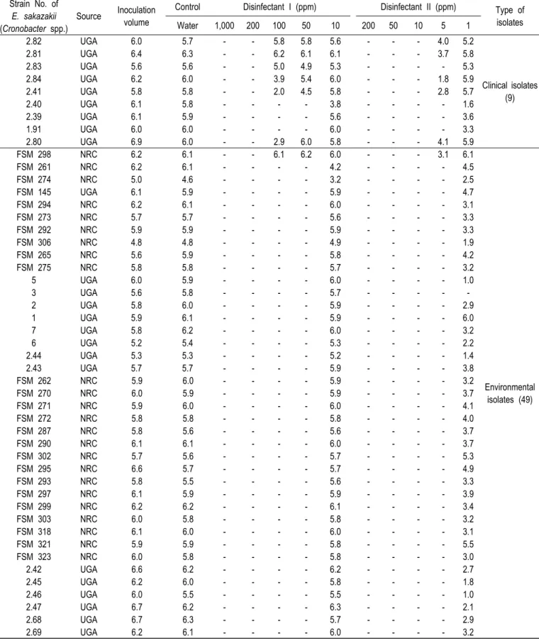
In this study, planktonic cells with an initial population of ca. 4.8 to 6.3 log CFU/mL were subjected to a sanitizer inactivation test. As shown in Table 1, trends in the inactivation of planktonic cells were exposed to 1 to 1,000 ppm disinfectant I and 1 to 200 ppm disinfectant II. Planktonic cells of E. sakazakii (Cronobacter spp.) were inactivated rapidly. Seven strains (6 clinical and 1 environmental) can survive in a 100 ppm concentration of disinfectant I, and at the most 5 ppm of disinfectant II. But 51 strains (3 clinical and 48 environmental) were not killed in 10 ppm concentration of disinfectant I and 1 ppm of disinfectant II in water, respectively (Table 1). In clinical E. sakazakii (Cronobacter spp.) strains, a 0–3.8 log CFU/mL reduction in cell number was noted after exposure to disinfectant I for 10 min at 100 ppm, and 1.7–4.2 log CFU/mL reduction was exposed to disinfectant II for 10 min at 5 ppm (Table 1). In environmental E. sakazakii (Cronobacter spp.) strains, a 0–1.4 log CFU/mL reduction in cell number was noted after exposure to disinfectant I for 10 min at 10 ppm, and 0–5.8 log CFU/mL reduction was exposed to disinfectant II for 10 min at 1 ppm (Table 1).
A greater decrease in the number of planktonic cells was also observed as compared to clinical E. sakazakii (Cronobacter spp.).
To compare with the concentration of chlorine in Gram-positive, two hundred milligrams per liter of chlorine is the maximum concentration allowed to be used in shell egg wash. Two hundred mg/liter of chlorinated water was also the most effective treatment in reducing the populations of Listeria and Salmonella (4.6 and 3.03 log CFU per shell egg, respectively for 5-min treatment; data not shown).
The results of other previous studies similar to those of this study was compared and reviewed as follows. Kuo et al. [20] did not recover no viable salmonellae after chlorinated water treatment (200 ppm) for 1 min, and only 50% of the inoculated shell eggs treated with a chlorinated (200 ppm) water wash were negative for Salmonella spp.
Also, in Gram-negative, Baker et al. [15] reported that Helicobacter pylori was significantly more resistant to chlorine than Escherichia coli. The difference between the two organisms was more pronounced at higher doses of chlorine [15]. Thus, while exposure to 0.1 ppm of chlorine for 1 min resulted in the 0.3 log reduction in viable Helicobacter pylori cells and the 0.9 log reduction in viable Escherichia coli cells, exposure to 0.20 ppm chlorine for 1 min was associated with the 1.8 log reduction in viable Helicobacter pylori cells and over 4.0 log reduction in viable Escherichia coli cells [15]. Shirai et al. [16] studied effects of chlorine, iodine, and QAC disinfectants on several exotic disease viruses. In general, QAC are good bacterial agents, and they are widely used for the disinfection of environmental surfaces [16]. However, precleaning of such surfaces is often necessary, because the effectiveness of QAC is reduced in the presence of soap and organic matter [16]. And Best et al. [21] reported efficacies of selected disinfectants against Mycobacterium tuberculosis, and also demonstrated that sodium hypochlorite required an available chlorine concentration of 10,000 ppm before an effective level of reduction could be obtained.
Our results showed that the clinically isolated E. sakazakii (Cronobacter spp.) was
more resistant to chlorine and QAC than the environmental isolated E. sakazakii (Cronobacter spp.; p<0.05; Table 1). In general, chlorine disinfectants are volatile, so it is already known that using a high concentration is good for the bactericidal effect [10,13,16–18,21]. A similar trend was also observed in this study. In particular, QAC showed a much stronger effect against E. sakazakii (Cronobacter spp.). Considering the improvement of the bactericidal effect on E. sakazakii (Cronobacter spp.), QAC is considered to be a very useful disinfectant.
Also, numerous studies conducted over the past 20 years have pointed out that microorganisms may have a variety of genetic and physiological mechanisms to respond to adverse or stressful conditions [22–26]. In particular, microorganisms under stress could exhibit the variety of changes, ranging from minor metabolic changes to more extreme alterations in cellular structure [22–26]. The physiological or structural changes resulting from exposure to moderate or sub-lethal stress allow an organism to survive high stress conditions that could be fatal [23,25,26]. For example, in the food manufacturing facility designed with conditions that inhibit the growth of pathogens and spoilage agents as much as possible, microorganisms could be sublethal when exposed to acid, cold, heat, nutrient depletion, and so on [22–26]. Thus, while these conditions may retard the growth of microorganisms, they may also induce stress- induced cellular changes that allow the organism to persist within these environments [22–26].
In fact, controlling the presence and growth of E. sakazakii (Cronobacter spp.) species have been very difficult for the food industry, and this difficulty has been attributed in part to the ability of E. sakazakii (Cronobacter spp.) to grow under high temperature or refrigeration condition [2,8,9,10,23]. Although the vehicle for E. sakazakii (Crono- bacter spp.) has not been identified in all cases, dried infant formula has been epi- demiologically identified as the source of E. sakazakii (Cronobacter spp.) [1,3,5,8,24,26].
Hence, control of E. sakazakii (Cronobacter spp.) in the food-manufacturing environ- ment has been a challenge despite the fact that this species is sensitive to commonly used chemicals such as QAC and chlorine-based chemical sanitizers. However, studies on the efficacy of fungicides against E. sakazakii (Cronobacter spp.) cultured under stress conditions are very scarce. Scheepe-Leberkuhne and Wagner [27] revealed that E.
sakazakii (Cronobacter spp.) produced viscous capsular material, and then the organism could form a biofilm on feeding equipment and contact surfaces. Also, Iversen et al.
[22] reported the biofilm formation of E. sakazakii (Cronobacter spp.) in infant formula milk on materials commonly used for infant-feeding equipments and work surfaces.
When grown in infant formula milk, E. sakazakii (Cronobacter spp.) adhered to silicon, latex and polycarbonate to a greater extent than to stainless steel [22]. Namely, these materials were commonly used for infant-feeding equipments and in preparation area [22].
Until now, there are a few reported data relating to the elimination of E. sakazakii
(Cronobacter spp.) biofilms by commercial disinfectants such chlorine, QAC, hydrogen
peroxide, ozone etc., as sanitizer. Recently, studies on the control of various micro-
organisms using several natural substances are being conducted with much interest [28].
Table 1. Effectiveness of disinfectant I and II treatment for inactivating E. sakazakii (Cronobacter spp.) in water after 10 min exposure
(Unit: log 10 CFU/mL)
Strain No. of E. sakazakii (Cronobacter spp.)
Source Inoculation volume
Control Disinfectant I (ppm) Disinfectant II (ppm) Type of isolates
Water 1,000 200 100 50 10 200 50 10 5 1
2.82 UGA 6.0 5.7 - - 5.8 5.8 5.6 - - - 4.0 5.2
Clinical isolates (9)
2.81 UGA 6.4 6.3 - - 6.2 6.1 6.1 - - - 3.7 5.8
2.83 UGA 5.6 5.6 - - 5.0 4.9 5.3 - - - - 5.3
2.84 UGA 6.2 6.0 - - 3.9 5.4 6.0 - - - 1.8 5.9
2.41 UGA 5.8 5.8 - - 2.0 4.5 5.8 - - - 2.8 5.7
2.40 UGA 6.1 5.8 - - - - 3.8 - - - - 1.6
2.39 UGA 6.1 5.9 - - - - 5.6 - - - - 3.6
1.91 UGA 6.0 6.0 - - - - 6.0 - - - - 3.3
2.80 UGA 6.9 6.0 - - 2.9 6.0 5.8 - - - 4.1 5.9
FSM 298 NRC 6.2 6.1 - - 6.1 6.2 6.0 - - - 3.1 6.1
Environmental isolates (49)
FSM 261 NRC 6.2 6.1 - - - - 4.2 - - - - 4.5
FSM 274 NRC 5.0 4.6 - - - - 3.2 - - - - 2.5
FSM 145 UGA 6.1 5.9 - - - - 5.9 - - - - 4.7
FSM 294 NRC 6.2 6.1 - - - - 6.0 - - - - 3.1
FSM 273 NRC 5.7 5.7 - - - - 5.6 - - - - 3.3
FSM 292 NRC 5.9 5.9 - - - - 5.9 - - - - 3.3
FSM 306 NRC 4.8 4.8 - - - - 4.9 - - - - 1.9
FSM 265 NRC 5.6 5.9 - - - - 5.8 - - - - 4.2
FSM 275 NRC 5.8 5.8 - - - - 5.7 - - - - 3.2
5 UGA 6.0 5.9 - - - - 6.0 - - - - 1.0
3 UGA 5.6 5.8 - - - - 5.7 - - - - -
2 UGA 5.8 6.0 - - - - 5.9 - - - - 2.9
1 UGA 5.9 6.1 - - - - 5.9 - - - - 6.0
7 UGA 5.8 6.2 - - - - 6.0 - - - - 3.2
6 UGA 5.2 5.4 - - - - 5.3 - - - - 2.2
2.44 UGA 5.3 5.3 - - - - 5.2 - - - - 1.4
2.43 UGA 5.7 5.7 - - - - 5.9 - - - - 3.8
FSM 262 NRC 5.9 6.0 - - - - 5.9 - - - - 3.2
FSM 270 NRC 6.0 5.9 - - - - 5.9 - - - - 3.7
FSM 271 NRC 5.9 6.0 - - - - 6.0 - - - - 4.1
FSM 272 NRC 5.8 5.8 - - - - 5.8 - - - - 4.0
FSM 287 NRC 5.8 5.6 - - - - 5.6 - - - - 3.7
FSM 290 NRC 6.1 6.1 - - - - 6.0 - - - - 3.7
FSM 302 NRC 5.7 5.6 - - - - 5.7 - - - - 5.3
FSM 295 NRC 6.6 5.7 - - - - 5.7 - - - - 4.9
FSM 293 NRC 5.8 5.5 - - - - 5.6 - - - - 3.3
FSM 297 NRC 6.1 5.9 - - - - 5.9 - - - - 3.9
FSM 299 NRC 6.2 6.2 - - - - 6.1 - - - - 3.4
FSM 303 NRC 6.0 5.8 - - - - 5.8 - - - - 3.2
FSM 318 NRC 6.1 6.0 - - - - 6.0 - - - - 3.1
FSM 321 NRC 5.9 5.9 - - - - 5.8 - - - - 5.5
FSM 323 NRC 6.0 5.8 - - - - 5.8 - - - - 3.0
2.42 UGA 6.6 6.2 - - - - 6.2 - - - - 2.7
2.45 UGA 6.2 6.0 - - - - 5.8 - - - - 1.8
2.46 UGA 6.0 5.5 - - - - 5.5 - - - - 1.0
2.47 UGA 6.7 6.2 - - - - 6.3 - - - - 2.1
2.68 UGA 6.7 6.3 - - - - 5.7 - - - - 2.9
2.69 UGA 6.2 6.1 - - - - 6.0 - - - - 3.2
Therefore, it is considered that various related studies using various natural substances that could inhibit or inactivate E. sakazakii (Cronobacter spp.) should be further conducted.
Conclusion
The results obtained through this study are analyzed as follows. It was demonstrated that the E. sakazakii (Cronobacter spp.) strain isolated from the clinical sample was more resistant to the disinfectants used in this study than the E. sakazakii (Cronobacter spp.) strain isolated from the environmental sample. In other words, the E. sakazakii (Cronobacter spp.) strain isolated from the clinical sample was shown to be 10-fold resistant to disinfectant I and also 5-fold to disinfectant II, respectively. Disinfectant II, known as a QAC, was shown to be more strong to inactive E. sakazakii (Cronobacter spp.) in water used to clean infant formula equipments than disinfectant I, because the QAC concentrations used in the present study were 200 times lower than the 200 ppm maximum level permissible on food contact surfaces without rinsing. Further study is strongly required to determine the effectiveness for eliminating E. sakazakii (Crono- bacter spp.) biofilm using by commercial disinfectants, and also study that the lethality of stress-adapted E. sakazakii (Cronobacter spp.) when the microorganism is cultured under ideal conditions.
Conflict of Interest
The authors declare no potential conflict of interest.
Acknowledgement
This work was supported by Korea Institute of Planning and Evaluation for Tech-
Table 1. ContinuedStrain No. of E. sakazakii (Cronobacter spp.)
Source Inoculation volume
Control Disinfectant I (ppm) Disinfectant II (ppm) Type of isolates
Water 1,000 200 100 50 10 200 50 10 5 1
2.70 UGA 6.3 5.6 - - - - 5.9 - - - - 3.8
Environmental isolates (49)
2.71 UGA 6.8 6.3 - - - - 6.4 - - - - 3.5
2.72 UGA 6.4 5.8 - - - - 5.7 - - - - 2.5
2.73 UGA 6.3 6.0 - - - - 6.2 - - - - 5.8
2.74 UGA 6.4 6.1 - - - - 6.1 - - - - 2.4
2.75 UGA 4.5 4.0 - - - - 4.1 - - - - -
2.76 UGA 6.2 5.6 - - - - 5.7 - - - - 4.4
2.77 UGA 5.9 5.6 - - - - 5.7 - - - - 3.1
2.78 UGA 5.9 5.8 - - - - 5.7 - - - - 2.5
2.79 UGA 6.2 5.9 - - - - 5.8 - - - - 2.6
Disinfectant I contained 6.15% sodium hypochlorite.
Disinfectant II contained both 2.25% n-alkyl dimethylbenzyl ammonium chloride and 2.25% n-alkyl ethylbenzyl ammonium chloride.
UGA, Dr. Jeffrey Kornacki, University of Georgia, Athens, GA; NRC, Dr. John Marugg, Nestle Research Center, Lausanne, Switwerland.