681
©The Korean Society of Food Science and Technology
국내에서 분리한 Cronobacter spp.(Enterobacter sakazakii)의 건조내성 특성
이은진·류태화·박종현*
경원대학교 식품생물공학과
Tolerance of Korean Cronobacter spp. (Enterobacter sakazakii) Isolates to Dessication
Eunjin Lee, Tae Hwa Ryu, and Jong-Hyun Park* Department of Food Science and Biotechnology, Kyungwon University
Abstract Cronobacter spp. (Enterobacter sakazakii) is known to be highly resistant to dry conditions than any other Enterobacteriaeae. In this study, one hundred and ten Korean Cronobacter isolates were characterized to find out their survival characteristics under conditions of desiccation and humidity. Thirty percentage strains of the isolates showed high resistance to desiccation exposed on the metal surface for eight hours by half survival of the initial number, whileas less than 10% strains showed dry sensitivity by less one log scale survival among seven log scales. Finally, more than 90%
of the strains consisted of dry-resistant and dry-intermediate groups. The same tendencies were evident in a 15-day exposure. Dry-resistant and intermediate strain groups showed 3 log scale survival among 5 log initial numbers in the powdered infant formula for 30 days, which were more resistant than on the above metal surface exposed. So, almost the isolate strains showed high resistance to dry condition. Dry-resistant and intermediate groups exposed on the metal surface formed a biofilm at the beginning, and the dry-sensitive group showed biofilm formation mainly only after a 7-day exposure. However, without a time difference in formation of biofilm, the dry-resistant and sensitive isolates seemed to similar biofilm formation activity. Most of the isolates showed very low survival at 75% relative humidity in 48 hours;
however, they showed high resistance by 60% survival at 40% relative humidity. The Cronobacter isolates showed high resistance to desiccation on the metal surface and in the powdered infant formula, but low survival at high relative humidity. Therefore, high humidity may be a control method for Cronobacter in food processing environments.
Key words:Cronobacter, Korean isolate dessication, humidity, resistance
서 론
Cronobacter spp.(Enterobacter sakazakii)는 유아에게 치명적인
영향을주는급성기회감염균으로주로생후 1개월이내의신생 아, 조산아, 저체중아, 유아에게서수막염(neonatal meningitis), 패 혈증(bacteremia), 신생아 괴사성장염(necrotizing enterocolitis) 등
의병을 일으키는것으로알려졌다(1-4). 매우드물게감염되지만
이세균에감염된영유아의치사율이 33-80% 이상인것으로보
고되어매우 치명적인세균으로간주된다(5). Cronobacter는원래
E. sakazakii로명명되어오다가 Iversen 등(6)에의하여 Cronobacter spp.로개명하도록제안되었다. 영유아의 식용으로사용되는조제
분유와이유식은비살균공정으로생산되는식품이므로 Cronobacter
에의해오염되기 쉽고실제 한국의영유아 식품에서도 오염되 어사회적으로많은파장을일으키기도하였다. Cronobacter는조
제분유혹은 유아식에서주로 검출되는것으로알려져 있으나그 외에도 토양, 쥐, 파리, 우유분말공장, 초콜릿공장과 집등주변 환경에서 자주 검출되는 미생물로 보고되었으며 식품공장을 포 함한다양한인공환경에서도분리되었다(7,8).
이와같이자연계에다양하게분포되어있고유아에게치명적 인영향을 주는급성 기회감염균이기때문에최근에 그에대한
연구가많이진행되고있다. Kandhai 등(7)에따르면조제분유및
이유식의 공정또는미리준비해놓은유아식의 Cronobacter 오
염으로 조산아 또는 신생아들에게서발병을 유발시키며 전문의 들은 미리 물에 타 놓은 분유가 문제 시 된다고 보고하였다. Cronobacter의 감염량은 103-105 CFU/g으로 예측하고 있는데 이
것은 뇌수막염병원성미생물인 Neisseria menigitidis, Escherichia coli O157:H7, Listeria monocytogens 4D 등의 감염량과 유사한 것으로 예측하고 있다(8,9). 국제기구인 International Commission for Microbiological Specification for Food(ICMSF)에서는 Crono- bacter 를 “Severe hazard for restricted population, life threaten- ing or substantial chronic sequelae or long duration.”, 즉한정된 사람들에게매우위험하며만성혹은장기간생명을위협한다고 보고하였으며 많은 전문가들의 의견을 수렴하기 위하여 FAO/
WHO는 영유아용 분말식품에 대한위해에 대한조사를 하도록
권고하고 있다(10). 이러한급식용의 조제분유, 이유식과더불어
선식또한유아의성장기용영양식으로사용되고있어 Cronobacter
*Corresponding author: Jong-Hyun Park, Department of Food Sci- ence and Biotechnology, Kyungwon University, Seongnam, Gyeo- nggi 461-701, Korea
Tel: 82-31-750-5523 Fax: 82-31-750-5501
E-mail: [email protected]
Received August 22, 2009; revised September 28, 2009;
accepted October 9, 2009
건조식품이나 환경등에서 오염이높게 보고되는것은 다른미 생물혹은다른 Enterobacteriacae 종에비하여삼투압과건조스
트레스에높은저항성을가지기때문인것으로알려지고있다(15).
또다른 원인으로는 Cronobacter의 biofilm 형성능력을 들수 있는데 biofilm은물리적 장벽을형성함으로써 UV light, 삼투압
스트레스, 열처리, 영양고갈, 산, 세정액, 항생제, 항체등의환경
스트레스에대하여저항성을높여준다(16). Lehner 등(17)은 56종 의Cronobacter중 23종의균주가유리표면에서, 33개균주가 air- solid 표면에서, 16종의균주가두가지표면에서모두 biofilm을
형성하는 것을 확인하였다. 열처리에 의한 제어 법은 Jung(18), Kim(19), Nazarowec-White 등(20,21)에의해 연구되었으며그외 의Cronobacter 제어연구로는 Nazarowec-White 등(22)의연구로 조제분유에서의오염되어있는 Cronobacter에감염되는 bacterioph- age를분리하여감염을방지하고자 하는방법도보고되었다.
본연구에서는국내의건조분말식품등에서분리하여보관중 인Cronobacter spp.의건조특성, 건조내성에따른 biofilm 형성능 력과상대습도조절조건에서 Cronobacter spp.의생존특성을연
구하고자하였다.
재료 및 방법
균주와 시약
본연구에서사용된 Cronobacter spp.는국내에서유통중인조 제분유, 이유식, 생식, 선식, 샐러드등비가열섭취식품 160종의 시료로부터분리, 동정되어당연구실의균주은행에 보관되어있
는 112균주를사용하였다(23). 생화학적, 생리학적 특성을비교하 기 위해 공시균주인 C. muytjensii ATCC 51329와 Cronobacter
NCTC 2949를사용하였으며 사용되는시약은특급시약을사용
하였다.
건조내성에 따른 Cronobacter 의 분류
공시균주인 C. muytjensii ATCC 51329과 Cronobacter NCTC 2949를포함하여 112개의건조분말식품유래분리균주를대상시
험균주로 정하였다. 배양액은 각 균주를 10 mL의 tryptic soy broth(TSB; Oxoid, Hampshire, UK)에서 37oC, 24시간동안 전배
양시켜각균주를 7 log CFU/mL의수준으로 초기균수를조정
하였다. 7×7 cm2의스텐인레스스틸에 20µL를떨어뜨린후 25oC,
상대습도 75%의 항온항습 인큐베이터(Daehan Scientific, Seoul, Korea)에서노출시킨 후 0, 4, 8, 24, 48시간째에 50µL 멸균생 리식염수로 20회씩 2회 pippeting하여 건조된 균체를 회수하여
조제분유내에서의 건조내성 측정
C. muytjensii ATCC 51329와 Cronobacter NCTC 2949를포함 한 112개의분리균주를대상으로 Cronobacter가검출되지않은 조제분유(Namyang, Seoul, Korea) 10 g의시료에 spiking 하여 30
일동안 저장하였다. 사용된균주는 10 mL의 TSB에서 37oC 24
시간동안전배양시킨균주를최종생균수가 5-6 log CFU/mL의 수준이되도록희석한후 100µL를조제분유에첨가하여뭉치지 않게 고루 섞은후실온에서저장하면서초기생균수와 30일후
의생균수를측정하여그변화를측정하였다.
건조에 따른 biofilm 형성분석
대상균주로는 건조내성에따른 Cronobacter의분류에서의 고 내성그룹과 저내성그룹의 대표적 세균주씩을 시험균주로 하였 다. Biofilm 측정방법으로는 crystal violet의 OD를측정하는 12- well plastic plate(Becton Dickinson Labware, Franklin Lakes,
NJ, USA)법을이용하여측정하였고 각그룹간평균과표준편차
를계산하여 표기하였다. TSB 10 mL에 Cronobacter를접종하여
37oC에서 하루 밤 전배양된 균체를 12-well tissue culture test plate의각 well에 2 mL씩 접종하여 다시 배양하였다. 일정시간 항온조에서 노출된 test plate의 부유세균과 배양액을 aspirator를
이용하여제거한 후빈 well을 2.5 mL 생리식염수로격렬하게 진
탕하여 3번 세척하였다. 잔여식염수를 제거한후 2 mL의 99%
methanol을각 well에분주하고 15분동안방치하여 biofilm을표 면에 부착시켰다. Methanol 제거후 완전히건조시키고 각 well
마다 2 mL의 1% crystal violet(Hucker, Sigma, St. Louis, MO, USA) solution 분주후 5분동안방치하여 biofilm을염색하였다
(24). Crystal violet solution을제거하고건조한후 2.5 mL 증류수 로 1회세척하고 33%(v/v) glacial acetic acid를 1.6 mL씩분주하
여 착색된 crystal violet을 용출시켜 570 nm에서 microtiter plate reader(Tecan, Sunrise, A Graham, Salzburg, Austria)로 흡광도를 측정하였다.
습도에 대한 Cronobacter의 내성분석
38개의분리균주(고내성균주 13, 중간내성균주 22, 저내성균주
3균주)와 2개의 공시균주 C. muytjensii ATCC 51329와 Crono-
bacter NCTC 2949를 대상균주로 선정하여 상대습도 변화에 따
른생존균수를분석하였다. 10 mL의 TSB에서 37oC, 24시간 동안 전배양시킨 각균주를 7 log CFU/mL의 수준으로 초기 균수를 조정하였다. 7×7 cm2의 스테인레스스틸에 20µL를 떨어뜨렸으며
25oC에서상대습도(relative humidity, RH) 75%, 40%의항온항습 인큐베이터에서 0, 4, 8, 24, 48시간 노출시킨후각각 50µL 멸 균생리식염수로 20회씩 2회 pippeting하여건조된 균체를 회수
하여 TSA에 도말한 후 37oC에서 24시간 동안회복 및 배양을 하여형성된집락을계수하였다.
상대습도에따른생균수의사멸을 imaging analysis하기위하여 공초점 레이저 주사현미경(confocal laser scanning microscope, CLSM, MRC-1024, Bio-Rad, Hertfordshire, UK)으로 촬영하였다
(25). 대상균주로공시균주인 C. muytjensii ATCC 51329를사용하
였으며 대조구로 TSB 배양액을 사용하였고 75%, 40% RH에서
24시간 동안 슬라이드그래스에서 건조시켰다. LIVE/DEAD® BacLightTM Bacterial Viability Kit(L7007, for microscopy, Molec- ular Probes, InvitrogenTM, Carlsbad, CA, USA)를사용하여균체를 형광으로 염색시켰다. 두 개의 molecular prove component A (SYTO 9 dye, 1.67mM/propidium iodide, 1.67mM)와 component B(SYTO 9 dye, 1.67 mM/propidium iodide, 18.3 mM)를 1:2.5로 혼합한후혼합용액과멸균 D.W.를 1:4로혼합하여건조표면에 가한 후 15분 동안 암소에서 반응시켰다. 형광 prove는 생균과
사균에특이하게결합되는것을두개의파장으로촬영한후두
이미지를 merging시켰다. 생균은 초록으로, 사균은 적색으로
imaging하게된다.
결과 및 고찰
건조내성에 따른 Cronobacter의 분류
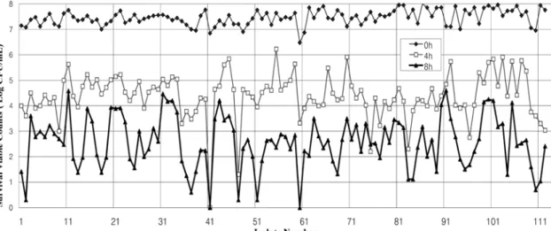
Cronobacter를 25oC와상대습도 75%에서 스테인레스스틸표면
에 8시간노출후의생존에대하여 Cronobacter NCTC 2949, C.
muytjensii ATCC 51329 두공시균주를포함한 112균주건조내성 실험을 수행하였다. 그결과 사용한 112개균주 중 7개 균주는
7 log CFU/mL 수준에서 8시간째에 1 log CFU/mL 생균수로떨
어지는 건조에 약한 저내성그룹(dry sensitive group)으로, 8시간
건조후 3 log CFU/mL 정도의생존을 보이는 37균주는고내성
그룹(dry tolerant group)으로, 그중간의생존율을보이는 68개균 주를중간내성그룹(dry intermediate group)으로분류하였다(Fig. 1).
국내식품에서 분리한 Cronobacter는건조에 대하여 90% 이상이 건조에대한중간정도의이상의내성을갖고있는것으로분석
되었고 건조에 약한 균주는 10% 이내의 적은 수를 보였다. 각
건조내성 그룹별로대표적 3균주씩을 선정하여 12-well microtit-
teplate의플라스틱 표면상에서의 15일의장기간상온방치시에
따른 생균수의변화를 측정하였을때건조 1일후부터는 세그 룹모두감소경향을보이나저내성그룹은고내성그룹에비해 3
log CFU/mL 이상사멸하였으며시간이지날수록감소추세를이
어갔다. 하지만 고내성그룹과중간내성그룹은 2일건조후생균 수가 증가하거나유지되는경향을보였다. 따라서단기 노출분류 에 의한중간내성그룹에 속하는 균주들도 장기적으로는 상당한 건조내성을보여주고있었다. 따라서금속과플라스틱표면건 조에서 단기 및 장기간의 건조노출에 대하여 식품에서 분리된
10% 미만의 균주만 건조에 약한것으로 나타났고 90% 이상의
Cronobacter 균주는건조에 강한저항성을 나타내는것으로 사료
된다(Fig. 2).
Fig. 1. Dry resistant pattern of 112 Cronobacter spp. at 25oC and 75% relative humidity according to each exposed times of 0, 4, and 8 hours.
Fig. 2. Viable count comparison of Cronobacter spp. for different resistant groups exposed on plastic surface at each time for 15 days. Nine strains of resistant (R-21, R-72, R-86), intermediate (I- 12, I-40, I-97), and sensitive strains (S-2, S-37, S-110) from each groups to desiccation were selected and tested.
조제분유에서의 건조내성 특성
Cronobacter가검출되지 않은 조제분유 시료에 5-6 log CFU/
mL의 수준이 되도록 C. muytjensii ATCC 51329와 Cronobacter NCTC 2949를포함한 112개의 분리균주를 spiking하여고루 섞 은후실온에서 보관하면서 30일후의 생균수변화를 분석하였 다. 금속과플라스틱표면에서의 건조특성에따른 분류와마찬가
지로 Cronobacter 건조고내성그룹과중간내성그룹은 30일이후
에도 5 log CFU/mL에서 3 log CFU/mL 이상의생균수를나타내
었다(Fig. 3). 오히려표면건조노출때보다더생존율이높아조
제분유에서는 더 내성이 증가함을 할 수가 있었다. Barron과 Forsythe(12)는건조상태의 조제분유에 Cronobacter 야생균주 10
가지를 7 log CFU/mL 수준으로 spiking하여 30개월간 저장실험 을한결과, 10균주 중한균주는 1년안에 모두사멸하였으나
두균주는 30개월까지 2 log CFU/mL 이상의 생균을 유지하고
있었으며나머지일곱균주는 20개월에서 30개월내에사멸하여 건조내성이크다고 보고하였다. 30개월에서 생존을보이는 두내 성균주의 사멸패턴으로는 4개월까지급속한 사멸곡선을 그리 다가 6개월 이후에안정적으로 2-3 log CFU/mL 수준을 유지한
것으로나타났다. 또한 Edelson-Mammel 등(13)은조제분유의높 은영양성분으로인해 700일이상 Cronobacter의생존이가능하 다고보고였다. 또한 Gurtler 등(26)은 Cronobacter건조시 Aw가 감소할수록생존률이높다고보고하였으며조제분유의 Aw는 0.2
로나타나 조제분유에오염된 Cronobacter가균자체의건조내성 특성과분유의낮은수분활성도와고영양적환경이복합적으로 작용하여높은 생존률을보인 것으로사료된다. 따라서대부분의
Cronobacter는조제분유 등의영양분이 많은건조분말 식품에서
생존율이 아주높아서이 세균의오염을 줄이는제어관리가 필 요함을알수있었다.
건조특성에 따른 biofilm 형성 특성
12-well plastic plate 법으로 건조환경에서의 biofilm 형성을고 내성그룹과저내성그룹에서 대표적인세균주씩선별하여측정하
였다(Fig. 4). 고내성그룹 균주의 biofilm 생성량은초기 12시간에
급속한 증가를보였으며 그후일주일까지는 시간이 지남에 따 라감소하는경향을보이다가 15일까지는다시증가하는경향을 보였다. 저내성그룹균주의경우에는건조영향에 따라균체의 사
멸에의해 biofilm 전체의 양이줄어들었고그중건조에적응
되어살아남은일부의균체가강하게 biofilm을생성한것으로보
이는데건조 15일에가까워질수록 biofilm양이고내성균주들과 거 의같아지는것을볼때에이와같은결과를 유추할수있다. 대
부분의세균들은물체의표면에부착하면 biofilm을생성하여환
경스트레스에 대한 저항성을 높이게 된다(27,28). Cronobacter도 스테인레스스틸, polyvinyl chloride, 실리콘, 락테스, 유리등에 부
Fig. 3. Survival of Cronobacter spp. spiked in powdered infant formula after a month.
Fig. 4. Comparison biofilm formation capability for dry- sentitive/tolerant strains of Cronobacter spp. by microtiter plate method. Mean values with SD for six representative strains (R-21, R-72, R-86, S-2, S-37 and S-110) from dry tolerant group and sensitive group were shown.
착하여 biofilm을생성하는것으로 보고되어있다(17,29). 이러한
biofilm은식품제조현장에서 이세균의제어를어렵게하는요인
이되기도하며특히살균소독제에대한저항성을높혀서이세
균에대한위해가능성이더높아지게되게한다(30,31). 그러므
로 biofilm은건조 스트레스에저항하는하나의 Cronobacter의생 육적응수단일 것으로보이며 고내성 균주와저내성 균주의건
조내성에 대한 biofilm생성의 시간적인 차이만 존재하고 대부분
의 Cronobacter는건조스트레스에대하여 biofilm을형성시키는 특성을가지고있는것으로보인다.
상대습도에 따른 Cronobacter의 생존특성
38개의 분리균주와 2개의공시균주로 건조 시상대습도에 따 른제어가가능한지알아보기 위한연구를 수행하였다. 스텐인레 스스틸위에 7 log CFU/mL 수준의 Cronobacter를접종한후항 온항습인큐베이터에서온도 25, 상대습도(relative humidity, RH) 75%와 40%로설정한후노출시간에따라 그생균수를측정하였 다. 75% RH의경우에는 8시간까지 7 log CFU/mL에서 5 log의 급격한감소를 보이다가 8시간 이후부터 감소기울기가 완만히
지나 24-48시간내에 모두사멸하는것으로나타났다. 반면 40%
RH의경우 8시간째약 2 log가사멸한후 24시간까지는 확연한
증가나감소없이비슷하게 5 log CFU/mL 정도의 생균수를유
지하였으며 48시간째에도 4-5 log CFU/mL 수준의생균수를 유
지하였다(Fig. 5).
상대습도에 따른 생균수의 사멸을 Backlight kit를 사용하여
confocal laser scanning microscope로 촬영한결과인 Fig. 6을보
면 (A)는대조구로 사용한 C. muytjensii ATCC 51329의배양액 에서빨간색으로 보이는사균(dead cell) 몇개를제외한모든세
포가초록으로살아있는세포임을보여준다. 25oC, 40% RH에서
24시간건조시킨 (B)의경우에는 생균:사균 비율이 약 1:1 정도
로나타나며 (C)의경우 25oC, 75% RH에서 24시간건조시킨결 과로몇개의 초록색으로 표시되는생체효소를 제외한 모든세
포가적색으로사멸한것을볼수있다. (D)는 37oC와고습에서
24시간동안 건조시킨 Cronobacter로써생균세포가 없이모두 적
색으로 보여 고습일수록 세포의 사멸이 증가함을 확인하였다. Gurtler 등(26)이 Cronobacter 건조 시 Aw가 높을수록 생존률이
낮다고보고한것과동일한결과였으며건조식품인조제분유등
의생산현장 환경에서오염된 Cronobacter의제어관리에활용이
가능하리라 사료된다. Mullane 등(32)은조제분유와그공정중의
Cronobacter의오염도를분석한결과분리된 Cronobacter의 70%
이상이공정중에서분리하였으며특히분유건조공정과포장공 정이 이들 세균의 주요 오염장소라는 것을 보고하였다. 따라서 이작업환경공간이주요한오염경로인것으로제시하였다. 그러 므로이와 같은상대습도와작업온도를 조절하여 Cronobacter의 오염경감에 기여할수있으리라 사료된다.
요 약
Cronobacter spp.(Enterbacter sakazakii)의건조저항성은다른장 내세균들보다강한특성을 가지고있는 것으로알려져있다. 본 연구는 국내식품으로부터 분리한 110개의 Cronobacter균주로부 터 건조내성 특성을 분석하고 여러상대습도에서의 생존특성을 연구하고자 하였다. 국내 식품에서 분리한 Cronobacter 110균주 중 8시간금속표면 건조노출에 대하여 초기균수의 약절반정도 생존하는 고내성그룹이 30%, 대부분이 사멸하는 저내성그룹이
약 10%를차지하여전체균주중중간정도이상의내성을가지
는균주가 90% 이상을차지하고있었다. 15일간장기건조노출
에경우도비슷한경향을보여주었다. 조제분유에서 Cronobacter
건조 노출의 경우고내성그룹과 중간내성그룹은 30일이후에도 5 log CFU/mL에서 3 log CFU/mL 이상의생균수를나타내었다.
오히려금속표면건조때보다더생존율이높아조제분유에서는 더내성이증가함을할수가있었다. 따라서국내식품에서 분리
Fig. 5. Viable count of Cronobacter spp. on stainless steel at 25oC under 40% and 75% relative humidity. Forty isolates including three dry-sensitive strains were exposed in the different relative
humidity and the viability was determined. Fig. 6. Image of confocal laser scanning microscope (CLSM) with dry-sensitive C. muytjensii ATCC 51329 for 24 hours.
Green, live cell; red, dead cell; (A), control, 37oC TSB overnight culture; (B), dry at 25oC with 40% relative humidity; (C), dry with 25oC in 75% relative humidity; (D), dry at 37oC with high humidity for 24 hours.
것으로보인다.
감사의 글
본연구는 2008년도농림기술관리센터의 지원에의해 이루어
진연구(108147-02-1-CG000)의일부로이에감사드립니다.
문 헌
1. Guillaume-Gentil O, Sonnard V, Kandhai MC. Marugg JD, Joos- ten H. A Simple and rapid cultural method for detection of Enterobacter sakazakii in environmental samples. J. Food Protect.
68: 64-69 (2005)
2. Arad I, Baras M, Gofin R, Bar-Oz B, Peleg O. Dose parity affect the neonatal outcome of very-low-birth-weight inrants? Eur. J.
Obstet. Gyn. R. B. 94: 283-289 (2001)
3. Farmer JJ, Asbury MA, Hickman FW, Brenner DJ. The Entero- bacteriaceae study group. Enterobacter sakazakii: A new species of “Enterobacteriaceae” isolated from clinical specimens. Int. J.
Syst. Bacteriol. 30: 369-58 (1980)
4. Jung MK, Park JH. Prevalence and thermal stability of Entero- bacter sakazakii from unprocessed ready-to-eat agricultural prod- ucts and powdered infant formulas. Food Sci. Biotechnol. 15:
152-155 (2006)
5. Iversen C, Forsythe SJ. Risk profile of Enterobacter sakazakii, an emergent pathogen associated with infant milk formula. Food Sci.
Technol. 14: 443-454 (2003)
6. Iversen C, Lehner A, Mullane N, Bidlas E, Cleenwerck I, Marugg J, Fanning S, Stephan R, Joosten H. The taxonomy of Enterobacter sakazakii: Proposal of a new genus Cronobacter gen. nov. and descriptions of Cronobacter sakazakii comb. nov., Cronobacter sakazakii subsp. sakazakii, comb. nov., Cronobacter sakazakii subsp. Malonaticus subsp. nov., Cronobacter turicensis sp. nov., Cronobacter muytjensii sp.nov., Cronobacter dublinensis sp. nov., and Cronobacter genomospecies 1. BMC Evol. Biol. 7:
64-67 (2007)
7. Gurtler JB, Kornacki JL, Beuchat LR. Enterobacter sakazakii: A colifrom of increased concern to infant health. Int. J. Food Microbiol. 104: 1-34 (2005)
8. Park JH, Jung MK. Food safety by Enterobacter sakazakii: Newly emergent pathogen from infant formula foods. Trends Agric. Life Sci. 3: 44-53 (2005)
9. International Commission on Microbiological Specification for Foods (ICMSF). Microbiological Testing in Food Safety Manage- ment. Vol. 7. Academic/Plenum Publisher, New York, NY, USA (2002)
10. Nazarowec-White M. Biological characterization of Enterobacter sakazakii. PhD thesis, University of Ottawa, Ottawa, Canada (1998)
11. Breeuwer P, Lardeau A, PeterzM, Joosten HM. Desication and
Aspect promoting environmental persistnace, J. Food Protect. 68:
2287-2294 (2005)
18. Jung MK, Park JH. Prevalence and thermal stability of Entero- bacter sakazakii from unprocessed ready-to-eat agricultural prod- ucts and powdered infant formulas. Food Sci. Biotechnol. 15:
152-155 (2006)
19. Kim SH, Park JH. Thermal resistance and inactivation of Entero- bacter sakazakii Isolates during rehydration of powdered infant formula. J. Microbiol. Biotechnol. 17: 364-368 (2007)
20. Nazarowec-White M, Farber JM. Thermal tolerance of Entero- bacter sakazakii on reconstituted dried- infant formula. Lett.
Appl. Microbiol. 24: 9-13 (1997)
21. Nazarowec-White M, McKeller RC, Piyasena P. Predicrive mon- eling of Enterobacter sakazakii inactivation in bovine milk during high-temperature shore-time pasterurization. Food. Res. Int. 32:
375-379 (1999)
22. Kim KP, Klumpp J, Loessner MJ. Enterobacter sakazakii bacte- irophage can prevent bacterial growth in reconstituted infant for- mula. Int. J. Food Microbiol. 115: 195-203 (2007)
23. KFDA. Monitoring of Food-borne Pathogens on Ready-to-Eat Sunsik. Korea Food & Drug Administration, Seoul, Korea. pp.
15-21 (2006).
24. Vasseur P, Vallet-Gely I, Soscia C, Genin S, Filloux A. The pel genes of the Pseudomonas aeruginosa PAK strain are involed at early and late stage of biofilm formation. Microbiology 151: 985- 997 (2005)
25. InvitrogenTM LIVE/DEAD® BacLightTM Bacterial Viability Kits.
Available from: http://probes.invitrogen.com/media/pis/mp07007.
pdf. Accessed Oct. 23, 2006.
26. Gurtler JB, Kornacki JL, Beuchat LR. Enterobacter sakazakii: A colifrom of increased concern to infant health. Int. J. Food Microbiol. 104: 1-34 (2005)
27. Kumar, CG, Anand SK. Significance of microbial biofilms in food industry: A review. Int. J. Food Microbiol. 42: 9-27 (1998) 28. Ryu, JH, Kim H, Beuchat LR. Attachment and biofilm formation
by Escherichia coli O157:H7 on stainless steel as influenced by exopolysaccharide production, nutrient availability, and tempera- ture. J. Food Protect. 67: 2123-2131 (2004)
29. Iversen C, Lane M, Forsythe SJ. The growth profile, thermotoler- ance, and biofilm formation of Enterobacter sakazakii grown in infant formula milk. Lett. Appl. Microbiol. 38: 378-382 (2004) 30. Frank JF, Ehlers J, Wicker L. Removal of Listeria monocytogenes
and poultry soil-containing biofilms using chemical cleaning and sanitizing agents under static conditions. Food Prot. Trends 23:
654-663 (2003)
31. Norwood DE, Gilmour A. The growth and resistance to sodium hypochlorite of Listeria monocytogenes in a steady-state multispe- cies biofilm. J. Appl. Microbiol. 88: 512-520 (2000)
32. Mullane NR, Whyte P, Wall PG, Quinn T, Fanning S. Application of pulsed-field gel electrophoresis to characterize and trace the prevalence of Enterobacter sakazakii in an infant formula pro- cessing facility. Int. J. Food Microbiol. 116: 73-81 (2007)