전방 경추 추간판 제거술 및 유합술 후 인접분절의 골극 형성에 대한 관여인자
Factors Affecting Adjacent Level Ossification Development after Anterior Cervical Discectomy and Fusion
정의섭 • 최병열 • 신충식
전주 예수병원 정형외과
목적: 전방 경추 추간판 제거술 및 유합술 후 인접분절 골화 형성의 발생에 영향을 미치는 인자에 대해서 알아보고자 하였다.
대상 및 방법: 전방 경추 추간판 제거술 및 유합술을 시행 받은 75명의 환자를 대상으로 하였다(외상 28명,퇴행성 경추질환 47명). 추시중 골화의 발생 시기 및 빈도를 확인하였고 외상군과 퇴행성 질환군에 따른 발생 빈도를 비교하여 연부조직 손상이 미치는 영향을 알고자 하였 다. 유합 범위, 나이, 수술 시간 등과의 연관성에 대해서도 조사하였다.
결과: 총 33예(44%)의 환자에서 골화가 관찰되었다. 술 후 1년 이내 관찰된 경우가 5예(15%), 1-2년 사이에 관찰된 경우가 10예(30%), 2-3년 사이에 관찰된 경우가 13예(40%), 3년 이후 관찰된 경우가 5예(15%)였다. 유합 범위만이 골화의 발생과 통계학적으로 의미있는 연 관을 보였다(p<0.001). 외상군과 질병군의 비교에서 골화의 발생빈도는 유의한 차이를 보이지 않았다(p=0.63).
결론: 인접분절 골화 형성은 술 후 관찰되기까지 상당기간의 추시가 필요하며 연부조직의 손상 정도보다는 유합에 따른 인접분절의 과도한 부하가 발생에 더 큰 영향을 미칠 것으로 생각된다.
색인단어: 경추, 전방 추간판 제거술 및 유합술, 인접분절 골화 형성
접수일 2012년 6월 1일 수정일 2012년 7월 13일 게재확정일 2012년 9월 21일 교신저자 최병열
전주시 완산구 서원로 365, 전주 예수병원 정형외과 TEL 063-230-8744, FAX 063-230-8819 E-mail docby@hanmail.net
Copyright © 2012 by The Korean Orthopaedic Association
“This is an Open Access article distributed under the terms of the Creative Commons Attribution Non-Commercial License (http://creativecommons.org/licenses/by-nc/3.0/) which permits unrestricted non-commercial use, distribution, and reproduction in any medium, provided the original work is properly cited.”
대한정형외과학회지:제 47권 제 6호 2012
서 론
경추 전방 감압 및 유합술은 퇴행성 병변에 의한 신경병증이나 척수병증의 치료에 널리 이용되고 있는 효과적인 치료방법이다.1-4) 비교적 드물긴 하지만 수술로 인해 발생하는 여러 가지 조기 및 후기 합병증에 대하여 보고되고 있다.
인접분절에서의 골화의 형성은 널리 알려진 후기 합병증 중 하 나이지만 골화 형성 자체가 실제 수술에 의한 영향으로 발생하는 지 확실치 않으며 그 임상적 의의 또한 불분명하다.2,5-9) 골화의 발 생을 피할 수 있는 방법은 없지만 수술 방법의 변화를 통하여 골 화의 발생 빈도를 줄여보기 위한 여러 가지 노력이 시행되어 왔
고 이러한 시도는 주로 세심한 연부조직 조작과 관련되어 있었 다.5-9) 하지만 골화 발생에 대하여 명확히 밝혀진 유발 인자가 없 고 실제 수술 중 발생하는 연부조직 손상이 골화 형성에 미치는 영향에 대해서도 분명히 논의된 바 없다.
저자들은 인접분절 골극의 발생시기 및 발생에 관여하는 인자 에 대하여 조사하였고 외상군과 질병군과의 골화 발생 빈도를 비 교하여 연부조직 손상이 골화의 발생에 영향을 주는지 알아보고 자 하였다.
대상 및 방법
1. 대상
2004년 4월부터 2008년 4월까지 본원에서 전방 유합술 및 전방 금 속판 고정술을 시행 받은 환자 중 최소 2년 이상 추시가 가능했 던 환자를 대상으로 하였다. 감염이나 종양에 의한 경우, 재수술,
전후방 유합술을 시행 받은 경우, 추시 중 불유합이 발생한 경우, 금속판이나 나사못의 고정 실패가 발생한 경우, 수술 전 유합 부 위의 인접분절에 이미 뚜렷한 골극이 형성된 경우는 본 연구에 서 제외하였다. 외상에 의한 수술 환자는 수술 소견상 전방 연부 조직(전종 인대, 경장근 등)의 손상이 확인된 경우를 대상으로 하 였다. 평균 연령은 51.65±10.97세이며, 평균 추시 관찰은 44.64±
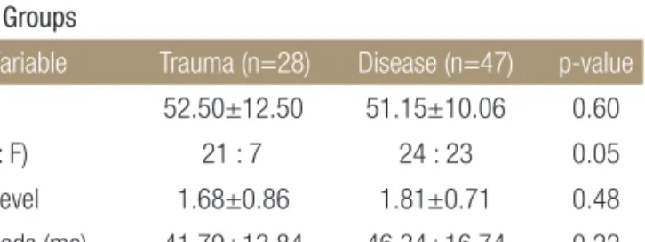
15.78개월, 남자 45명, 여자 30명이었다. 총 75명의 환자 중 외상으 로 인해 수술을 시행했던 경우가 28예, 퇴행성 경추 질환과 동반 된 신경근증 및 척수증에 대해 수술한 경우가 47예였다. 외상군과 질병군 간에 나이, 성별, 추시기간, 유합 범위의 유의한 통계학적 차이는 관찰되지 않았다(Table 1).
2. 수술 방법
전 예에서 Smith와 Robinson10)의 전방 도달법을 이용하였다. 전종 인대와 추간판을 제거한 후 Casper 개대기를 이용하여 기존의 추 간판 간격에 추가로 2-3 mm 정도의 인접 추체를 확장하고 병변 추체 주위의 골극과 남은 추간판 및 상하 추체의 연골 종판을 후 종 인대 전방까지 완전히 제거하여 척수의 감압을 시도하였고 필
요한 경우에는 후종 인대도 일부 제거하였다. 좌측 장골 능에서 최소 침습적으로 채취한 해면골을 케이지(Polyethyletherketone cage, Stryker spine, South Allendale, NJ, USA)에 채운 후 추체 사 이에 삽입하였고, 자가 물림 나사 및 금속판(Maxima plate, U & I corporation, Euijeongbu, Korea)을 이용하여 인접 추체에 고정하 였다. 수술 후 Philadelphia 경부 보조기를 4주간 착용하도록 하였 고 이후에는 연성 경부 보조기를 2주간 추가로 착용하도록 권장 하였다.
3. 방법
인접분절 골화의 발생에 대한 평가는 술 후 3개월, 6개월, 9개월, 12개월 이후 6개월 간격으로 시행된 경추부 측면 단순 방사선 사 진을 통하여 이루어졌다. 금속판과 인접분절 추간판 간의 거리는 두측과 미측에서 각각 측정하여 분석하였다(Fig. 1). 모든 측정은 척추외과 전문의 2명이 각각 2회씩 측정하여 평균을 구하였다. 후 종인대 골화 및 황색인대의 골화의 존재는 술 전 전산화단층촬영 을 통하여 확인하였다. 통계학적 분석에는 독립표본 T-검정 및 Fisher's exact test를 이용하였고 유의수준은 0.05 이하로 설정하였 다.
결 과
75예 중 33예(44%)에서 추시중 유합 인접분절의 골화를 관찰할 수 있었다. 유합 근위 분절에서의 발생이 20예, 원위 분절에서의 발생이 10예, 근위부와 원위부 양측에서 발생한 경우가 3예였다.
골화가 처음 관찰된 시기는 술 후 평균 26.3개월(6-80개월)이었 다. 술 후 1년 이내에 관찰된 경우가 5예(15%), 1-2년 사이에 관찰 된 경우가 10예(30%), 2-3년 사이에 관찰된 경우가 13예(40%), 3년 이후 관찰된 경우가 5예(15%)였다.
골화 발생군의 평균 유합 범위는 2.15±0.71 분절로 비발생 Table 1. Patient Characteristics between the Trauma Group and the
Disease Groups
Variable Trauma (n=28) Disease (n=47) p-value
Age (yr) 52.50±12.50 51.15±10.06 0.60 Sex (M : F) 21 : 7 24 : 23 0.05 Fusion level 1.68±0.86 1.81±0.71 0.48 F/U periods (mo) 41.79±13.84 46.34±16.74 0.22 Duration of Op (min) 86.11±27.04 89.09±22.75 0.61 Values are presented as mean±standard deviation or number. M, male;F, female; F/U, follow-up; Op, operation.
Figure 1. Radiographic parameters on lateral plane radiographs of the cervical spine. The plate-to-disc distances were measured from the tips of the plate to the (A) cephalad and (B) to the caudal, respectively.
군 1.45±0.67 분절에 비하여 통계학적으로 의미 있게 많았다(p
<0.001). 그 외 나이, 추시기간, 수술시간, 남녀비, 후종인대 골화 증, 황색인대 골화증의 존재여부는 골화의 발생과 유의한 상관관 계를 보이지 않았다(Table 2).
외상군에서는 28예 중 11예(39%), 질병군에서는 47예 중 22예 (47%)에서 골화를 관찰할 수 있었고 발생 빈도에 있어서 양 군 간 의 의미 있는 통계학적 차이는 없었다(p=0.63) (Figs. 2-4).
유합 근위 분절에서 골화가 관찰된 23예에서 금속판과 인접분 절 추간판 간의 간격은 2.92±1.8 mm로 비발생군(3.34±2.17 mm) 과 통계학적으로 유의한 차이는 없었다(p=0.4209). 유합 원위 분 절에서 골화가 관찰된 13예에서 금속판과 인접분절 추간판 간의 간격은 4.3±2.56 mm로 이 또한 비발생군(5.23±1.82 mm)과의 통 계학적으로 의미 있는 차이는 없었다(p=0.1343).
Table 2. Affecting Factors in Development of Ossification in Adjacent Level
Variable ALOD (+) (n=33) ALOD (-) (n=42) p-value
Age (yr) 53.88±10.61 49.90±11.06 0.12 Sex (M : F) 23 : 10 22 : 20 0.15 Fusion level 2.15±0.71 1.45±0.67 <0.01 F/U periods (mo) 44.09±17.26 45.07±14.71 0.79 Duration of Op (min) 93.33±22.26 83.76±25.25 0.09OPLL (+) 4 4 0.72
OYL (+) 0 1 >0.99
Values are presented as mean±standard deviation or number. ALOD, adjacent level ossification development; M, male; F, female; F/U, follow-up; Op, operation; OPLL, ossification of posterior longitudinal ligament; OYL, ossification of yellow ligament.
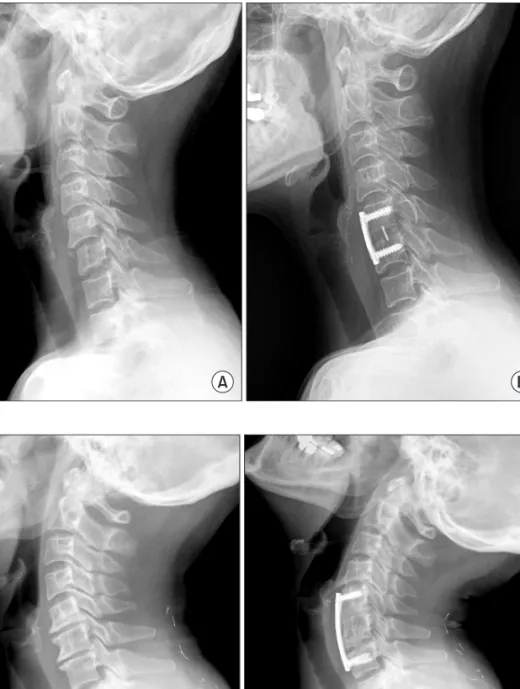
Figure 2. A 61-year-old female patient had anterior cervical discectomy and fusion with Polyethyletherketone (PEEK) cage (Stryker spine, USA) packed with autogenous iliac bone and plate stabiliz- ation for unilateral facet dislocation at C5- 6. (A) Preoperative lateral radiograph. (B) Last follow-up radiograph (postoperative 57 months), it shows no change in adjacent levels.
Figure 3. A 46-year-old female patient had anterior cervical discectomies and fusions with cages and plate for degenerative cervical spondylosis associated with disc herniation at C5-6, 6-7. (A) Preoperative lateral radiograph. (B) Last follow-up radiograph (postoperative 49 months), it shows ossification in cranial adjacent disc space.
외상군에서 척수손상이 발생한 경우는 18예(64%)로 모두 불 완전 척수손상이었다. 이 중 인접분절 골화가 발생한 경우는 9예 로 척수손상과 골화의 발생과는 통계학적 연관을 보이지 않았다 (p=0.2264).
고 찰
경추 전방 감압 및 유합술 후 지연형 합병증으로 발생하는 인접분 절의 골화 발생은 정확한 발병 기전은 밝혀지지 않았지만 일반적 으로 골화가 발생하지 않는 전방 종인대 부위에 골화가 진행되는 이소성 골형성의 일종으로 설명되고 있다.9,11) 이소성 골형성은 외 상, 인공관절 치환술, 중추신경계 손상 후 발생되는 것으로 보고되 고 있으며 대개 외상이나 수술 후 2에서 12주 후부터 관찰되기 시 작하여 성숙까지는 1-2년의 시간이 걸리는 것으로 알려져 있다.
발생의 위험인자는 명확하지 않지만 골주위 연부조직 손상이 심 하고 운동이 제한된 상태에서 더 쉽게 발생한다고 하였다.12-16) 저자들의 경우 골화가 처음 관찰된 시기는 술 후 평균 26.3개월 이었고 이는 일반적인 이소성 골형성의 발현시기와 큰 차이를 보 였다. Yang 등8)은 금속판을 이용하지 않은 전방 경추 유합술 후 인접분절에서 관찰된 골극은 모든 예에서 6개월에서 12개월 사 이에 관찰되었다고 하였고 Park 등11)은 금속판을 이용한 전방 경 추 유합술 후 인접분절에 발생한 골화의 경우 술 후 12개월 내에 나타나지 않은 경우 추시 중에 더 진행될 가능성은 적다고 하였 다. 저자들의 경우 인접분절의 골화가 처음 관찰된 경우가 술 후 2년 이후였던 경우가 절반 이상(56%)이었다. 골화의 발생 시기에 대해서는 이전의 보고와 비교하여 좀 더 장기간의 추시가 필요한 것으로 보인다.
수술과 관계된 인접분절 골화의 발생 위험 인자에 대하여 여러 보고가 있었고 이는 전종인대 및 경장근의 과도한 박리, Caspar 핀에 의한 전방 종인대의 자극 등 주로 술 중 발생하는 연부조직 손상과 관련된 것이었다.5,8,17) 저자들은 연부조직 손상이 인접분 절 골화 형성에 미치는 영향을 확인하기 위해 퇴행성 질환군과 외상군에서의 발생빈도를 비교하였다. 퇴행성 질환군의 경우 술 중 발생하는 연부조직 손상을 줄이기 위해 전종 인대는 병변 부 위 추간판 높이만큼만 제거하였고 상 하 연결부위는 남겨두었으 며 경장근의 박리도 최소화하였다. 반면 외상군의 경우 수술 소 견상 대부분 전종 인대나 경장근의 심한 손상이 관찰되었고 이는 유합 분절뿐 아니라 근위부 및 원위부로도 손상이 이어져 있는 경우가 많았다. 술 중 연부조직 손상이 더 심했던 외상군에서 골 화의 발생이 더 증가할 것으로 생각되었으나 조사 결과 양 군 간 의 유의한 차이는 보이지 않았다.
금속판을 이용한 전방 고정술은 전방 유합술에 있어서 전만 의 유지 및 유합률의 향상에 있어서 장점이 있지만 이와 관련된 adjacent level ossification development (ALOD) 발생에 대한 여러 보고가 있었다.5-9) 특히 금속판과 인접분절 추간판 간의 거리는 ALOD의 발생과 밀접한 연관이 있는 것으로 보고되고 있지만 본 연구에서는 이전의 연구에 부합되는 결과를 얻을 수 없었다.
경추간 유합술은 인접분절에 가해지는 응력 및 추간판의 압력 을 증가시키는 것으로 보고되어 있으며 이는 유합 범위가 증가할 수록 그 영향이 더 커지는 것으로 알려져 있다.18-21) 본 연구에서는 인접분절의 골화 발생에 영향을 미치는 인자로 유합 범위만이 유 의한 상관 관계를 보였다. 유합 범위의 증가에 따른 응력의 증가 가 인접분절의 전방 종인대에 장기간 반복적인 자극을 주어 골화 발생을 야기하였을 것으로 생각된다. 다만 인접분절의 퇴행성 변 Figure 4. A 40-year-old male patient had anterior cervical discectomies and fusions with cages and plate for degenerative cervical spondylosis associated with disc herniation at C5-6, 6-7. (A) Preoperative lateral radiograph. (B) After follow-up of 15 months it shows no definite change in adjacent level. (C) Lateral radiograph at 34 months after surgery shows ossification in caudal adjacent disc space.
화는 일반적으로 운동 범위의 증가와 함께 불안정성을 동반하여 나타나지만20,22,23) 인접분절의 골화 발생은 오히려 운동을 감소시 켜11) 인접분절을 안정화시키는 양상을 보인다. 따라서 인접분절 의 골화 발생은 인접분절의 퇴행성 변화와는 별개의 현상으로 해 석되어야 한다고 생각되며 여기에 대해서는 임상적 결과의 비교 와 함께 다시 논의가 필요할 것으로 보인다.
결 론
금속판을 이용한 전방 감압 및 유합술 후 인접분절의 골화를 관 찰하기까지는 이전의 보고와 비교하여 수술 후 상당 기간의 추시 가 필요하였다. 골화의 발생 빈도는 외상군과 질병군간의 비교에 서 차이를 보이지 않았고 유합 범위만이 골화의 발생에 영향을 미치는 것으로 관찰되었다. 따라서 인접분절 골화의 발생에 있어 술 중 발생하는 연부조직 손상정도보다는 유합에 따른 인접분절 의 생역학적 변화가 더 큰 영향을 미치는 것으로 생각한다.
참고문헌
1. Bohlman HH, Emery SE, Goodfellow DB, Jones PK. Robin- son anterior cervical discectomy and arthrodesis for cervi- cal radiculopathy. Long-term follow-up of one hundred and twenty-two patients. J Bone Joint Surg Am. 1993;75:1298-307.
2. Braunstein EM, Hunter LY, Bailey RW. Long term radiograph- ic changes following anterior cervical fusion. Clin Radiol.
1980;31:201-3.
3. Gore DR, Sepic SB. Anterior cervical fusion for degenerated or protruded discs. A review of one hundred forty-six pa- tients. Spine (Phila Pa 1976). 1984;9:667-71.
4. Goto S, Mochizuki M, Kita T, et al. Anterior surgery in four consecutive technical phases for cervical spondylotic myelop- athy. Spine (Phila Pa 1976). 1993;18:1968-73.
5. Goffin J, van Loon J, Van Calenbergh F, Plets C. Long-term results after anterior cervical fusion and osteosynthetic stabili- zation for fractures and/or dislocations of the cervical spine. J Spinal Disord. 1995;8:500-8.
6. Kim KS. Long-term effects on the cervical spine after anterior locking plate fixation. J Korean Neurosurg Soc. 2001;30:493- 500.
7. Park JB, Cho YS, Riew KD. Development of adjacent-level ossification in patients with an anterior cervical plate. J Bone Joint Surg Am. 2005;87:558-63.
8. Yang JY, Song HS, Lee M, Bohlman HH, Riew KD. Adjacent level ossification development after anterior cervical fusion
without plate fixation. Spine (Phila Pa 1976). 2009;34:30-3.
9. Park JY, Zhang HY, Oh MC. New technical tip for anterior cervical plating: make hole first and choose the proper plate size later. J Korean Neurosurg Soc. 2011;49:212-6.
10. Smith GW, Robinson RA. The treatment of certain cervical- spine disorders by anterior removal of the intervertebral disc and interbody fusion. J Bone Joint Surg Am. 1958;40:607-24.
11. Park JB, Watthanaaphisit T, Riew KD. Timing of development of adjacent-level ossification after anterior cervical arthrodesis with plates. Spine J. 2007;7:633-6.
12. Modabber MR, Jupiter JB. Reconstruction for post-trau- matic conditions of the elbow joint. J Bone Joint Surg Am.
1995;77:1431-46.
13. Garland DE, Orwin JF. Resection of heterotopic ossification in patients with spinal cord injuries. Clin Orthop Relat Res.
1989;(242):169-76.
14. Hasegawa M, Ohashi T, Uchida A. Heterotopic ossification around distal femur after total knee arthroplasty. Arch Orthop Trauma Surg. 2002;122:274-8.
15. Sperling JW, Cofield RH, Rowland CM. Heterotopic os- sification after total shoulder arthroplasty. J Arthroplasty.
2000;15:179-82.
16. Purtill JJ, Eng K, Rothman RH, Hozack WJ. Heterotopic os- sification. Incidence in cemented versus cementless total hip arthroplasty. J Arthroplasty. 1996;11:58-63.
17. Mähring M. Segment changes in the cervical spine following cervical spondylodeses of unstable injuries. Unfallchirurgie.
1988;14:247-58.
18. Nagata H, Schendel MJ, Transfeldt EE, Lewis JL. The effects of immobilization of long segments of the spine on the adjacent and distal facet force and lumbosacral motion. Spine (Phila Pa 1976). 1993;18:2471-9.
19. Döhler JR, Kahn MR, Hughes SP. Instability of the cervical spine after anterior interbody fusion. A study on its incidence and clinical significance in 21 patients. Arch Orthop Trauma Surg. 1985;104:247-50.
20. Fuller DA, Kirkpatrick JS, Emery SE, Wilber RG, Davy DT. A kinematic study of the cervical spine before and after segmen- tal arthrodesis. Spine (Phila Pa 1976). 1998;23:1649-56.
21. Garrido BJ, Wilhite J, Nakano M, et al. Adjacent-level cervical ossification after Bryan cervical disc arthroplasty compared with anterior cervical discectomy and fusion. J Bone Joint Surg Am. 2011;93:1185-9.
22. Aota Y, Kumano K, Hirabayashi S. Postfusion instability at the
adjacent segments after rigid pedicle screw fixation for degen- erative lumbar spinal disorders. J Spinal Disord. 1995;8:464- 73.
23. Kolstad F, Nygaard ØP, Leivseth G. Segmental motion adja- cent to anterior cervical arthrodesis: a prospective study. Spine (Phila Pa 1976). 2007;32:512-7.
Factors Affecting Adjacent Level Ossification Development after Anterior Cervical Discectomy and Fusion
Eo-Sub Jeong, M.D., Byeong-Yeol Choi, M.D., and Chung-Sik Shin, M.D.
Department of Orthopedic Surgery, Presbyterian Medical Center, Jeonju, Korea
Purpose: The purpose of this study is to analyze the affecting factors of adjacent level ossification development (ALOD) after anterior
cervical discectomy and fusion.Materials and Methods: This study enrolled 75 patients who underwent anterior cervical discectomy and fusion and were
followed-up for more than two years. Twenty-five patients were related with trauma and 47 patients were diagnosed as degenerative cervical disorder. We assessed the incidence, location and timing of ALOD, and compared the incidence of ossification between trauma and degenerative disease groups to know the effect of soft tissue damage. We also reviewed the correlation between the development of ossification at adjacent level and the factors, such as fusion level, age, operation time, duration of follow-up, and the presence of ossification of posterior longitudinal ligament (OPLL), as well as ossification of yellow ligament (OYL).Results: Ossification developed in 33 patients (44%). Five cases (15%) were detected during the first year after surgery, 10 (30%)
cases detected during the second year after surgery, 13 (40%) between second and third year, and 5 (15%) cases of more than three years after surgery. Only the fusion level was related with the development of ossification statistically (p<0.001). Age, operation time, duration of follow-up, sex ratio, presence of OPLL, and OYL were not related with the incidence of ossification significantly. There was no significant difference in the incidence of ALOD between the trauma group and degenerative disease group (p=0.3625).Conclusion: To detect ALOD, it needs a long time for follow-up after surgery. We thought that ALOD is affected by excessive loading
at the adjacent level after fusion rather than severity of the soft tissue damage.Key words: cervical vertebrae, anterior discectomy and fusion, adjacent level ossification development
Received June 1, 2012 Revised July 13, 2012 Accepted September 21, 2012 Correspondence to: Byeong-Yeol Choi, M.D.
Department of Orthopedic Surgery, Presbyterian Medical Center, 365, Seowon-ro, Wansan-gu, Jeonju 561-750, Korea
TEL: +82-63-230-8744
FAX: +82-63-230-8819 E-mail: docby@hanmail.netCopyright © 2012 by The Korean Orthopaedic Association
“This is an Open Access article distributed under the terms of the Creative Commons Attribution Non-Commercial License (http://creativecommons.org/licenses/by-nc/3.0/) which permits unrestricted non-commercial use, distribution, and reproduction in any medium, provided the original work is properly cited.”
The Journal of the Korean Orthopaedic Association Volume 47 Number 6 2012