소아 부신성백질이영양증은 성염색체 열성의 유전성 대사 고신호강도를 보여 뇌실, 뇌조, 뇌구주위 등 뇌척수액과 인접
F L A I R기법을 이용한 소아 부신성백질이영양증의 자기공명영상 소견 : T2 강조영상과의 비교1
황아실이・서정진・정광우・정태웅・정용연・강형근・국 훈2・우영종2・황태주2
목적 : 소아 부신성백질이영양증에서 fluid attenuated inversion recovery(FLAIR) 영상과 T 2 강조영상소견을 비교 분석하여, 진단에 있어서 FLAIR 영상의 효용성을 평가해보고, 자기공 명영상소견과 임상증상과 연관성을 알아보고자 하였다.
대상 및 방법 : 생화학적 방법을 통해 부신성백질이영양증으로 확진된 6명( 6-17세, 평균연
령 1 0 . 2세)의 남아 환아를 대상으로, 1.5T MR기기를 사용하여 F L A I R ( T R / T E / T I = 1 0 0 0 4 / 1 2 3 / 2 2 0 0 )영상과 T2 강조 급속스핀에코( T R / T E = 4 0 0 0 / 1 0 4 )영상을 얻었다. FLAIR 영상에서 병변부위의 발견(detection), 명확도(conspicuity), 파급범위( e x t e n t )를 2명의 방사선과 의사가 합의하에 T2 강조영상과 시각적으로 비교하였다. 정량적으로는 병변/뇌척수액 대조도 (contrast), 병변/뇌척수액 대조도 대 잡음비(contrast to noise ratio(CNR)), 병변/백질 대조 도, 병변/백질 대조도 대 잡음비를 FLAIR 영상과 T2 강조영상에서 비교하였다. 또한 자기 공명영상에서 나타난 병변부위를 크게 시신경계, 청신경계, 운동계로 나누어 임상증상과 비 교하였으며 임상증상이 없었던 환아에서도 고신호강도를 보이는 백질 부위가 있는지 알아 보았다.
결과 : 시각적 평가에서, 병변부위의 발견은 6예중 2예( 3 3 % )에서 FLAIR 영상이 T2 강조
영상보다 우수하였고, 4예에서는 비슷하였다. 병변부위의 명확도와 파급정도는 6예 모두 FLAIR 영상에서 T2 강조영상보다 우수하였다. 정량적인 평가에서 병변/뇌척수액 대조도와 병변/뇌척수액 C N R은 FLAIR 영상이 T2 강조영상보다 우월하였으며 병변/백질 대조도와 병변/백질C N R은 T2 강조영상보다 열등하였다. 1예에서는 FLAIR 영상에서 고신호강도의 병변부위 내 저신호강도가 보여 병변부위 내 뇌연화성 변화를 한 부분을 구분하여 볼 수 있었다. 자기공명영상에서 시신경계, 청신경계, 운동계의 침범부위와 임상증상과 비교시, 임 상증상이 있었던 4예 모두에서 영상소견과 임상증상이 부합하였는데, 이중 1예에서는 운동 계 증상이 없었으나 추체계를 포함한 운동계에 고신호강도를 보였다. 또한 임상증상이 없었 던 2예에서도 자기공명영상에서 고신호강도를 보이는 백질병변을 발견할 수 있었다.
결론 : F L A IR 영상은 소아 부신성백질이양증을 진단하는데 있어서 T2 강조영상보다 비슷
하거나 더 나은 영상을 제공하였으며, 자기공명영상에서 나타난 병변부위는 임상 증상과 잘 부합되었고 또한 증상이 없었던 환아에서도 자기공명영상에서 병변부위를 발견할 수 있었다.
척수액이 신호가 없어지거나 매우 약화되어 뇌실주변의 백질 의 병변이 더욱 더 명확히 나타나서 병변의 발견율이 높아진 다는 보고가 있다(8).
이에 저자들은 후두엽을 중심으로한 뇌측실 주위 백질부위 를 침범하는 것으로 알려져 있는 소아 부신성백질이영양증에 서 FLAIR 영상과 T2 강조영상소견을 비교 분석하여 진단에 있어서 FLAIR 영상의 효용성을 평가해보고, 자기공명영상소 견과 임상증상과 연관성을 알아보고자 한다.
대상 및 방법
1 9 9 6년 1월부터 1 9 9 8년 6월까지 자기공명영상을 시행한 6명 의 소아 부신성백질이영양증 환자를 대상으로 하였는데, 이들 모두 혈장의 장쇄 지방산(very long chain fatty acid)의 증 가를 확인함으로서 진단되었다. 연령분포는 6 - 1 7세였고, 평균 연령은 1 0 . 2세였으며, 6명 모두 남아였다.
자기공명영상은 1.5T MR기기(Signa Horizon, GE Medical Systems, Milwaukee, USA)를 이용하여 시상면과 축상면의 T 1 강조영상을 얻었고, 축상면의 급속 스핀에코 T2 강조영상과 FLAIR 영상을 얻 었 다 . 촬 영 기 법 은 T1 강조영상은 TR/TE=450-500/8-14msec, T2 강조영상 TR/TE=4000/ 104m- sec, FLAIR 영상은 T R / T E / T I = 1 0 0 0 4 / 1 2 3 / 2 2 0 0 m s e c로 하였으 며, matrix size는192x256, FOV는 2 2 c m으로 하였다.
먼저 시각적인 평가로서 FLAIR 영상과 T2 강조영상 각각 에서 병변의 뚜렷한 정도를 4등급으로 나눠 1 )보이지 않는 병 변, 2)있는 듯 없는 듯한 병변(equivocal), 3)병변의 존재가 분 명하지만 덜 뚜렷한 병변, 4) 병변의 존재가 뚜렷하고 명확한 병변중 하나로 평가하여, 병변부위의 발견(detection), 파급범 위( e x t e n t )정도와 동일한 병변의 두 영상에서의 명확도( c o n- s p i c u i t y )를 2명의 방사선과 의사가 합의하에 비교 분석하였다.
객관적인 정량적 평가로서 FLAIR 영상과 T2 강조영상 각 각에서 정상백질, 뇌척수액, 병변, 배경의 신호강도를 크기가 0 . 2 - 0 . 8 c m2인 원형 c u r s e r로 2 - 3회 측정하여 평균값을 구하였다.
이 평균값으로부터 각 영상에서 병변/뇌척수액 대조도( l e- sion/CSF contrast)=(병변의 신호강도-뇌척수액 신호강도) / 뇌척수액 신호강도로, 병변/뇌척수액 대조도 대 잡음비( l e- sion/CSF contrast to noise ratio(CNR))=(병변의 신호강도-뇌 척수액 신호강도) /배경신호강도의 표준편차로, 병변/백질 대 조도(lesion/WM contrast)=(병변의 신호강도-백질 신호강 도) /백질의 신호강도, 병변/백질 대조도 대 잡음비(lesion to WM CNR)=(병변의 신호강도-백질 신호강도) /배경신호강도 의 표준편차로 구하여 FLAIR 영상과 T2 강조영상간의 통계 적 차이가 있는지를 Exact Wilcoxon test로 알아보았다.
자기공명영상에서 나타난 병변부위를 크게 시신경계, 청신 경계, 운동계로 나누어, 임상증상이 있었던 경우에서 영상소견 과 임상증상과의 연관성을 알아보았고 임상증상이 없었던 환 아에서도 고신호강도를 보이는 백질 부위가 있는지 알아보았 다.
결 과
시각적 평가(Table 1)에서 병변부위의 발견은 6예중 2예 ( 3 3 . 3 % )에서 뇌실주변부, 뇌구주변, 뇌저조 주변 부위 등의 뇌 척수액과 인접한 부위의 백질 병변에서 FLAIR 영상이 T2 강 조영상보다 우월하였으며(Fig.1), 4예( 6 6 . 7 % )에서는 비슷하였 다. 병변부위의 파급정도와 명확도는 6예 모두 FLAIR 영상에 서 T2 강조영상보다 우월하였다. 특히 뇌척수액과 인접한 뇌실 주변부의 백질, 뇌구주변의 대뇌반구의 변두리부위, 우회조 (ambient cistern), 상소뇌조(supeior cerebellar cistern), 뇌
Table 1. Visual Assessment of Lesion between FLAIR and T2WI F L A I R > T 2 W I F L A I R = T 2 W I F L A I R < T 2 W I
D e t e c t i o n 2 4 0
E x t e n t 6 0 0
C o n s p i c u i t y 6 0 0
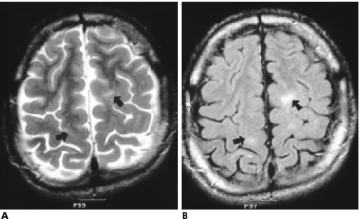
Fig. 1. A 17-year-old boy with visual and motor symptoms.
A) On axial T2-weighted image, the le- sions(arrows) are masked by high s i g- nal intensity of CSF. B) Axial F L A I R image shows linear high signal le- sions(arrows) along superior parietal subcortical white matter.
A B
저조(basal cistern)와 인접한 뇌간과, 뇌교각 부위( c e r e b r o p o n- tine angle area)에서 잘 볼 수 있었다(Fig.2).
정량적인 평가(Table 2)에서는 FLAIR 영상에서 병변/뇌척 수액 대조도와 병변/뇌척수액 C N R이 T2 강조영상보다 통계
적으로 우월하였고 병변/백질 대조도와 병변/백질 C N R은 T 2 강조영상보다 열등하였다( P<0 . 0 5 ) .
1예에서는 T2 강조 영상에서 모두 고신호강도를 보인 병변 부위가 FLAIR 영상에서는 고신호강도의 병변부위 내 저신호 강도가 보여 병변부위 내 뇌연화성 변화를 한 부분을 구분하 여 볼 수 있었다( F i g . 3 ) .
자기공명영상에서 시신경계, 청신경계, 운동계의 침범부위와 임상증상과 비교시(Table 3), 임상증상이 있었던 4예 모두에서 영상소견과 임상증상이 일치하였는데, 이중 1예에서는 운동계 증상이 없었으나 추체계를 포함한 운동계에 고신호강도를 보 였다. 임상증상이 없었던 2예에서도 자기공명영상에서 고신호 강도를 보이는 백질의 병변을 발견할 수 있었다(Fig.4). 1예에 서는 병변부위가 후두엽을 포함한 뇌후방의 백질, 시신경계, 그리고 내포후각 부위였고 또 다른 1예에서는 주로 추체계를 Table 2. Quantitative Assessment of FLAIR and T2WI in the
Lesion of Adrenoleukodystrophy(n=6)
F L A I R T 2 W I p - v a l u e Lesion/CSF contrast 1 9 . 7 2 0 . 1 9 0 . 0 1 6 Lesion/CSF CNR 5 4 . 7 4 2 2 . 5 8 0 . 0 3 Lesion/WM contrast 0 . 9 9 1 . 7 2 0 . 0 1 6 Lesion/WM CNR 2 7 . 6 1 6 6 . 6 6 0 . 0 1 6 Note : CNR=contrast to noise ratio, WM=white matter
CSF=cerebrospinal fluid
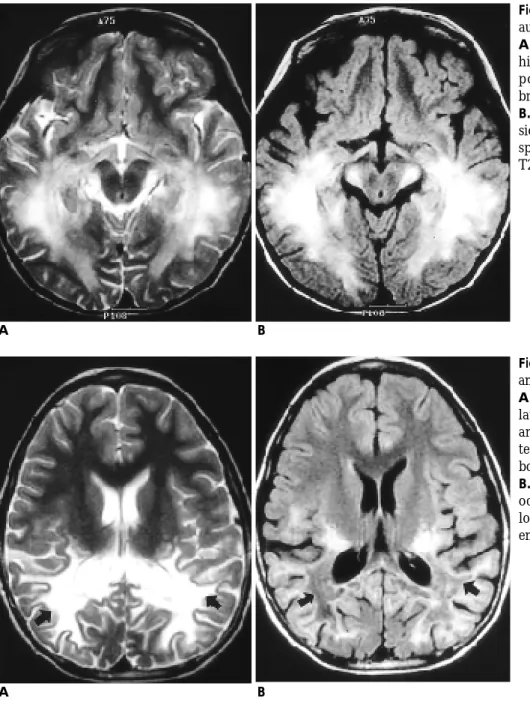
Fig. 2. A 6-year-old boy with visual, auditory, and motor symptoms.
A. Axial T2-weighted image shows high signal intensities in bilateral tem- poro-occipital white matter and brachium of inferior colliculus.
B. On FLAIR image, the high signal le- sions appear to be more discrete, con- spicuous, and extensive than those of T2-weighted image.
A B
Fig. 3. A 9-year-old boy with visual and auditory symptoms.
A. Axial T2-weighted image shows bi- lateral confluent high signal intensity areas in temporo-occipital white mat- ter(arrows) and both lateral geniculate bodies.
B. On FLAIR image, bilateral temporo- occipital white matter lesions appear low signal intensity, which indicating
포함한 운동계였다.
고 찰
부신성백질이영양증은 성염색체 열성 유전질환으로 p e r o x i-
some 내에 있는 효소(Liganoceroyl-coASH ligase) 결핍에 의하여 장쇄 포화지방산(very long chain fatty acid)이 대뇌 조직에 침 착하여 백질의 진행성 탈수초화를 일으키는 질환이다(1-7). 소 아 부신성백질이영양증의 병변은 후두엽의 심부 백질에서 시 작하여 난형중심(centrum semiovale)이나 내포(internal capsule)
Table 3. Correlation of Clinical Symptoms and Lesions on MRI
Clinical Symptoms
Visual Pathway
A u d i t o r y P a t h w a y
Motor Pathway
White matter in cerebral hemisphere ( m a i n l y
parieto-occipital area)
Impaired hearing Impaired vision E x o t r o p i a Decreased i n t e l l i g e n c e Optic radiation Lateral geniculate b o d y
Medial geniculate b o d y
Auditory radiation
Internal capsule ( P o s t . l i m b ) Cerebral peduncle Pyramidal tract
+
Impaired hearing Impaired vision Gait disturbance D e m e n t i a
Optic radiation Lateral geniculate b o d y
Lateral lemniscus Brachium of inferi- or colliculus Medial geniculate b o d y
Auditory radiation Internal capsule ( P o s t . l i m b ) Cerebral peduncle Pyramidal tract
+
Impaired vision Progressive gait d i s t u r b a n c e R t . h e m i p a r e s i s
Optic radiation Lateral geniculate b o d y
-
Internal capsule ( P o s t . l i m b ) Cerebral peduncle Pyramidal tract
+
Impaired hearing Impaired vision Q u a d r i p l e g i a Attention deficit
Optic radiation Lateral geniculate b o d y
Medial geniculate b o d y
Auditory radiation
Internal capsule ( P o s t . l i m b ) Cerebral peduncle Pyramidal tract
+
A s y m p t o m a t i c
Optic radiation Lateral geniculate b o d y
-
Internal capsule ( P o s t . l i m b )
+
A s y m p t o m a t i c
-
-
Internal capsule ( P o s t . l i m b ) Cerebral peduncle Pyramidal tract
+
Case No 1 * 2 3 4 5 6
A g e / S e x 9 / M 6 / M 1 7 / M 1 0 / M 7 / M 1 2 / M
Note :+ - High signal intensity in white matter of cerebral hemisphere, mainly parieto-occipital area on T2WI and FLAIR image
*- Patient without motor symptom reveals high signal intensity in motor pathway on MRI
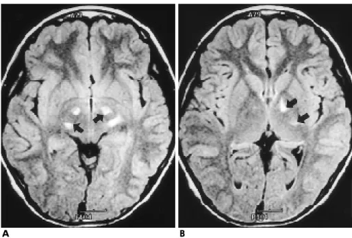
Fig. 4. A asymptomatic 12-year-old boy with adrenoleukodystrophy.
A , B) Axial FLAIR images demonstrate high signal intensities in both cerebral peduncles (arrows) and in the posteri- or limb of left internal capsule (ar- r o w s ) .
A B
를 통하여 전방으로 진행하는 것이 보통이고 드물게 전두엽을 주로 침범한 병변에서는 후방으로 병변이 진행하는 것으로 알 려져 있다(1-7). 저자들의 경우에서도 증상이 있었던 4예와 증 상이 없었던 2예에서 병변은 후두엽의 심부 백질에서 시작하 여 난형중심(centrum semiovale)이나 내포(internal capsule)를 통하여 전방으로 진행하였다.
이러한 소아 부신성백질이영양증에서 변성을 일으킨 백질부 위는 T2 이완시간이 증가되어 T2 강조영상에서 정상 뇌조직의 신호강도보다 고신호강도로 보인다(2). 그러나 T2 강조영상에 서는 뇌척수액보다 뇌실질의 신호가 빨리 소실되고, 이에 대해 상대적으로 강한 신호강도를 보이는 뇌척수액의 움직임이 인 공음영을 형성하고 부분 용적 효과를 나타내어 영상의 질이 저하된다(8-10). FLAIR 영상은 1 8 0 o의 반전 펄스( i n v e r s i o n pulse), 긴 반전시간(inversion time: TI)과 에코시간(echo time:
T E )을 이용해 뇌척수액의 신호를 없애거나 매우 약화시키고, heavily T2 강조영상을 만들어 냄으로써 병변조직이 가장 밝은 고신호강도를 나타나게하여 병변의 인지도를 증가시키는 방법 이다. 따라서 뇌척수액과 인접한 대뇌반구의 변두리, 기저부 뇌조 주위(basal cistern), 뇌간(brain stem), 회질-백질의 경계부, 뇌실 주위(periventricular) 병변의 진단에 유용하다. 여러 연구 에서 이러한 FLAIR 영상이 뇌경색, 다발성 경화증 등 탈수초 질환, 염증성 질환, 두부손상, 뇌지주막하 출혈, 뇌종양등의 광 범위한 뇌 질환에서 민감성이 높다고 하였다(8-12). 특히 뇌백 질 질환에 있어서 뇌실 주변의 백질 병변은 까맣게 나타나는 뇌척수액과 저음영으로 보이는 백질을 배경으로 더욱 더 명확 하게 보일 수 있다. 여러 연구에서 다발성경화증을 포함한 다 양한 백질질환에서 FLAIR 영상의 효용성에 대해 언급하고 있 는데, 대부분의 경우에서 T2 강조영상과 비교하였을 때, 병변 의 명확성이나 범위, 갯수, 병변대 잡음비등이 FLAIR 영상에 서 같거나 보다 나은 결과를 나타내었고(10-11), 저자들의 연구 에서도 FLAIR 영상에서 T2 강조영상보다 시각적으로 병변이 더 뚜렷이 나타났으며 또한 높은 병변/뇌척수액 대조도와 병 변/뇌척수액 C N R을 보여 이와 일치한 결과를 보였다.
FLAIR 영상에서 T2 강조영상보다 대뇌각(cerebral peduncle), 추체계(pyramidal tract), 측모대(lateral lemniscus)등의 뇌기저 조 주변부, 뇌간부위의 백질병변이 더 잘 보였는데 이는 뇌척 수액에서 나오는 신호강도를 없애 뇌척수액으로 인한 잡상이 감소되고 병변과 정상조직의 대조도가 크게 향상되었기 때문 으로도 생각되지만 또한 긴 T E를 사용한 FLAIR 영상에서는 정상적으로 많은 백질의 신경로( t r a c t )가 고신호강도를 보일 수 있기 때문으로도 생각된다. 정상적으로 고신호강도를 보일
할수 있다고 하였다. 이 연구에서도 1예(Fig. 3)에서는 T2 강조 영상에서 모두 고신호강도를 보인 병변부위가 FLAIR 영상에 서는 오래되어 뇌연화성 변화를 한 부분이 저신호강도를 보여 병변부위내 뇌연화성 변화를 한 부분을 구분하여 볼 수 있었 다.
김 등( 6 )은 소아 부신성백질이영양증에서 자기공명영상에서 나타난 백질의 병변부위가 임상증상과 잘 부합된다고 하였고, 이 연구의 결과도 거의 일치하였지만 1예에서는 운동계에 신 경학적 증상이 없었으나 영상소견에서 운동계에 고신호강도 병변이 있었는데, 이는 복잡한 뇌기능에 대한 여러 작용으로 인한 것인지 아니면 임상증상이 발현하기 전 이상소견이 나타 난 것인지 연구가 필요하다. Aubourg등( 1 3 )은 나중에 증상이 나타난 환자에서 아직 증상이 없었을 때 시행한 자기공명영상 에서 백질 병변을 발견할수 있어 임상증상이 나타나기 전에도 자기공명영상 소견으로 질병의 진행과정을 예측할 수 있을 것 이라고 주장하였다. 저자들의 증례에서도 임상증상이 아직 나 타나지 않았던 2예에서 시신경계, 운동계, 그리고 후두엽 백질 에서 이상소견을 볼 수 있었다. 따라서 이 환자들에서도 고신 호강도의 병변의 영상소견으로 질병과정을 예측할 수 있을 것 으로 생각된다.
결론적으로, 비록 적은 수를 대상으로 하였지만 FLAIR 영 상은 소아 부신성백질이양증을 진단하는데 있어서 T2 강조영 상보다 비슷하거나 더 나은 영상을 제공하였으며, 자기공명영 상에서 나타난 병변부위는 임상증상과 잘 부합되었고 또한 증 상이 없었던 환아에서도 자기공명영상에서 병변부위를 발견할 수 있었다.
참 고 문 헌
1. Kumar AJ, Rosenbaum AE, Naidu S, Wener L, et al. Adrenoleu- kodystrophy : correlating MR Imaging with CT. R a d i o l o g y 1987 ; 165 : 497-504
2. Jensen ME, Sawyer RW, Braun IF, Rizzo WB. MR imging appear- ance of childhood adrenoleukodystrophy with auditory, visual, and motor pathway involvement. R a d i o G r a p h i c s 1990 ; 10 : 53-66 3. Uchiyama M, Hata Y, Tada S. MR imaging of adrenoleukodystro-
phy. N e u r o r a d i o l o g y 1991 ; 33 : 25-29
4. Aubourg P, Adamsbaum C, Lavallard-Rousseau MC, Lemaitre A, et al. Brain MRI and electrophysiologic abnormalities in preclinical and clinical adrenomyeloneuropathy. N e u r o l o g y 1992 ; 42 : 85-91 5. 유지숙, 김기중, 고태성, 이백희등. 부신백질이영양증의임상적, 생
화학적소견및뇌자기공명영상소견 . 대한소아신경학회지 1993 ; 1: 50-63
6. 김태경, 김인원, 김우선, 연경모. 소아부신성백질이영양증의자기
arachnoid hemorrhage with fluid-attenuated inversion recovery pulse sequence. R a d i o l o g y 1995 ; 196 : 773-777
11. 이상현, 장기현, 박흥석, 심정석등. 뇌질환의진단에있어서 F L A I R MR Sequence의임상적유용성. 대한방사선의학회지1997 ; 37 : 1-7 12. 공근영, 최우석, 김의종. 뇌경색의급속 FLAIR MR 영상소견: T 2강
조영상과의비교 . 대한방사선의학회지1997 ; 37 : 9-15
13. Aubourg P, Sellier N, Chaussain JL, Kalifa G. MRI detects cerebral involvement in neurologically asymptomatic patients with a- drenoleukodystrophy. N e u r o l o g y 1989 ; 39 : 1619-1621
J Korean Radiol Soc 1999;40:5 91- 5 9 6
Address reprint requests to : Asiry Hwang, M.D., Department of Diagnostic Radiology, Chonnam University Medical School
#8, Hak-dong, Dong-ku, Kwangju, 501-757, Korea Tel. 82-62-220-5745 Fax. 82-62-226-4380
MR Imaging with Fluid Attenuated Inve rsion Re c overy Sequence of Childhood Ad re n o l e u ko d ys t ro p hy :
Comparison with T2 Weighted Spin Echo Imaging
1Asiry Hwang, M.D., Jeong-Jin Seo, M.D., Gwa n g - Woo Jeong, Ph.D., Ta e - Woong Chung, M.D., Yo n g - Yeon Jeong, M.D., Heoung-Keun Kang, M.D.,
Hoon Kook, M.D.2, Young-Jong Woo, M.D.2, Tai-Joo Hwang, M.D.2
1Department of Diagnostic Radiology, Chonnam University Medical School
2Department of Pediatrics, Chonnam University Medical School
Purpose : The purpose of this study was to evaluate the usefulness of FLAIR(Fluid Attenuated Inversion Recovery) MR imaging in childhood adrenoleukodystrophy by comparing with those of T2-weighted FSE imaging, and to correlate MRI finidings with clinical manifestations.
Materials and Methods : Axial FLAIR images(TR/TE/TI=10004/123/2200) and T2-weighted FSE images(TR/TE=4000/104) of brain in six male patients(age range : 6-17 years, mean age : 10.2 years) with biochemically confirmed adrenoleukodystrophy were compared visually by two radiologists for detection, conspicuity, and the extent of lesion. Quantitatively, we compared lesion/CSF contrast, lesion/CSF contrast to noise ratio(CNR), lesion/white matter(WM) contrast, and lesion/WM CNR between FLAIR and T2 weighted image. We correlated MR findings with clinical manifestations of neurologic symptoms and evaluated whether MRI could detect white matter lesions in neurologically asymptomatic patients.
Results : Visual detection of lesions was better with FLAIR images in 2 of the 6 cases and it was equal in the remainders. Visual conspicuity and detection of the extent of lesion were superior on FLAIR images than T2-weighted images in all 6 cases. In the quantitative assessment of lesions, FLAIR was superior to T2- weighted image for lesion/CSF contrast and lesion/CSF CNR, but was inferior to T2 weighted image for lesion/WM contrast and lesion/WM CNR. In one case, FLAIR images distinguished the portion of encephalomalacic change from lesions. MR findings of adrenoleukodystrophy were correlated with clinical manifestations in symptomatic 4 cases, and also detected white matter lesions in asymptomatic 2 cases.
Conclusion : MR imaging with FLAIR sequence provided images that were equal or superior to T2- weighted images in the evaluation of childhood adrenoleukodystrophy. MRI findings were well correlated with clinical manifestations and could detect white matter lesions in neurologically asymptomatic adrenoleukodystrophy patients.
Index words : Brain, MR Brain, diseases Brain, white matter