Distal radius fractures are one of the most frequently treated upper limb injuries1) that are often associated with complications such as impaired mobility and stiffness of
noninvolved finger joints mainly due to posttraumatic and post-immobilization edema of the hand.2-5) Early initiation of joint motion and preservation of adjacent joint motion can be important to prevent finger stiffness after distal radius fractures and should be taken into consideration in selecting treatment and immobilization methods.6,7)
To the best of our knowledge, there are no detailed descriptions or established guidelines regarding proper location of immobilization devices at the distal end of the wrist for preservation of finger motion. In the meantime, the distal volar end of wrist immobilization has been com-
The Effect of Distal Location of the Volar Short Arm Splint on the Metacarpophalangeal
Joint Motion
Joon Yub Kim, MD, Dong Wook Sohn, MD, Ho Youn Park, MD*, Jeong Hyun Yoo, MD, Joo Hak Kim, MD, Myung Gon Jung, MD, Jae Ho Cho, MD
Department of Orthopedic Surgery, Myongji Hospital, Seonam University College of Medicine, Goyang,
*Department of Orthopedic Surgery, Uijeongbu St. Mary’s Hospital, College of Medicine, The Catholic University of Korea, Uijeongbu, Korea
Background: The goals of this study were to compare maximal metacarpophalangeal joint (MCPJ) flexion angles after applica- tion of a volar short arm splint at 3 different locations and verify the relations between the three different physical and radiological locations.
Methods: Forty dominant hands of healthy subjects were analyzed in the study. We defined a transverse skin folding line as a line drawn from the radial aspect of the thenar crease to the ulnar aspect of the distal transverse palmar crease. The distal end of the volar short arm splint was applied on 3 parallel locations to this line. Location A was on this transverse skin folding line; location B was 1 cm proximal to location A; and location C was 1 cm distal to location A. Two orthopedic surgeons measured the maximal MCPJ flexion angles of each finger except the thumb with the application of a volar short arm splint at 3 different locations as well as without a splint as a control. Radiological locations of the 3 different distal ends of the volar short arm splint were also as- sessed by anteroposterior radiographs of the wrist.
Results: When the splint was applied at location A and C, the maximal MCPJ flexion angle decreased to a mean of 83° (91% of control value) and 56° (62% of control value), respectively (compared to the control, p < 0.001). At location B, the maximal MCPJ flexion angle was a mean of 90° (99% of control value); no significant difference was observed compared to the control or without the splint (p = 0.103). On radiography, the average length from the metacarpal head to the distal end of the splint at all fingers de- creased in the order of location B, A, and C (29 mm, 19 mm, and 10 mm, respectively; p < 0.001).
Conclusions: We recommend applying the distal end of a volar short arm splint at proximal 1 cm to the transverse skin folding line to preserve MCPJ motion perfectly, which is located at distal 44% of the whole metacarpal bone length radiologically.
Keywords: Metacarpophalangeal joint, Joint range of motion, Splint
Copyright © 2016 by The Korean Orthopaedic Association
This is an Open Access article distributed under the terms of the Creative Commons Attribution Non-Commercial License (http://creativecommons.org/licenses/by-nc/4.0) which permits unrestricted non-commercial use, distribution, and reproduction in any medium, provided the original work is properly cited.
Clinics in Orthopedic Surgery • pISSN 2005-291X eISSN 2005-4408 Received November 29, 2015; Accepted April 2, 2016
Correspondence to: Dong Wook Sohn, MD
Department of Orthopedic Surgery, Myongji Hospital, Seonam University College of Medicine, 55 Hwasu-ro 14beon-gil, Deogyang-gu, Goyang 10475, Korea
Tel: +82-31-810-6539, Fax: +82-31-810-6900 E-mail: dwsohn@mjh.or.kr
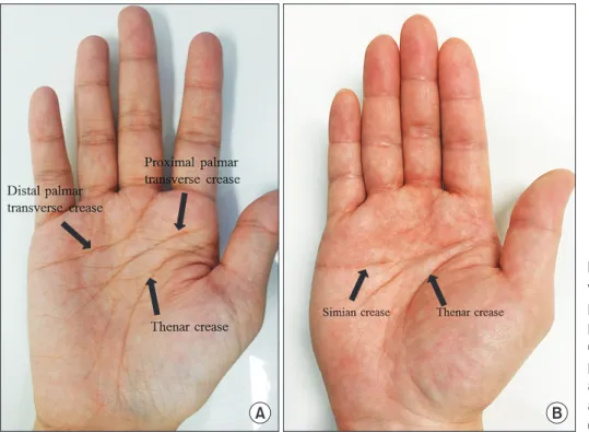
monly located just proximal to the midpalmar region or proximal to the metacarpophalangeal joint (MCPJ) in or- der not to limit MCPJ motion.8-10) However, the definition of midpalmar region is vague and there are too diverse individual palmar crease patterns to use as a reference point during the application of a splint (Fig. 1). Therefore, it would be helpful to establish the proper physical and radiological locations of the distal end of wrist immobili- zation. In this study, we aimed to determine the suitable distal location of wrist immobilization with a short arm splint, the simplest form of wrist immobilization. Radio- logical evaluation is not mandatory to identify the distal location of a splint; however, it could give us additional in- formation on whether the distal end of a splint is properly located.
We hypothesized that there would be a constant ref- erence line for application of the volar short arm splint and radiological findings regarding the distal locations of the volar short arm splint would be informative. The goals of this study were to compare maximal MCPJ flexion angles after application of a volar short arm splint at three differ- ent locations and verify the relationship between the 3 dif- ferent physical and radiological locations.
METHODS
Forty healthy volunteers (20 males and 20 females) with a median age of 31 years (range, 21 to 49 years) were re-
cruited for the study. The exclusion criteria were patients with a history of trauma to the hand and wrist and ail- ments such as arthritis, flexor tenosynovitis, trigger finger, and Dupuytren’s disease in the hand and wrist that could limit the range of motion of the fingers.
A transverse line that forms on the palm during MCPJ flexion between the radial end of the thenar crease and the ulnar end of the distal transverse palmar crease (transverse skin folding line) was drawn. Location A was designated at the center of this line that is usually located between the distal and proximal transverse palmar creases.
Location B was determined at 1 cm proximal to location A. Location C was determined at 1 cm distal to location A, which is usually located between the distal transverse palmar crease and the proximal digital crease. We applied a volar short arm splint on the dominant hand of the study subjects at each location (Fig. 2).
The volar short arm splint was constructed to a size of 4 inch × 15 inch (10 cm × 38 cm). The distal end of the splint was cut to be parallel to the palmar digital crease of each patient and the distal center of the splint was applied to the 3 designated locations. A 4-inch (10-cm) elastic bandage was then applied over the splint to cover the splint completely and prevent movement of the splint.
Two orthopedic surgeons measured the MCPJ flex- ion angle using a manual goniometer. At first, maximal flexion angle of the MCPJ was measured at each finger without application of the splint as control. Measurements
A B
Fig. 1. The crease of midpalmar region is variable and there are diverse individual palmar crease patterns. The transverse palmar creases are of varying patterns depending on the individual as some people do not have separated proximal and distal transverse palmar creases (A) and instead have one transverse palmar crease that is called a simian crease (B).
were repeated after application of the splint at each of the 3 locations; the subjects were instructed to flex their fingers until there was any resistance during flexion.
The locations of the distal end of the volar short
arm splint were evaluated on anteroposterior radiographs of the wrist. The distance from the metacarpal head to the distal end of the splint was measured for every metacarpal bone of all subjects and the ratio to the whole metacarpal bone length was calculated in percentage (Fig. 3).
A sample size calculation was performed with an 80% power to detect a minimum difference of 5° in MCPJ flexion angle assuming a standard deviation of 10°. Al- though the minimum requirement was 33 patients, seven more patients were added to the cohort; therefore, a total of 40 subjects were enrolled in this study.
The Wilcoxon signed-rank test was used to compare the maximal flexion angle of the MCPJ and the radio- graphic location that was measured as the ratio of the dis- tance between the metacarpal head and the distal end of the splint to the total metacarpal bone length. The interra- ter reliability of the measurements was evaluated with use of the intraclass correlation coefficient (ICC). A p-value of less than 0.05 was considered statistically significant. All statistical analyses were conducted with SPSS ver. 16 (SPSS Inc., Chicago, IL, USA).
RESULTS
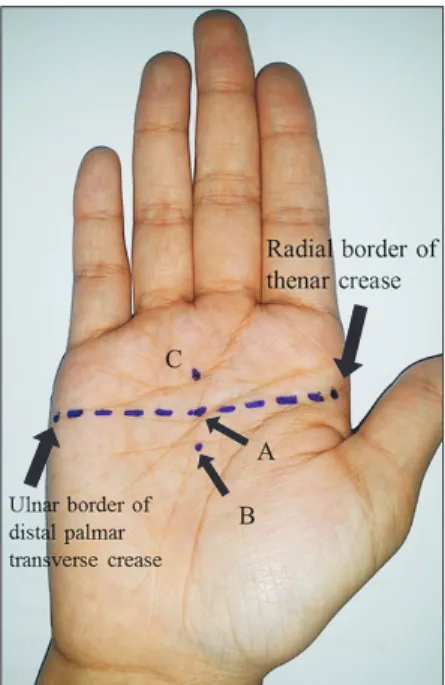
The maximal MCPJ flexion angles according to the 3 dif- ferent distal locations of the volar short arm splint were measured by two observers who were orthopedic surgeons (Table 1). The ICC of the interrater reliability for the mea- surement of maximal MCPJ flexion angle at each location was over 0.80 (excellent agreement) in every measure- ment of the fingers. When the splint was applied at loca- Fig. 2. Three different locations for distal end of the volar short arm
splint were tested. A transverse skin folding line was marked with dashed line between the radial border of the thenar crease and the ulnar border of the distal palmar transverse crease. Location A was marked on the transverse skin folding line, location B was defined as 1 cm vertically proximal to location A, and location C was defined as 1 cm vertically distal to location A.
A B C
Fig. 3. (A) Location A. (B) Location B. (C) Location C. The distance from the metacarpal head to the distal end of the splint (a) and the whole length of the metacarpal bone (b) were measured for every metacarpal bone except for the thumb on the wrist anteroposterior X-ray according to location A, B, and C.
tion A and C, the maximal MCPJ flexion angle decreased to a mean of 83° (91% of control value) and 56° (62% of control value), respectively (compared to the control, p
< 0.001). At location B, the maximal MCPJ flexion angle was a mean of 90° (99% of control value); no significant difference was observed compared to the control (p = 0.103). There was also significant difference in the average maximal MCPJ flexion angles among location A, B, and C (location A vs. location B, location B vs. location C, and location A vs. C, all p < 0.001).
In the comparison of maximal MCPJ flexion angles among fingers, the maximal MCPJ flexion angles of the little finger at location A and C were significantly lower than those of index, middle, and ring fingers (p < 0.001) (Table 1).
The results of radiological evaluation of the distance from the metacarpal head to the distal end of the splint at each finger are described in Table 2. The average length from the metacarpal head to the distal end of the splint at all fingers decreased in the order of location B, A, and C (29 mm, 19 mm, and 10 mm, respectively; p < 0.001). The av-
erage ratio of each location to the whole metacarpal bone length was 44% at location B, 29% at location A and 15%
at location C (p < 0.001).
DISCUSSION
The current study demonstrated that the MCPJ flexion was limited to 91% of the control when the distal end of the volar short arm splint was located at the transverse skin folding line of the palm. The MCPJ flexion was well preserved when the distal end of the volar short arm splint was located at 1 cm proximal to the transverse skin fold- ing line. Since the influence of 91% maximal MCPJ flexion angle compared to the control on finger stiffness has never been reported, we cannot confirm if the decrease is within an acceptable range. However, we believe that the ideal location of the distal end of the volar short arm splint for complete preservation of MCPJ flexion is more proximal to the transverse skin folding line, not on the transverse skin folding line.
At location A and C, the little finger was more limit-
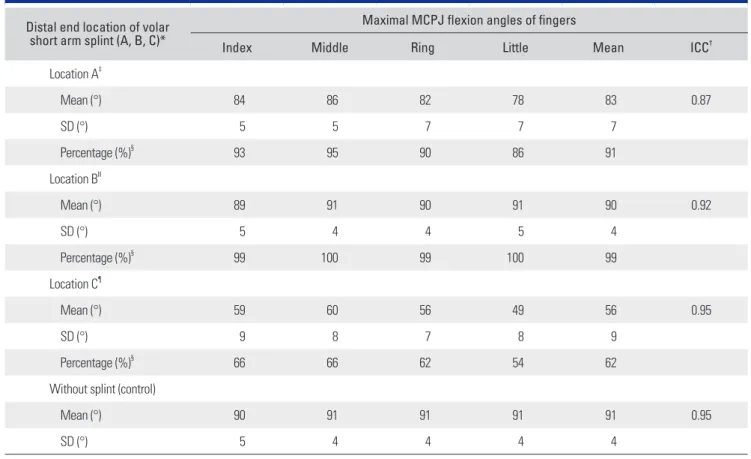
Table 1. Maximal MCPJ Flexion Angle of Fingers According to the Distal End Location of the Volar Short Arm Splint Distal end location of volar
short arm splint (A, B, C)*
Maximal MCPJ flexion angles of fingers
Index Middle Ring Little Mean ICC†
Location A‡
Mean (°) 84 86 82 78 83 0.87
SD (°) 5 5 7 7 7
Percentage (%)§ 93 95 90 86 91
Location BΙΙ
Mean (°) 89 91 90 91 90 0.92
SD (°) 5 4 4 5 4
Percentage (%)§ 99 100 99 100 99
Location C¶
Mean (°) 59 60 56 49 56 0.95
SD (°) 9 8 7 8 9
Percentage (%)§ 66 66 62 54 62
Without splint (control)
Mean (°) 90 91 91 91 91 0.95
SD (°) 5 4 4 4 4
MCPJ: metacarpophalangeal joint, ICC: intraclass correlation coefficient.
*40 Subjects for each. †Intraclass correlation coefficient for interrater reliability. ‡Location A: center of transverse skin folding line. §Percentage of the MCPJ flexion angle compared to control (%). ΙΙLocation B: 1 cm proximal to location A. ¶Location C: 1 cm distal to location A.
ed in MCPJ flexion than other fingers. Therefore, we pos- tulate that a splint with more removed design at the little finger would be preferable for maximal MCPJ flexion.
On the radiological evaluation, the average distance from the metacarpal head to the distal end of the volar short arm splint at location A and B was 19 mm and 29 mm, respectively. The average ratio of the distance to the whole metacarpal bone length was 29% at location A and 44% at location B. Based on these results, we suggest that the distal end of the volar short arm splint should be placed proximal to 29 mm (44%) from the metacarpal head to the whole length of metacarpal bone radiologically to preserve the MCPJ flexion angle perfectly.
Bugbee and Botte11) reported that anatomy text- books and reports paid little attention to the relationships between skin creases of the hand and underlying anatomic structures; therefore, there is a lack of quantitative assess- ments of these relationships and a surprising lack of agree- ment in the descriptions provided. They also reported that a line joining the radial and ulnar borders of the transverse palmar creases usually marked the level of the metacarpal
necks, not the MCPJs. In our study, location A was located at a line joining the radial border of the thenar crease and ulnar border of the transverse palmar crease and the radiological result supports the previous report that the transverse palmar crease corresponds to the metacarpal neck. However, the transverse palmar creases are of vary- ing patterns depending on the individual as some people do not have a separated proximal and distal transverse pal- mar crease and instead have one transverse palmar crease that is called a simian crease. Therefore, we cannot estab- lish the proximal transverse palmar crease or the distal palmar crease as a single reference line. The thenar crease also has a variable morphology and its vertical appearance might not be useful as a reference line for application of a volar short arm splint.
In order to determine the proper distal location of the volar short arm splint, we have to define a reference lo- cation for MCPJ flexion that is identifiable on the surface of the hand. In this respect, we recognize that the central part of the palmar and thenar creases is more variable and the peripheral part of them is likely to be more constant. A Table 2. Radiographic Distance from the Metacarpal Head to the Distal End of the Splint and the Ratio to the Whole Metacarpal Bone Length
Distal end location of volar short arm splint (A, B, C)*
Distance from metacarpal head to distal end of splint
Index Middle Ring Little Total ICC†
Location A‡
Mean (mm) 22 22 17 14 19 0.97
SD (mm) 6 5 5 1 6
Percentage (%)§ 31 32 28 24 29
Location BΙΙ
Mean (mm) 32 32 27 24 29 0.96
SD (mm) 6 6 5 4 6
Percentage (%)§ 44 46 44 42 44
Location C¶
Mean (mm) 14 14 9 5 10 0.96
SD (mm) 6 5 6 6 7
Percentage (%)§ 19 19 14 8 15
Whole metacarpal§ bone length
Mean (mm) 72 69 61 56 65 0.95
SD (mm) 5 5 4 3 8
ICC: intraclass correlation coefficient.
*40 Subjects for each. †Intraclass correlation coefficient for interrater reliability. ‡Location A: center of transverse skin folding line. §Percentage of the metacarpophalangeal joint flexion angle compared to control (%). ΙΙLocation B: 1 cm proximal to location A. ¶Location C: 1 cm distal to location A.
line joining the radial and ulnar borders of the transverse palmar creases or the radial border of the thenar crease and the ulnar border of the distal transverse palmar crease is the constant skin folding line of the MCPJ, which usu- ally marks the level of the metacarpal neck. Therefore, we defined this line as transverse skin folding line and used it as a reference in this study. We believe it could be a useful anatomic landmark for similar surface anatomy and place- ment studies in the future.
There are some limitations in our study. First, MCPJ flexion angle measurements were performed in normal healthy subjects without any trauma; thus, there is a pos- sibility of observing different results from subjects with hand or wrist injuries. Second, this is a time zero study and we do not know whether the restriction of MCPJ flexion by the volar short arm splint might affect MCPJ stiffness.
Third, the resistance that restricted further flexion of the
MCPJ is a subjective value that could be different among subjects. Finally, we need to assess the impact of distal location of the volar short arm splint on finger stiffness in the clinical setting and various locations should be exam- ined for optimal placement of the volar short arm splint after distal radius fracture surgery.
In summary, we recommend that the distal end of the volar short arm splint should be located at minimum 1 cm proximal to the transverse skin folding line to preserve MCPJ motion perfectly, which is located at distal 44% of the whole metacarpal bone length on the radiograph.
CONFLICT OF INTEREST
No potential conflict of interest relevant to this article was reported.
REFERENCES
1. Singer BR, McLauchlan GJ, Robinson CM, Christie J. Epi- demiology of fractures in 15,000 adults: the influence of age and gender. J Bone Joint Surg Br. 1998;80(2):243-8.
2. Warwick D, Field J, Prothero D, Gibson A, Bannister GC.
Function ten years after Colles' fracture. Clin Orthop Relat Res. 1993;(295):270-4.
3. Gutow AP. Avoidance and treatment of complications of dis- tal radius fractures. Hand Clin. 2005;21(3):295-305.
4. Dekkers M, Soballe K. Activities and impairments in the early stage of rehabilitation after Colles' fracture. Disabil Re- habil. 2004;26(11):662-8.
5. Belloti JC, Santos JB, Atallah AN, Albertoni WM, Faloppa F. Fractures of the distal radius (Colles' fracture). Sao Paulo Med J. 2007;125(3):132-8.
6. Teunis T, Bot AG, Thornton ER, Ring D. Catastrophic thinking is associated with finger stiffness after distal radius fracture surgery. J Orthop Trauma. 2015;29(10):e414-20.
7. Broos PL, Fourneau IA, Stoffelen DV. Fractures of the distal radius: current concepts for treatment. Acta Orthop Belg.
2001;67(3):211-8.
8. Levy J, Ernat J, Song D, Cook JB, Judd D, Shaha S. Outcomes of long-arm casting versus double-sugar-tong splinting of acute pediatric distal forearm fractures. J Pediatr Orthop.
2015;35(1):11-7.
9. Bong MR, Egol KA, Leibman M, Koval KJ. A comparison of immediate postreduction splinting constructs for control- ling initial displacement of fractures of the distal radius: a prospective randomized study of long-arm versus short-arm splinting. J Hand Surg Am. 2006;31(5):766-70.
10. Kim JK, Kook SH, Kim YK. Comparison of forearm rota- tion allowed by different types of upper extremity immobili- zation. J Bone Joint Surg Am. 2012;94(5):455-60.
11. Bugbee WD, Botte MJ. Surface anatomy of the hand: the relationships between palmar skin creases and osseous anatomy. Clin Orthop Relat Res. 1993;(296):122-6.