INTRODUCTION
Alzheimer’s disease (AD) is the most common form of de- mentia and is clinically characterized by global deficits in cog- nition.
1Despite differences in etiology, AD and vascular de- mentia often coexist and have overlapping risk factors and
pathologies.
2In addition to old age, the risk factors for AD in- clude hypertension, peripheral arterial disease, cardiovascular diseases, diabetes, and smoking, and the mechanisms underly- ing AD appear to be closely associated with vascular factors.
3Nicergoline is an ergoline derivative that is used for the treat- ment of dementia and other age-related cognitive deficits.
4While nicergoline was initially developed as a vasodilator and main- ly prescribed for cerebrovascular diseases, it has a wider spec- trum of pharmacological and clinical properties leading to its use in various forms of dementia including AD. Several ran- domized controlled trials have investigated the therapeutic
Changes in Regional Cerebral Perfusion after Nicergoline Treatment in Early
Alzheimer’s Disease: A Pilot Study
Jooyeon J. Im,
1* Hyeonseok S. Jeong,
1* Jong-Sik Park,
2YoungSoon Yang,
3Seung-Hee Na,
2Jin Kyoung Oh,
1Yong-An Chung,
1In-Uk Song
2Departments of 1Radiology and 2Neurology, Incheon St. Mary’s Hospital, College of Medicine, The Catholic University of Korea, Seoul, Korea
3Department of Neurology, Veterans Hospital, Seoul Medical Center, Seoul, Korea
Background and Purpose Nicergoline is an ergoline derivative that is used to treat cognitive deficits in cerebrovascular disease and vari- ous forms of dementia. Although therapeutic effects of nicergoline have been established, little is known about its effects on cerebral perfusion in Alzheimer’s disease (AD). The aim of this study was to examine the role of nicergoline in regional cerebral blood flow (rCBF) of AD patients using technetium-99m hexa-methyl-propylene-amine-oxime single photon emission computed tomography (SPECT).
Methods Sixteen patients with early AD underwent a comprehensive clinical assessment including cognitive testing and SPECT scans before and after nicergoline treatment. Nicergoline (30 mg twice daily) was administered for an average duration of 1.5 years. Clinical and cognitive functioning was assessed using the Mini-Mental State Examination, Clinical Dementia Rating (CDR), CDR-Sum of Boxes, Global Deterioration Scale, Barthel Activities of Daily Living Index, Instrumental Activities of Daily Living, and Geriatric Depression Scale.
Results Nicergoline treatment induced changes in the severity of dementia, cognitive function, activities of daily living, and depressive symp- toms, which were not statistically significant. During the follow-up, the patients showed significant increases in their relative rCBF in the superior frontal gyrus, precentral gyrus, and postcentral gyrus.
Conclusions Nicergoline treatment improves perfusion of the frontal and parietal regions in early AD patients. It is possible that the increased perfusion in the superior frontal gyrus may be related to the mechanisms that delay or prevent progressive deterioration of cognitive functions in AD.
Key Words Alzheimer disease, nicergoline, regional cerebral blood flow, single photon emission computed tomography, cognition.
Received: November 22, 2017 Revised: December 1, 2017 Accepted: December 1, 2017
Correspondence: In-Uk Song, MD, PhD, Department of Neurology, Incheon St. Mary’s Hospital, College of Medicine, The Catholic University of Korea, 56 Dongsu-ro, Bupyeong-gu, Incheon 21431, Korea
Tel: +82-32-280-5010, Fax: +82-32-280-5244, E-mail: [email protected]
*These authors contributed equally to this work.
cc This is an Open Access article distributed under the terms of the Cre- ative Commons Attribution Non-Commercial License (http://creative- commons.org/licenses/by-nc/4.0) which permits unrestricted non-com- mercial use, distribution, and reproduction in any medium, provided the ori- ginal work is properly cited.
DND
Print ISSN 1738-1495 / On-line ISSN 2384-0757
Dement Neurocogn Disord 2017;16(4):104-109 / https://doi.org/10.12779/dnd.2017.16.4.104
ORIGINAL ARTICLE
DND
efficacy of nicergoline in patients with dementia and demon- strated that nicergoline treatment improved or prevented the deterioration of cognitive symptoms in AD patients,
5-7as well as in patients with senile dementia, vascular, or mixed type.
8-11Nicergoline plays a role in the molecular and cellular patho- physiology of dementia. In in vitro and animal studies, nicergo- line has been reported to act as an α1-adrenoceptor antagonist, resulting in vasodilation and blood flow increase,
12choliner- gic neurotransmission,
13-15enhanced noradrenaline and do- pamine turnover,
16cerebral metabolic activity,
17,18and neu- roprotection.
19-21Moreover, nicergoline has been shown to mediate neuronal signal transduction by modulating phos- phoinositide pathway, protein kinase C (PKC) translocation, and PKC-mediated α-secretase processing of amyloid precur- sor protein implicated in the pathophysiology of AD.
22-24While the clinical effects and potential mechanisms of nicer- goline on AD have been studied, its effects on the brains of AD patients remain unclear. A single study used electroen- cephalography to measure neural activities correlated with nicergoline treatment in senile dementia of Alzheimer type and multi-infarct dementia.
11The aim of this study was to elucidate the effects of nicergoline on regional cerebral blood flow (rCBF) in early AD patients using single photon emis- sion computed tomography (SPECT).
METHODS
Participants
Sixteen patients with AD were recruited at Incheon St.
Mary’s Hospital (Incheon, Korea). The diagnosis of AD was made according to the Diagnostic and Statistical Manual of Mental Disorders-IV criteria,
25and the National Institute of Neurological and Communicative Disorders and Stroke and the Alzheimer’s Disease and Related Disorders Association criteria.
26Patients with a history of head trauma, epilepsy, stroke, mixed or vascular dementia, radiological findings on magnetic resonance imaging (MRI), or other neurological or psychiatric disorders were excluded from the study.
The study was approved by the Institutional Review Board of the Incheon St. Mary’s Hospital. Written informed consent was obtained from all participants.
Nicergoline administration
Patients received oral nicergoline at a dose of 30 mg twice daily for 1.5 years on average. All patients were also undergo- ing treatment with acetylcholinesterase inhibitors (AChEI) for AD at the time of the study. Safety assessments including ad- verse events, physical examinations, monitoring of vital signs, electrocardiography, and laboratory tests were performed.
Clinical assessment
All patients underwent a comprehensive clinical assess- ment including a detailed medical history and neurological examination, by board-certified neurologists. Global cogni- tive functioning was evaluated with the Mini-Mental State Ex- amination (MMSE).
27Assessments for dementia severity in- cluded the Clinical Dementia Rating (CDR),
28CDR-Sum of Boxes (CDR-SB), and Global Deterioration Scale (GDS).
29The Barthel Activities of Daily Living Index (Barthel ADL In- dex)
30and Instrumental Activities of Daily Living (IADL)
31were used to evaluate functional status. The Geriatric Depres- sion Scale (GDS-Depression) was used to assess depressive symptoms.
32Image acquisition and analysis
SPECT images were acquired with a dual-head gamma camera (Discovery NM640, GE Healthcare, Milwaukee, WI, USA) at the baseline and follow-up visits. Participants were scanned approximately 40 min after a bolus intravenous in- jection of 1110 MBq of technetium-99m hexamethylpropyl- ene amine oxime. All images were corrected for attenuation and reconstructed in a 128×128 matrix with a voxel size of 3.9×3.9×3.9 mm using filtered back projection.
Data were analyzed using Statistical Parametric Mapping (SPM) 12 (Wellcome Department of Cognitive Neurology, Institute of Neurology, London, UK). All SPECT images were registered and spatially normalized to the SPM SPECT template (Montreal Neurological Institute, McGill University, Montreal, Canada) using a 12-parameter affine transforma- tion and nonlinear warping with 25-mm cutoff and 16 itera- tions. Images were then re-sliced with a voxel size of 2×2×2 mm and smoothed with a 16-mm full-width half-maximum Gaussian kernel. After spatial normalization, a voxel-based intensity of the images was normalized to the mean value of the cerebellum using the Automated Anatomical Labeling atlas.
33-35A paired t-test was used to examine changes in regional per- fusion in the follow-up scans compared with the baseline on a voxel-by-voxel basis. Regions with the voxel subsets exceed- ing a threshold of p<0.001 and a cluster size of 100 or more contiguous voxels were reported as significant.
Statistical analysis
The Shapiro-Wilk test was used to determine the normal- ity of distribution for each variable. Changes in continuous variables between the baseline and follow-up visits were per- formed with a paired t-test or Wilcoxon signed rank sum test.
A two-tailed p value of less than 0.05 was regarded as statisti-
cally significant. All analyses were conducted with Stata 13
(Stata Corp., College Station, TX, USA).
Jooyeon J. Im et al.
Nicergoline Treatment in Alzheimer’s Disease
RESULTS
Demographic and clinical characteristics of the participants are listed in Table 1. Sixteen patients (6 males and 10 females) with early AD were included in the study. The mean age at the baseline was 77.0±6.4 years. The mean duration between baseline and follow-up assessments was 1.5±0.5 years. The CDR scores remained unchanged during the study period.
Moreover, changes in MMSE (t=-0.11, p=0.91), CDR-SB (t=
-1.84, p=0.09), GDS (z=-1.00, p=0.32), IADL-C (t=-0.26,
p=0.80), IADL-P (t=-1.16, p=0.26), Barthel ADL Index (z=1.84, p=0.07), and GDS-Depression (z=-0.57, p=0.57) scores from baseline to follow-up were not significant.
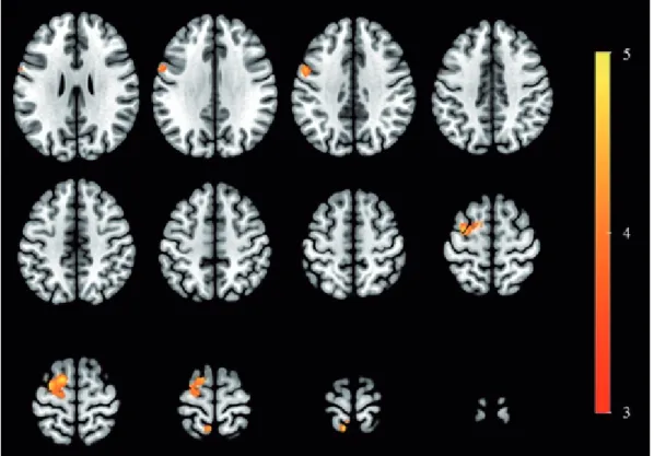
The results of image analysis showed significant increases in relative rCBF of the left superior frontal gyrus (t=4.90, p<
0.001, cluster size=465 voxels), left postcentral gyrus (t=5.16,
p<0.001, cluster size=112 voxels), and the left precentral gyrus(t=4.56, p<0.001, cluster size=105 voxels) at follow-up com- pared with baseline (Fig. 1 and Table 2). There were no signif- icant decreases in relative rCBF.
DISCUSSION
In the present study, we investigated the effects of nicergo- line on cerebral perfusion in early AD patients. We found that nicergoline treatment increased the relative rCBF in the left superior frontal gyrus, left postcentral gyrus, and left precen- tral gyrus. No significant differences in clinical measures were found at follow-up compared with baseline. Previous longi- tudinal studies investigating AD patients demonstrated a global reduction in rCBF
36and in cognitive and neuropsychiat-
ric performance over a one-year period.
37,38Deterioration in cerebral perfusion and cognitive performance represent the natural course of the disease. Our results suggest that nicergo- line may have beneficial effects on AD by interfering with the degenerative processes.
Apart from the medial temporal lobe, which has been wide- ly implicated in the pathophysiology of early AD,
36,39,40previ- ous neuroimaging studies of normal aging, mild cognitive impairment (MCI), and AD also found structural and func- tional deficits in the frontal and parietal regions. Using MRI, cross-sectional studies of normal aging have reported a non- linear and regional atrophy within the brain, and the prefrontal cortex declined more rapidly than the other brain regions.
41-43A longitudinal study showed that gray matter atrophy was more prominent in the frontal and parietal cortices than tem- poral and occipital cortices in healthy older adults.
44In func- tional imaging studies using positron emission tomography and SPECT, studies have demonstrated a reduction in glucose metabolism and blood flow in the frontal and parietal regions in patients who progressed from MCI to AD.
45,46Further- more, regional decreases in cerebral metabolism and perfu- sion in the frontal and temporo-parietal regions were found in the early stages of AD.
47-50Our findings suggest that nicergoline treatment improved the brain perfusion in the frontal and parietal regions of early AD patients. In particular, the largest cluster was found in the superior frontal gyrus, which is associated with higher cog- nitive functions such as working memory.
51Moreover, our study found increased cerebral perfusion in the precentral and postcentral gyri, which correspond to the primary motor and sensory cortices. While sensory and motor dysfunctions
Table 1. Demographic and clinical characteristics of the participantsCharacteristics Baseline (mean±SD or n) Follow-up (mean±SD or n) Test*
Age (year) 77.0±6.4
Sex (male:female) 6:10
MMSE 20.3±4.4 20.4±4.2 t=-0.11, p=0.91
CDR
0.5 14 14
1.0 2 2
CDR-SB 2.6±1.4 2.9±1.8 t=-1.84, p=0.09
GDS 3.6±0.7 3.7±0.7 z=-1.00, p=0.32
IADL-C 16.8±10.6 17.5±10.9 t=-0.26, p=0.80
IADL-P 13.3±9.0 15.2±10.2 t=-1.16, p=0.26
Barthel ADL Index 19.6±0.8 18.3±2.3 z=1.84, p=0.07
GDS-Depression 15.7±10.8 15.7±9.0 z=-0.57, p=0.57
*Paired t-test or Wilcoxon signed rank sum test.
ADL: Activities of Daily Living, CDR: Clinical Dementia Rating, CDR-SB: Clinical Dementia Rating-Sum of Boxes, GDS: Global Deterioration Scale, GDS-Depression: Geriatric Depression Scale, IADL: Instrumental Activities of Daily Living-Current Performance (C) and Potential Performance (P), MMSE: Mini-Mental State Examination, SD: standard deviation.
DND
in AD have received relatively little attention in the field of AD research, recent evidence suggests that sensory and mo- tor changes may precede the cognitive symptoms associated with AD pathogenesis.
52Taken together, it is possible that in- creased perfusion in the frontal and parietal regions may de- lay or prevent progressive deterioration of cognitive functions in AD.
Limitations of the study include the relatively small sam- ple size and the lack of a control group. Moreover, cognitive evaluation with a more comprehensive neuropsychological battery may have facilitated the detection of subtle changes in cognitive function during the study period. Finally, it is possi-
ble that the rCBF changes observed in our study may have been affected by AChEI. However, previous SPECT studies involv- ing AD patients treated with long-term AChEI therapy showed no increases in rCBF
53-55or the effects were mainly localized to the frontal lobe.
56,57In conclusion, this was the first SPECT study, to our knowl- edge, to examine cerebral perfusion changes associated with nicergoline treatment in early AD patients. Nicergoline treat- ment resulted in a favorable outcome involving cerebral per- fusion during early AD. Moreover, the clinical symptoms of AD remained stable without deterioration during the study period. Further studies involving larger sample sizes and pla- cebo-controlled design are required to confirm, and elucidate the treatment effects and the underlying neural mechanisms of nicergoline in AD.
Conflicts of Interest
The authors have no financial conflicts of interest.
Acknowledgements
This research was supported by the National Research Foundation of Korea (NRF) funded by the Ministry of Science and ICT (2017R1C1B2011802).
REFERENCES
1. Scarpini E, Scheltens P, Feldman H. Treatment of Alzheimer’s dis- Fig. 1. Brain areas showing significantly increased regional cerebral blood flow in nicergoline-treated Alzheimer’s disease patients compared with baseline. Images are shown in neurological convention. Color bar represents the voxel-level t-values.
Table 2. Changes in regional cerebral blood flow after nicergoline treatment
Region t p Coordinates*
(x, y, z) Cluster size (voxels) Baseline < follow-up
Left superior frontal gyrus 4.90 <0.001 -16, 2, 66 465 Left postcentral gyrus 5.16 <0.001 -6, -50, 74 112 Left precentral gyrus 4.56 <0.001 -56, 4, 36 105 Baseline > follow-up
None
*The coordinates refer to the Montreal Neurological Institute coordinate system.
Jooyeon J. Im et al.
Nicergoline Treatment in Alzheimer’s Disease
ease: current status and new perspectives. Lancet Neurol 2003;2:539- 2. Gorelick PB. Risk factors for vascular dementia and Alzheimer dis-547.
ease. Stroke 2004;35(11 Suppl 1):2620-2622.
3. Skoog I, Kalaria RN, Breteler MM. Vascular factors and Alzheimer disease. Alzheimer Dis Assoc Disord 1999;13 Suppl 3:S106-S114.
4. Winblad B, Carfagna N, Bonura L, Rossini BM, Wong EH, Battaglia A. Nicergoline in dementia: a review of its pharmacological proper- ties and therapeutic potential. CNS Drugs 2000;14:267-287.
5. Bracco L, Bonura ML, Battaglia A. Six-month, multicentre, double- blind trial of nicergoline in the treatment of mild to moderate AD and its 12-month follow-up [abstract]. Neurosci Lett 1999;552:18.
6. Crook TH. Nicergoline in the treatment of probable Alzheimer’s dis- ease. Preliminary results of a double-blind, randomized, placebo-con- trolled study [abstract]. J Neurol Sci 1997;150:S18.
7. Winblad B, Bonura ML, Rossini BM, Battaglia A. Nicergoline in the treatment of mild- to- moderate Alzheimer’s disease. Clin Drug Inves- tig 2001;21:621-632.
8. Arrigo A, Moglia A, Borsotti L. A double-blind, placebo-controlled, crossover trial with nicergoline in patients with senile dementia [ab- stract]. Int J Clin Pharmacol Res 1982;2(4 Suppl 1):33-41.
9. Battaglia A, Bruni G, Ardia A, Sacchetti G. Nicergoline in mild to moderate dementia. A multicenter, double-blind, placebo-controlled study. J Am Geriatr Soc 1989;37:295-302.
10. Nappi G, Bono G, Merlo P, Borromei A, Caltagirone C, Lomeo C, et al. Long-term nicergoline treatment of mild to moderate senile demen- tia: results of a multicentre, double-blind, placebo-controlled study.
Clin Drug Investig 1997;13:308-316.
11. Saletu B, Paulus E, Linzmayer L, Anderer P, Semlitsch HV, Grünberg- er J, et al. Nicergoline in senile dementia of Alzheimer type and multi- infarct dementia: a double-blind, placebo-controlled, clinical and EEG/
ERP mapping study. Psychopharmacology (Berl) 1995;117:385-395.
12. Winblad B, Fioravanti M, Dolezal T, Logina I, Milanov IG, Popescu DC, et al. Therapeutic use of nicergoline. Clin Drug Investig 2008;28:
533-552.
13. Carfagna N, Di Clemente A, Cavanus S, Damiani D, Gerna M, Salmoi- raghi P, et al. Modulation of hippocampal ACh release by chronic nicergoline treatment in freely moving young and aged rats. Neurosci Lett 1995;197:195-198.
14. McArthur RA, Carfagna N, Banfi L, Cavanus S, Cervini MA, Fariello R, et al. Effects of nicergoline on age-related decrements in radial maze performance and acetylcholine levels. Brain Res Bull 1997;43:305- 15. Ogawa N, Asanuma M, Hirata H, Kondo Y, Kawada Y, Mori A. Cho-311.
linergic deficits in aged rat brain are corrected with nicergoline. Arch Gerontol Geriatr 1993;16:103-110.
16. Moretti A, Carfagna N, Caccia C, Carpentieri M. Effect of ergolines on neurotransmitter systems in the rat brain. Arch Int Pharmacodyn Ther 1988;294:33-45.
17. Le Poncin-Lafitte M, Grosdemouge C, Duterte D, Rapin JR. Simul- taneous study of haemodynamic, metabolic and behavioural sequelae in a model of cerebral ischaemia in aged rats: effects of nicergoline.
Gerontology 1984;30:109-119.
18. Miccheli A, Puccetti C, Capuani G, Di Cocco ME, Giardino L, Calzà L, et al. [1-13C]Glucose entry in neuronal and astrocytic intermedi- ary metabolism of aged rats. A study of the effects of nicergoline treatment by 13C NMR spectroscopy. Brain Res 2003;966:116-125.
19. Caraci F, Chisari M, Frasca G, Canonico PL, Battaglia A, Calafiore M, et al. Nicergoline, a drug used for age-dependent cognitive impair- ment, protects cultured neurons against beta-amyloid toxicity. Brain Res 2005;1047:30-37.
20. Mizuno T, Kuno R, Nitta A, Nabeshima T, Zhang G, Kawanokuchi J, et al. Protective effects of nicergoline against neuronal cell death in-
duced by activated microglia and astrocytes. Brain Res 2005;1066:78- 21. Sortino MA, Battaglia A, Pamparana F, Carfagna N, Post C, Canonico 85.
PL. Neuroprotective effects of nicergoline in immortalized neurons.
Eur J Pharmacol 1999;368:285-290.
22. Caputi A, Di Luca M, Pastorino L, Colciaghi F, Carfagna N, Wong E, et al. Nicergoline and its metabolite induce translocation of PKC iso- forms in selective rat brain areas. Neurosci Res Commun 1998;23:159- 23. Carfagna N, Cavanus S, Damiani D, Salmoiraghi P, Fariello R, Post C. 167.
Modulation of phosphoinositide turnover by chronic nicergoline in rat brain. Neurosci Lett 1996;209:189-192.
24. Cedazo-Minguez A, Bonecchi L, Winblad B, Post C, Wong EH, Cow- burn RF, et al. Nicergoline stimulates protein kinase C mediated al- pha-secretase processing of the amyloid precursor protein in cultured human neuroblastoma SH-SY5Y cells. Neurochem Int 1999;35:307- 25. American Psychiatric Association. Diagnostic and Statistical Manual 315.
of Mental Disorders (DSM-IV-TR). Washington, DC: American Psy- chiatric Association, 2000.
26. McKhann G, Drachman D, Folstein M, Katzman R, Price D, Stadlan EM. Clinical diagnosis of Alzheimer’s disease: report of the NINCDS- ADRDA Work Group under the auspices of Department of Health and Human Services Task Force on Alzheimer’s Disease. Neurology 1984;34:939-944.
27. Kang Y, Na DL, Hahn S. A validity study on the Korean Mini-Mental State Examination (K-MMSE) in dementia patients. J Korean Neurol Assoc 1997;15:300-308.
28. Morris JC, Heyman A, Mohs RC, Hughes JP, van Belle G, Fillen- baum G, et al. The Consortium to Establish a Registry for Alzheimer’s Disease (CERAD). Part I. Clinical and neuropsychological assess- ment of Alzheimer’s disease. Neurology 1989;39:1159-1165.
29. Choi SH, Na DL, Lee BH, Hahm DS, Jeong JH, Jeong Y, et al. The validity of the Korean version of Global Deterioration Scale. J Korean Neurol Assoc 2002;20:612-617.
30. Wade DT, Collin C. The Barthel ADL Index: a standard measure of physical disability? Int Disabil Stud 1988;10:64-67.
31. Kang SJ, Choi SH, Lee BH, Kwon JC, Na DL, Han SH, et al. The reliability and validity of the Korean Instrumental Activities of Daily Living (K-IADL). J Korean Neurol Assoc 2002;20:8-14.
32. Jung IK, Kwak DI, Shin DK, Lee MS, Lee HS, Kim JY. A reliability and validity study of geriatric depression scale. J Korean Neuropsy- chiatr Assoc 1997;36:103-112.
33. Tzourio-Mazoyer N, Landeau B, Papathanassiou D, Crivello F, Etard O, Delcroix N, et al. Automated anatomical labeling of activations in SPM using a macroscopic anatomical parcellation of the MNI MRI single-subject brain. Neuroimage 2002;15:273-289.
34. Pickut BA, Dierckx RA, Dobbeleir A, Audenaert K, Van Laere K, Ver- vaet A, et al. Validation of the cerebellum as a reference region for SPECT quantification in patients suffering from dementia of the Al- zheimer type. Psychiatry Res 1999;90:103-112.
35. Soonawala D, Amin T, Ebmeier KP, Steele JD, Dougall NJ, Best J, et al. Statistical parametric mapping of (99m)Tc-HMPAO-SPECT im- ages for the diagnosis of Alzheimer’s disease: normalizing to cerebel- lar tracer uptake. Neuroimage 2002;17:1193-1202.
36. Kogure D, Matsuda H, Ohnishi T, Asada T, Uno M, Kunihiro T, et al.
Longitudinal evaluation of early Alzheimer’s disease using brain per- fusion SPECT. J Nucl Med 2000;41:1155-1162.
37. Benoit M, Robert PH, Staccini P, Brocker P, Guerin O, Lechowski L, et al. One-year longitudinal evaluation of neuropsychiatric symptoms in Alzheimer’s disease. The REAL.FR Study. J Nutr Health Aging 2005;9:95-99.
38. Burns A, Jacoby R, Levy R. Progression of cognitive impairment in
DND
Alzheimer’s disease. J Am Geriatr Soc 1991;39:39-45.
39. Masdeu JC, Zubieta JL, Arbizu J. Neuroimaging as a marker of the onset and progression of Alzheimer’s disease. J Neurol Sci 2005;236:
55-64.
40. Scahill RI, Schott JM, Stevens JM, Rossor MN, Fox NC. Mapping the evolution of regional atrophy in Alzheimer’s disease: unbiased analysis of fluid-registered serial MRI. Proc Natl Acad Sci U S A 2002;
99:4703-4707.
41. Coffey CE, Wilkinson WE, Parashos IA, Soady SA, Sullivan RJ, Pat- terson LJ, et al. Quantitative cerebral anatomy of the aging human brain: a cross-sectional study using magnetic resonance imaging. Neu- rology 1992;42(3 Pt 1):527-536.
42. Fox NC, Schott JM. Imaging cerebral atrophy: normal ageing to Al- zheimer’s disease. Lancet 2004;363:392-394.
43. Raz N, Gunning FM, Head D, Dupuis JH, McQuain J, Briggs SD, et al. Selective aging of the human cerebral cortex observed in vivo: dif- ferential vulnerability of the prefrontal gray matter. Cereb Cortex 1997;
7:268-282.
44. Resnick SM, Pham DL, Kraut MA, Zonderman AB, Davatzikos C.
Longitudinal magnetic resonance imaging studies of older adults: a shrinking brain. J Neurosci 2003;23:3295-3301.
45. Drzezga A, Lautenschlager N, Siebner H, Riemenschneider M, Wil- loch F, Minoshima S, et al. Cerebral metabolic changes accompany- ing conversion of mild cognitive impairment into Alzheimer’s disease:
a PET follow-up study. Eur J Nucl Med Mol Imaging 2003;30:1104- 1113.
46. Encinas M, De Juan R, Marcos A, Gil P, Barabash A, Fernández C, et al. Regional cerebral blood flow assessed with 99mTc-ECD SPET as a marker of progression of mild cognitive impairment to Alzheimer’s disease. Eur J Nucl Med Mol Imaging 2003;30:1473-1480.
47. Chase TN, Foster NL, Fedio P, Brooks R, Mansi L, Di Chiro G. Re- gional cortical dysfunction in Alzheimer’s disease as determined by positron emission tomography. Ann Neurol 1984;15 Suppl:S170-S174.
48. Friedland RP, Budinger TF, Koss E, Ober BA. Alzheimer’s disease:
anterior-posterior and lateral hemispheric alterations in cortical glu- cose utilization. Neurosci Lett 1985;53:235-240.
49. Metter EJ, Riege WH, Kameyama M, Kuhl DE, Phelps ME. Cere- bral metabolic relationships for selected brain regions in Alzheimer’s, Huntington’s, and Parkinson’s diseases. J Cereb Blood Flow Metab 1984;4:500-506.
50. Perani D, Di Piero V, Vallar G, Cappa S, Messa C, Bottini G, et al.
Technetium-99m HM-PAO-SPECT study of regional cerebral perfu- sion in early Alzheimer’s disease. J Nucl Med 1988;29:1507-1514.
51. du Boisgueheneuc F, Levy R, Volle E, Seassau M, Duffau H, Kink- ingnehun S, et al. Functions of the left superior frontal gyrus in hu- mans: a lesion study. Brain 2006;129(Pt 12):3315-3328.
52. Albers MW, Gilmore GC, Kaye J, Murphy C, Wingfield A, Bennett DA, et al. At the interface of sensory and motor dysfunctions and Al- zheimer’s disease. Alzheimers Dement 2015;11:70-98.
53. Kimura N, Kumamoto T, Masuda T, Hanaoka T, Okazaki T, Arakawa R. Evaluation of the regional cerebral blood flow changes during long-term donepezil therapy in patients with Alzheimer’s disease us- ing 3DSRT. J Neuroimaging 2012;22:299-304.
54. Nobili F, Koulibaly M, Vitali P, Migneco O, Mariani G, Ebmeier K, et al. Brain perfusion follow-up in Alzheimer’s patients during treatment with acetylcholinesterase inhibitors. J Nucl Med 2002;43:983-990.
55. Ushuijima Y, Okuyama C, Mori S, Kubota T, Nakai T, Nishimura T.
Regional cerebral blood flow in Alzheimer’s disease: comparison be- tween short and long-term donepezil therapy. Ann Nucl Med 2006;20:
425-429.
56. Shimizu S, Hanyu H, Iwamoto T, Koizumi K, Abe K. SPECT follow- up study of cerebral blood flow changes during Donepezil therapy in patients with Alzheimer’s disease. J Neuroimaging 2006;16:16-23.
57. Shimizu S, Kanetaka H, Hirose D, Sakurai H, Hanyu H. Differential effects of acetylcholinesterase inhibitors on clinical responses and ce- rebral blood flow changes in patients with Alzheimer’s disease: a 12-month, randomized, and open-label trial. Dement Geriatr Cogn Dis Extra 2015;5:135-146.