미만성대형 B세포림프종과 동반된 Henoch-Sch ö nlein 자반증 1예
부산대학교 의학전문대학원 내과학교실, 1진단검사의학교실
김일두ㆍ이승근ㆍ이혜정ㆍ조우성ㆍ최영진ㆍ신호진ㆍ정주섭ㆍ조군제ㆍ이은엽
1
A Case of Henoch-Schönlein Purpura Associated with Diffuse Large B Cell Lymphoma
Il Du Kim, M.D., Seung Geun Lee, M.D., Hye Jeong Lee, M.D., Woo Sung Jo, M.D., Young Jin Choi, M.D., Ho Jin Shin, M.D., Joo Seop Chung, M.D., Goon Jae Cho, M.D. and Eun Yup Lee, M.D.1 Departments of Internal Medicine and 1Laboratory Medicine, School of Medicine, Pusan National University, Busan, Korea
A 69-year-old female was referred to our institution due to abdominal pain and palpable purpura on both buttocks and legs. A skin biopsy of her purpura revealed granulocyte infiltration and leucocytoclasia around the arterioles and venuoles at the dermis, as well as an elevated serum immunoglobulin A level, hematuria and proteinuria. Therefore she was diagnosed with Henoch-Schönlein purpura. She had been diagnosed with diffuse large B cell lymphoma after a biopsy of her left inguinal lymph node 12 years ago and received 6 cycles of CHOP (cyclophosphamide, doxorubicin, vincristine and prednisone) chemo- therapy, which was followed by a complete remission. Abdominal and chest CT revealed multiple lymph node enlargement and bowel wall thickening at the ileocecal area, and lesions were observed in a whole body PET CT scan. Recurrence of the diffuse large B cell lymphoma was confirmed by a biopsy of the ileocecal area via colonoscopy. The purpura was improved after oral prednisolone therapy and etopo- side, oxaliplatin and ifosfamide salvage combination chemotherapy was used to treat the lymphoma.
(Korean J Hematol 2007;42:162-166.)
Key Words: Henoch-Schönlein purpura, Diffuse large B cell lymphoma, Malignancy, Vasculitis
162
Correspondence to:Joo Seop Chung, M.D.
Department of Internal Medicine, School of Medicine, Pusan National University
10, Ami-dong 1-ga, Seo-gu, Busan 602-739, Korea Tel: +82-51-254-3127, Fax: +82-51-240-7673 E-mail: [email protected]
접수:2007년 2월 27일, 수정:2007년 4월 20일 승인:2007년 4월 24일
교신저자:정주섭, 부산시 서구 아미동 1가 10
602-739, 부산대학교병원 내과 Tel: 051-254-3127, Fax: 051-240-7673 E-mail: [email protected]
서 론
Henoch-Schönlein 자반증(Henoch-Schönlein purpura, HSP)은 주로 둔부와 하지에서 촉지되는 자반과 관절 통, 복통, 사구체신염을 특징으로 하는 전신적 혈관염 증후군이다. 면역글로불린 A를 포함한 면역복합체의 침착이 병인이며 주로 4세에서 7세 사이의 소아에서 많이 발생하고 부신피질스테로이드와 면역억제제를
투여하면 비교적 쉽게 치료가 가능하다. 유발요인으로 는 상기도 감염, 약물, 음식, 곤충자상 등이 있으며 소 아와 달리 성인에서는 흔히 발생하지는 않는다. 성인 에서 악성종양이 HSP의 원인으로 드물게 보고되고 있 으며 이 중 비호지킨림프종은 3예가 보고된 바 있다.
이에 저자들은 양측 하지에서 촉지되는 자반과 복통으 로 내원하여 임상증상과 혈청검사 및 피부 조직검사를 시행하여 HSP로 진단된 환자에서 컴퓨터단층촬영, 양 전자방출단층촬영 및 조직검사를 통해 재발된 미만성
Fig. 1. Cutaneous finding shows multiple palpable purpura on both legs.
Fig. 2. Abdominal computed tomography shows the en- largement of multiple mesenteric lymph nodes and mild thickening of bowel wall at ileocecal area.
대형B세포림프종이 동반된 것이 확인된 1예를 보고하 는 바이다.
증 례
환 자: 69세, 여자
주 소: 복통, 양측 하지에서 촉지되는 자반 현병력: 2006년 8월 복통과 양측 하지에서 촉지되는 자반을 주소로 타 병원 방문 후 본원 응급실을 방문하 였다.
신체검사: 내원 당시 혈압 140/70mmHg, 맥박 분당 84회, 호흡수 분당 20회, 체온 36oC였으며, 의식은 명료 하였고 지남력 장애는 관찰되지 않았다. 외견상 경한 만성 병색을 보였으나 빈혈이나 황달, 경정맥의 돌출 이나 림프절의 비대는 관찰되지 않았다. 흉부 청진에 서 심잡음 및 양 폐야의 수포음은 청진되지 않았다. 복 부 진찰에서 장음이 감소되어 있었고 간과 비장은 촉 진되지 않았으며 우하복부에 경한 압통이 관찰되었으 며 경직은 관찰되지 않았다. 상하지 진찰에서 양 하지 에 다발성으로 촉지되는 자반과 함께 부종이 관찰되었 으며(Fig. 1), 양측 슬관절통이 있었다.
검사실 소견: 일반혈액검사에서 백혈구가 16,500×
109/L (중성구 87.5%, 림프구 4.0%, 단핵구 4.9%, 호산 구 0.1%)로 상승되어 있었으며, 혈색소 11.6g/dL, 혈소 판 414,000×109/L였다. 혈청생화학검사에서 AST 30 IU/L, ALT 30IU/L, ALP 544IU/L, LDH 464 IU/L, 총단 백 6.3g/dL, 알부민 2.9g/dL, 총빌리루빈 0.65mg/dL, 직 접빌리루빈 0.20mg/dL, 칼슘 8.9mg/dL, 인 5.4mg/dL, 콜레스테롤 162mg/dL, 혈중요소질소 9.9mg/dL, 크레아 티닌 0.7mg/dL였고, 혈청전해질은 나트륨 139.1mmol/L,
칼륨 4.29mmol/L, 클로라이드 102.3mmol/L였다. 혈청 C-반응단백은 14.07mg/dL로 상승되어 있었다. 혈청 면 역글로불린A는 1,124.4mg/dL로 상승되어 있었다. 소 변검사에서 혈뇨 및 단백뇨가 관찰되었다.
과거력: 12년 전에 우측 서혜부 림프절 종대가 있어 타 병원에서 조직검사로 미만성대형B세포림프종(sta- ge IIIa)으로 진단받은 후 6주기의 CHOP (cyclophos- phamide, doxorubicin, vincristine, prednisone) 항암화학 요법 후 완전관해된 과거력이 있었다. 이외의 특이 병 력은 없었으며 지속적으로 복용하고 있는 약은 없었다.
사회력: 특이 소견 없음.
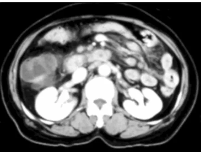
방사선학적 소견: 복부 컴퓨터단층촬영에서 복부 내 다발성 림프절 종대 및 회맹장연결부 장벽의 비후 소 견이 관찰되었다(Fig. 2). 흉부 컴퓨터단층촬영에서도 다발성 림프절 종대가 관찰되었으며 폐의 우하엽에 림 프종의 침범소견을 보였다(Fig. 3). 전신 양전자방출단 층촬영에서 경부, 흉부와 복부의 다발성 림프절 및 회 맹장 연결부에 포도당 섭취 증가 소견이 관찰되었다 (Fig. 4).
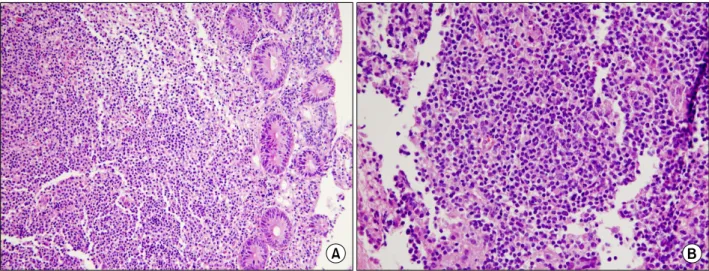
병리조직학적 소견: 피부 조직검사에서 진피혈관 내 표재성 혈관 주위의 중성구, 림프구, 조직구 침윤과 핵 진 등의 혈관염의 소견을 보였다(Fig. 5). 골수 조직검 사에서는 림프종의 침범은 관찰되지 않았다. 대장내시 경을 통한 회맹장연결부의 조직검사에서 비정상적인 림프구의 광범위한 증식 소견을 보여 림프종으로 진단 되었다(Fig. 6).
경 과: 환자는 HSP 진단하에 부신피질스테로이드 투여 후 자반의 현저한 감소를 보였다. 복부와 흉부의
Fig. 3. Chest computed tomography shows supraclavicular, bilateral paratracheal, subcarinal, right hilar lymph nodes enlarge- ment, bilateral pleural effusion and lung involvement of lymphoma at right lower lobe.
Fig. 4. Whole body positron emission tomography shows lymphoma involvement in right tonsil, both level IIA lymph nodes of the neck, left supraclavicular lymph node, lower lobe of right lung, spleen and ileocecal valve, portahepatis, portocaval, paraaortic, and aortocaval lymph nodes, lymph nodes around ileocolic artery and jejunum, retroperitoneal lymph nodes around right psoas muscle, right common iliac lymph node, right internal and external iliac lymph no- des, and both inguinal lymph nodes.
Fig. 5. Pathologic finding of skin biopsy shows neutrophils infiltration and leucocytoclasia around arterioles and ven- uoles at dermis (H&E, ×400).
컴퓨터단층촬영, 전신 양전자방출단층촬영과 대장내 시경을 통한 조직검사의 소견으로 미만성대형B세포림 프종의 재발로 진단하여 etoposide, oxaliplatin, ifosfa- mide 항암화학요법치료를 받았지만 약 2개월 후 병의
진행으로 사망하였다.
고 찰
본 증례는 성인에서의 HSP가 악성종양, 특히 미만성 대형B세포림프종과 관련되어 발생한 경우이다. HSP는 하지와 둔부에서 촉지되는 자반, 복통, 혈뇨, 관절통을 나타내는 전신적인 혈관염으로 주로 4세에서 7세 사이 의 소아에서 나타나지만 성인에서도 나타날 수 있다.1) HSP는 임상양상이 다양하나 1990년 미국류마티스 학회에 따르면 촉지되는 자반, 20세 이전의 발병연령,
Fig. 6. Pathologic finding of ileocecal area biopsy via colonoscopy shows atypical lymphoid cell proliferation in lamina propria (A: H&E, ×200) and large neoplastic cells with vesicular nuclei (B: H&E, ×400).
복통, 세동맥 또는 세정맥 벽의 과립구 침윤 중 2가지 이상이 만족되면 HSP의 진단에 있어 87%의 민감도와 특이도를 나타낸다.2) 본 환자의 경우 촉지되는 자반, 복통, 혈뇨 및 단백뇨, 관절통, 피부조직검사에서 혈관 벽의 중성구 침윤 및 핵진 등의 혈관염 소견을 보여 HSP로 진단할 수 있었다.
250명의 성인 Henoch-Schönlein 신염 환자들을 대상 으로 한 Pillebout 등의 보고에 따르면, HSP는 소아에 비해 성인에서 좋지 않은 예후를 보였으며 사망원인 중 악성종양이 27%로 가장 높은 비율을 차지하였다.3) 본 증례에서는 소변검사에서 단백뇨와 혈뇨가 있어 신장침범을 의심할 수 있었으나 신조직 검사는 시행하 지 않아 Henoch-Schönlein 신염의 발생 여부는 확인되 지 않았다.
악성종양과 관련된 HSP는 세계적으로 31증례가 보 고되었으며4) 국내 보고는 아직 없다. 대부분 고형종양 이었으며 그 중 폐암 8예, 전립선암 5예, 신장암 2예, 직장암, 유방암, 신경초종, 소장암, 위암이 각각 1예가 보고되었고 혈액학적 질환으로는 다발성골수종 5예, 비호지킨림프종 3예, 호지킨림프종 2예, 골수형성이상 증후군이 각각 1예가 보고되었다.4-10)
악성종양이 혈관염을 유발하는 기전은 아직 명확하 지 않으나 여러 가지 가설들이 제시되고 있다. 악성종 양세포가 신생항원으로 작용하여 유사한 혈관항원에 대한 면역반응을 유발하고, 비정상적인 항체를 생성할 수 있다. 또한 면역복합체의 제거를 감소시키고, 면역 글로불린 M에서 A로의 변화를 초래하는 비정상적인 림프구를 유발할 수 있으며, 악성세포 자체 또는 종양
색전의 배출을 통해 유발되는 염증성 사이토카인의 비 정상적 생성을 통해 혈관염의 발생에 기여한다고 알려 져 있다.5,6,11-13) 악성종양은 고점성상태를 초래하여 혈 관내피세포의 손상을 유발하고 면역복합체 침착을 증 가시켜 HSP 발생의 한 요소로 작용할 수도 있다.14) 악 성종양의 치료도 HSP의 발생에 영향을 줄 수 있는데, 항암화학요법 또는 방사선치료가 세포표면항원을 변 화시키거나 악성종양세포를 파괴시켜 세포 내에 존재 하던 새로운 항원에 대한 노출을 유발하는 것을 생각 할 수 있다.6) 또한 골수증식질환 혹은 림프증식질환에 서와 같이 면역이 감소되어 있는 질환의 경우에는 일 반적인 검사에서 찾아내지 못한 세균 혹은 바이러스 감염이 HSP의 원인으로 작용할 수 있다.7,12)
피부와 전신 혈관염은 비록 드문 경우지만 림프증식 질환과 연관되어 나타나는 것으로 알려져 있으며 그 중 가장 연관성이 높은 것은 한랭글로불린혈증과 림프 성림프종 및 Waldenstrom 거대글로불린혈증, 결절다 발동맥염과 털세포백혈병, 중추신경계의 육아종혈관 염과 호지킨림프종 등이 있으며 본 증례와 같이 HSP 와 비호지킨림프종이 동반된 경우는 림프증식질환 중 에서도 매우 드문 것으로 알려져 있다.15)
본 증례에서 보고된 바와 같이 다른 특별한 유발요 인이 없이 성인에서 HSP가 진단된 경우 동반된 악성 종양의 가능성을 항상 염두에 두어야 하며, HSP 이외 의 전신적 혈관염이 성인에서 발생한 경우에도 림프증 식질환 및 악성종양이 동반되었을 가능성이 있으므로 여기에 대한 확인검사가 필요할 것으로 생각된다.
요 약
저자들은 양측 하지에서 촉지되는 자반과 복통으로 내원하여 임상증상과 혈청검사 및 피부 조직검사를 통 해 HSP로 진단된 후 컴퓨터단층촬영, 양전자방출단층 촬영 및 대장내시경을 통한 조직검사를 통해 재발된 미만성대형B세포림프종이 동반된 것이 확인된 1예를 보고하는 바이다.
참 고 문 헌
1) Roth DA, Wilz DR, Theil GB. Schönlein-Henoch syndrome in adults. Q J Med 1985;55:145-52.
2) Mills JA, Michel BA, Bloch DA, et al. The American College of Rheumatology 1990 criteria for the classi- fication of Henoch-Schönlein purpura. Arthritis Rhe- um 1990;33:1114-21.
3) Pillebout E, Thervet E, Hill G, Alberti C, Vanhille P, Nochy D. Henoch-Schönlein purpura in adults:
outcome and prognostic factors. J Am Nephrol 2002;
13:1271-8.
4) Zurada JM, Ward KM, Grossman ME. Henoch- Schönlein purpura associated with malignancy in adults. J AM Acad Dermatol 2006;55(5 Suppl):S65- 70.
5) Hayem G, Gomez MJ, Grossin M, Meyer O, Kahn MF. Systemic vasculitis and epithelioma. A report of three cases with a literature review. Rev Rhum Engl Ed 1997;64:816-24.
6) Garcias VA, Herr HW. Henoch-Schönlein purpura associated with cancer of prostate. Urology 1982;19:
155-8.
7) Arrizabalaga P, Saurina A, Sole M, Blade J. Henoch- Schönlein IgA glomerulonephritis complicating mye- loma kidneys: case report. Ann Hematol 2003;82:
526-8.
8) Day C, Savage CO, Jones EL, Cockwell P. Henoch- Schönlein nephritis and non-Hodgkin’s lymphoma.
Nephrol Dial Transplant 2001;16:1080-1.
9) Ng JP, Murphy J, Chalmers EM, Hogg RB, Cum- ming RL, Peebles S. Henoch-Schönlein purpura and Hodgkin’s disease. Postgrad Med J 1988;64:881-2.
10) Blanco R, Gonzalez-Gay MA, Ibanez D, et al. He- noch-Schönlein purpura as clinical presentation of a myelodysplastic syndrome. Clin Rheumatol 1997;16:
626-8.
11) Pertuiset E, Liote F, Launay-Russ E, Kemiche F, Cerf-Payrastre I, Chesneau AM. Adult Henoch-Schö- nlein purpura associated with malignancy. Semin Arthritis Rheum 2000;29:360-7.
12) Greer JM, Longley S, Edwards NL, Elfenbein GJ, Panush RS. Vasculitis associated with malignancy.
Experience with 13 patients and literature review.
Medicine (Baltimore) 1988;67:220-30.
13) Kurzrock R, Cohen PR, Markowitz A. Clinical mani- festations of vasculitis in patients with solid tumors.
A case report and review of the literature. Arch Intern Med 1994;154:334-40.
14) Magro CM, Crowson AN. A clinical and histologic study of 37 cases of immunoglobulin A-associated vasculitis. Am J Dermatopathol 1999;21:234-40.
15) Wooten MD, Jasin HE. Vasculitis and lymphoproli- ferative diseases. Semin Arthritis Rheum 1996;26:
564-74.