Correspondence to:Soung Soo Kim, Ph.D.
Department of Biochemistry, College of Science, Yonsei University
134 Sinchon-dong, Seodaemun-gu, Seoul 120-749, Korea Tel: +82-2-2123-2698, Fax: +82-2-362-9897
E-mail: [email protected]
접수:2005년 10월 20일, 수정:2005년 11월 24일 승인:2005년 12월 13일
교신저자:김승수, 서울시 서대문구 신촌동 134
120-749, 연세대학교 이과대학 생화학과 Tel: 02-2123-2698, Fax: 02-362-9897
E-mail: [email protected]
siRNA Targeting Vascular Endothelial Growth Factor and Recombinant Human Prothrombin Kringle 2
Inhibits Leukemia-induced Angiogenesis
Bum Joon Kim and Soung Soo Kim
Department of Biochemistry, College of Science, Yonsei University, Seoul, Korea
Background: Vascular endothelial growth factor (VEGF) plays a role in the development of cancer and the progression of liquid tumors such as chronic lymphatic leukemia, non-Hodgkin lymphomas, and multiple myeloma. VEGF also triggers endothelial cells to secrete hematopoietic growth factors such as interleukin-6 (IL-6); this in turn promotes further leukemia growth, thereby contributing to a paracrine loop between the leukemia and the endothelial cells.
Methods: We transfected a small interfering RNA (siRNA) targeting VEGF into K562 cells in order to investigate the role of VEGF in the development of leukemic cancer. After the conditioned media (CM) of the K562 was cells added to the human umbilical endothelial cell (HUVEC) culture media, we compared the proliferation and tube formation of the HUVECs. Recombinant human prothrombin kringle2 (K2), which is a known angiogenic inhibitor, was also treated onto the HUVECs, and we then examined the level of IL-6 to determine the paracrine interaction between the leukemic and endothelial cells.
Results: RT-PCR and western blot analysis demonstrated that the siRNA efficiently down regulated the expression of VEGF in the K562 cells. When the CM of the K562 cells was added to the HUVEC culture, the proliferation of the HUVECs was stimulated. The proliferation of the HUVEC induced by the CM from the siRNA-VEGF K562 cells was diminished, compared with that of the vector control K562 cells.
K2 reduced not only the proliferation of the HUVECs, but also the secretion of IL-6 by the HUVEC.
Conclusion: The siRNA strategy is an alternative tool for inhibiting leukemia-induced angiogenesis. A combinated therapy with angiogenesis inhibitor K2 increases the efficiency. K2 modulates the production of IL-6, which may affect the paracrine interaction between leukemia and endothelial cells. (Korean J Hematol 2005;40:211-218.)
Key Words: Vascular endothelial growth factor, Leukemia, Angiogenesis, Interleukin-6, Kringle
211
INTRODUCTION
VEGF is a cytokine essential for the vascu- logenesis associated with normal embryonic de- velopment and for the angiogenesis associated with wound healing, cancers, and a variety of other important pathologies.1) It is well recog- nized that VEGF promotes tumor angiogenesis and that secretion of bioactive VEGF by cancer cells may be directly involved in tumor pro- gression.2) There are increasing evidences indi- cating that VEGF plays a role in cancer de- velopment and progression not only in solid tumors but also in liquid tumors such as chronic lymphatic leukemia, non-Hodgkin lymphomas, and multiple myeloma.3)
A number of reports have demonstrated that modulating the expression of VEGF in tumor cells resulted in either stimulation or inhibition of tumor angiogenesis and tumor growth. Schuch et al. showed that delivery of VEGF using microencapsulation technology enhanced tumor growth and vascularization, whereas treatment with a VEGF antagonist soluble NRP-1 inhibited tumor angiogenesis and growth.4) The expression of VEGF in tumor xenografts in nude mice has been shown to stimulate tumor growth and tumor angiogenesis.5) Down-regulated VEGF protein expression by antisense RNA in colon cancer cells resulted in inhibition of endothelial cell proliferation in tumor angiogenesis in vivo.6) Si- milarly, transfection of antisense VEGF effec- tively down-regulated VEGF secretion from head and neck squamous cell carcinoma cells and also inhibited the in vivo tumorigenicity of brain tumor cells.7,8)
VEGF also exert effects on cells in a manner unrelated to its angiogenic activity. VEGF in- duced both a time- and dose-dependent increase in IL-6 production from microvascular endo- thelial cells.9) Since IL-6 act as a survival factor for myeloma cells by preventing tumor cell apoptosis, it is conceivable that endothelial cells
may, in response to VEGF, release cytokines capable of sustaining tumor growth through paracrine loop.
In this report, we demonstrate that down- regulation of VEGF expression in leukemic cell line K562 cells using siRNA inhibits tumor angio- genesis. We also show that VEGF, secreted in CM of K562 cells, promotes the production of IL-6 by HUVEC. Since K2, reported as an angiogenesis inhibitor,10,11) reduces the secretion of IL-6 by HUVEC, K2 may play a role in the paracrine interaction between leukemia and endothelial cells.
MATERIALS AND METHODS
1. Cell culture
K562 was cultured in RPMI-1640 with 10%
fetal bovine serum (FBS), penicillin (100U/mL), and streptomycin (100ug/mL). HUVEC was grown in EBM-2 medium (Clonetics, Walkersville, MD, USA) with 10ng/mL epidermal growth factor, 1ug/mL hydrocortisone, 10ug/mL bovine brain extract, 2% fetal bovine serum (FBS), 50ug/ mL gentamicin and 50ng/mL Amphotericin B.
2. siRNA
We designed and synthesized complementary oligonucleotides against VEGF mRNA with 5' single strand overhangs for ligation into the pSilencerTM 1.0-U6 (Ambion, Austin, TX, USA) vector (5'-gatcccAAATGTGAATGCAGACCAAA GttcaagagaCTTTGGTCTGCATTCACATTTtttttt ggaaa-3'; 3'-agcttttccaaaaaaAAATGTGAATGCAG ACCAAAGtctcttgaaCTTTGGTCTGCATTCACA TTTgg-5'). In vitro transfection was performed using LipofectAMINE PLUS (Invitogen, Carls- bad, CA, USA) according to the manufacturers instructions. HUVECs were plated at a density of 8×10⁴ cells per well in 24-well plates. After 20h and 70~80% confluence, the cells were trans- fected with siRNAs in serum-free medium using LipofectAMINE PLUS. 0.8μg of siRNA was di- luted with Opti-MEM (50μL) in a small sterile
tube. After immediate mixing, the Lipofect- AMINE reagent (2μL) in Opti-MEM (50μL) was added, and the mixture was left at room temper- ature for 20 min. The HUVEC culture medium was replaced by fresh medium and the siRNA- lipid complex (100uL) was added. After incuba- tion for 4h at 37°C, 1mL of EBM-2 medium with 10% FBS was added. After 1 day, culture media changed to selective media containing 500U/mL of hygromycin to select cells containing the siRNA- VEGF vector.
3. Semiquantitative Reverse Transcription- PCR Analysis for VEGF Gene Expression Total RNA was obtained from cells using Trizol reagent (Invitorgen, Carlsbad, CA, USA).
Reverse transcription-PCR was performed with the isolated total RNA (1ug) using Access RT- PCR system (Promega, Madison, MI, USA) accord- ing to the manufacturers instructions. The fol- lowing primer pairs were used. Sense 5'-CCCT GATGAGATCGAGTACATCTT-3' and antiense 5'-GCCTCGGCTTGTCACATTTT-3'.
4. Conditioned medium (CM)
1.5×10⁶ K562 cells were cultured in serum-free RPMI-1640. After 72h, the culture supernatants were collected and cell debris was removed by centrifugation at 15,000×g for 5min at 4oC.
5. Western blotting
CM of K562 cells were concentrated using Centricon YM-10 (Millipore, Billerica, MA, USA).
Protein samples were separated by SDS-PAGE, transferred onto nitrocellulose membrane. After blocking, membrane was incubated with anti- EGF antibody. Bound antibodies were detected using peroxidase-coupled secondary antibodies, followed by developing using ECL plus (Amer- sham, Uppsala, Sweden).
6. Recombinant prothrombin kringle 2 (K2) K2 was expressed and purified according to the
protocol previously established in our labora- tory.10,11)
7. Endothelial cell proliferation assays
HUVECs (5×10³/well) were plated in 96 well plates in RPMI-1640 with 50% (v/v) CM. After 72h, 50uL of a 2mg/mL solution of MTT [3-(4,5- imethylthiazol-2-yl)-2,5-diphenyltetra-zolium bromide] was added to the wells and incubated at 37oC for 3h. The supernatant was removed and the reaction was stopped with dimethylsulfoxide (100uL/well). The plates were placed on a shaker for 1min and the absorbance was determined on a plate reader at 570nm.
8. Tube formation assay
After the growth medium was replaced with serum-free RPMI-1640, supplemented with 10 unit/mL heparin and antibiotics, the HUVECs were incubated for 24 more hours. The cells were then trypsinized and resuspended in serum-free medium (1×10⁵ cells/mL) in the presence of 50%
(v/v) CM with or without of 5ug/mL K2. Before cells were plated, the wells of 96-well tissue culture plates were evenly coated with 50uL/well growth factor-reduced Matrigel (Becton Dickin- son Labware, Bedford, MA, USA), which was allowed to solidify at 37oC for 30min, according to the manufacturers instructions. The cell sus- pension was then plated (200uL/well) onto the surface of the Matrigel and incubated at 37oC.
The cells were photographed after 12h.
9. Stimulation of HUVEC and IL-6 measure- ment
HUVECs were stimulated with 50% (v/v) CM of K562 cells in the presence or absence of 10ug/
mL K2. After cells were removed by centrifuga- tion, IL-6 concentrations in culture supernatants were determined by commercial enzyme-linked immunosorbent assay (R&D systems, Minnea- polis, MN, USA) according to the manufacturers instructions.
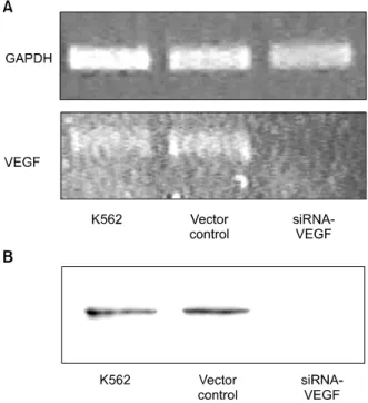
Fig. 1. Vascular endothelial growth factor (VEGF) expression in K562 cells. Control or siRNA to VEGF was transfected into K562 cells. (A) Total RNA was extracted from the cells and VEGF transcripts were analyzed by RT-PCR. Glyceraldehyde- 3-phosphate dehydrogenase (GAPDH) was used as a con- trol. (B) CM of K562 cells was analyzed by western blotting.
The blot showed that VEGF secretion of K562 cell was significantly decreased by transfection of siRNA targeting VEGF.
Fig. 2. Endothelial cell proliferation. HUVECs were cultured in 96 well plates at 5103 cells per well with or without 50%
(v/v) CM of K562 cells in the presence (□) or absence (■) of K2 for 72h. The proliferation of HUVECs was measured by MTT assay. Results represent the mean±SD (n=3).
RESULTS
1. Down-regulation of endogenous VEGF ex- pression by siRNA to VEGF
RT-PCR analysis was performed on total RNA from either siRNA-VEGF K562 cells or vector control K562 cells to assess the levels of VEGF mRNA expression. The results showed that the expression of VEGF mRNA was inhibited in K562 cells transfected with siRNA to VEGF, as compared to K562 cells transfected with vector alone, indicating that siRNA down-regulated the expression of endogenous VEGF (Fig. 1A). Wes- tern blotting was also performed to detect the secreted VEGF by K562 cells. The blot showed that VEGF secretion of K562 cell was signifi- cantly decreased by transfection of siRNA tar- geting VEGF (Fig. 1B).
2. Effects of CM of K562 cells on the proli- feration of HUVECs
Transfection of siRNA to VEGF into K562 cells resulted in decreased VEGF expression and secretion, thereby a decreased capacity of the resultant CM to enhance the proliferation of HUVECs. To evaluate this effect, 50% (v/v) CM of K562 cells was added to the growth media of HUVEC. The proliferation of HUVECs induced by the CM from siRNA-VEGF K562 cells was decreased, compared with CM from vector control K562 cells (Fig. 2).
3. Effects on tube formation of HUVECs HUVECs, plated on growth factor-reduced Matrigel, formed capillary-like structures in the absence of serum or exogenous growth factors.
HUVECs were inhibited to form capillary-like structures in the presence of 5ug/mL K2. The ability of serum-starved HUVECs to form capillary- like structures was partially restored by CM of K562 cells (Fig. 3). CM from siRNA- VEGF K562 cells were less effective than CM from vector control K562 cells, demonstrating that the acti- vating effect was due to VEGF expression.
Fig. 4. Production of interleXkin-6 (IL-6) by HUVECs.
HUVECs were cultured with 50% (v/v) CM of K562 cells in the presence (□) or absence (■) of 5 ug/mL K2 for 24h.
The culture supernatant was centrifuged and IL-6 con- centrations were determined by ELISA. Results represent the mean±SD (n=3).
Fig. 3. Tube formation assay. HUVECs were serum-starved for 24 h and cultured on growth factor-reduced Matrigel in the presence or absence of 50% (v/v) CM of K562 cells for 12h. Representative images were shown at ×40 magnifi- cation.
4. Effects of VEGF and K2 on IL-6 secretion by HUVECs
K562 cell-derived VEGF induced a time-depen- dent increase in IL-6 secretion by HUVECs (data not shown), implying there are paracrine interac- tions between myeloma and endothelial cells. CM from siRNA transfected K562 cells showed diminished IL-6 expression, indicating that IL-6 production by HUVECs was crossly related to exogenous VEGF concentrations (Fig. 4). IL-6 secretion was also inhibited in the presence of 10ug/mL K2.
DISCUSSION
Since Perez-Atayde et al. reported that bone marrow biopsies from children with acute lym- phoblastic leukemia showed significantly higher microvessel densities than control specimens,12) a number of investigators have indicated that leu- kemia cells induce angiogenesis in the bone marrow and that leukemia might be angiogenesis-depen- dent.13,14) VEGF, originally cloned from a human leukemia cell line HL60, has been found in many hematologic malignancies. An increased VEGF level has also been shown to be a predictor of unfavorable outcome in acute myeloid leukemia
patients.15) In an animal model using xenotrans- planted human leukemia and lymphoma cell lines, a strong correlation was observed between the amount of VEGF produced in vitro by cul- tured cells and the efficiency of tumor engraf- tment.16) VEGF is also expressed and secreted by myeloma cell lines and plasma cells isolated from bone marrow of patients with multiple myeloma.9) In addition, circulating levels of VEGF have been shown to correlate with overall survival and event-free survival in non-Hodgkins lymphomas, and with progression-free survival in chronic lymphocytic leukemia.17,18) It has recently been demonstrated that down-regulation of VEGF in leukemia cells resulted in inhibition of tumor angiogenesis and tumor growth. Introduction of antisense VEGF into K562 cells resulted in the decrease of cell proliferation and migration.19) It is well known that the introduction of siRNA into a mammalian cell triggers the degradation of the endogenous mRNA to which the siRNA hybridizes.20) This mechanism is highly sequence- specific and allows to turn off the expression of a target protein. Previously, it was reported that a siRNA to VEGF successfully inhibited the secretion and expression of VEGF in PC-3, hu- man prostate carcinoma cells, leading to the po- tent suppression of tumor growth in its xenograft model.21) In this report, we evaluated the effect of siRNA targeting VEGF in K562 leukemia cells on tumor angiogenesis. The proliferation of HUVECs induced by the CM from siRNA-VEGF K562 cells was decreased, compared with CM from vector control K562 cells (Fig. 2). The tube formation assay also indicated that siRNA to VEGF decreased the expression of VEGF in K562 cells (Fig. 3). Nevertheless, it was not sufficient to block angiogenesis by down-regulation of leukemia-derived VEGF with siRNA, indicating other angiogenic factors were also secreted by leukemia cells. The angiogenesis of HUVECs was significantly inhibited by combination with K2.
Accumulating data suggest the existence of a paracrine loop between leukemia cells and
endothelial cells that plays a significant role in supporting further growth of both cell popu- lations. In this paracrine loop, VEGF secreted by leukemia cells may stimulate endothelial cell proliferation and promote them to secret more hematopoietic growth factors, such as IL-6, which in turn enhance further leukemia growth.22) IL-6, a multifunctional cytokine, is produced by a number of cells and plays a role in the defense mechanism of a host.23) Higher serum IL-6 levels were reported in patients with multiple myeloma, renal cell carcinoma, and ovarian cancer, com- pared with normal subjects.24-26) Serum IL-6 level was recently reported to correlate with the disease status of gastric carcinoma and was demonstrated to decrease after tumor resection and increase when gastric carcinoma recurred.27) It was also shown that the treatment of IL-6 resulted in a significant induction of VEGF mRNA in various cell lines including human epidermoid carcinoma cell line, rat skeletal muscle myoblast, and glioma cells.28) Significant correlation between serum IL-6 and VEGF levels have been reported in a variety of advanced cancers. We here showed that VEGF, secreted in leukemia CM, promoted the production of IL-6 by HUVEC and the produced IL-6 level was decreased by adding K2 into growth media of HUVEC (Fig. 4). Since IL-6 may stimulate endothelial cell proliferation by enhancing VEGF production, the decreased level of IL-6, caused by K2, seems to be crossly related to the decreased proliferation of HUVEC.
Numerous antiangiogenic agents are currently in clinical trials. Monotherapies in these assays have been disappointing, and several companies have abandoned drugs targeting VEGF or its receptors.29) Since resistance could be responsible for the failures of these promising agents to consistently curtail human tumor growth, com- binational therapies are required for elimination of tumor.30,31) For example, the joining of the antiangiogenic protein endostatin with an anti- sense strategy against the epidermal growth fac- tor receptor (expressed on a fraction of human
tumor cells) has been shown to produce syner- gistic inhibitory effects on tumor growth.32) In this study, we show that the combination of si- RNA targeting VEGF and K2 can significantly inhibit tumor angiogenesis. In vivo transfection of siRNA targeting VEGF should be studied fur- ther to define the more effective set of treatment to tumor growth.
요 약
배경: 혈관내피성장인자(VEGF)는 종양신생혈관 증식을 비롯한 혈관형성과정에서 중요한 역할을 하 며, 만성림프모구백혈병, 비호지킨림프종, 다발골수 종 등 비고형성 암의 성장에도 영향을 미친다는 많 은 증거들이 보고되어 있다. 또한 혈관내피성장인 자는 내피세포로 하여금 IL-6와 같은 조혈성장인자 들의 생산, 분비를 촉진함으로써 백혈병의 증식 속 도가 더욱 빨라지게 한다. 이것은 암세포와 내피세 포간에 주변분비고리를 통한 상호작용을 통해 암증 식속도가 가속된다는 것을 의미한다.
방법: VEGF가 백혈병발생에 미치는 영향을 알아 보기 위해 siRNA를 사용하여 K562 백혈병세포로부 터 VEGF 생성을 억제시켰다. K562 세포배양액을 HUVEC 세포배양액에 넣어준 후 HUVEC세포의 증 식과 관형성과정의 변화를 조사하였다. 신혈관형성 억제인자로 알려진 재조합 사람 프로트롬빈 크링글 2 (K2)도 함께 HUVEC세포에 처리하여 HUVEC세 포로부터 생성, 분비되는 IL-6의 양을 측정함으로써 암세포와 혈관내피세포 사이에 발생하는 상호작용 을 살펴보았다.
결과: RT-PCR과 웨스턴블로트 분석 결과 siRNA 는 K562세포의 VEGF 생성을 효율적으로 억제한다 는 사실을 알 수 있었다. K562세포의 세포배양액을 HUVEC 세포배양액에 넣어주면 HUVEC세포의 증 식이 촉진되는데, siRNA로 VEGF 생성을 억제시킨 경우 대조군인 정상 K562세포의 경우보다 그 증식 효과가 감소되었다. 또한 K2를 처리한 경우 HUVEC 세포의 증식이 억제됨은 물론 IL-6의 분비도 억제 되었다.
결론: siRNA를 이용하여 VEGF 생성을 억제하는 것은 백혈병에 의해 유발되는 신생혈관생성을 억제 하는 새로운 방법이 될 수 있다. 이는 K2와의 병용 치료에 의해 그 효과가 배가 된다. K2는 HUVEC세
포의 IL-6생성을 억제함으로써 IL-6에 의한 백혈병 세포의 VEGF 생성 역시 억제될 수 있다.
ACKNOWLEDGEMENTS
This work was supported by Grant No. R01- 2005-000-10179-0 from the Basic Research Pro- gram of the Korea Science and Engineering Foundation and by the Brain Korea 21 project.
REFERENCES
1) Senger DR, Van de Water L, Brown LF, et al.
Vascular permeability factor (VPF, VEGF) in tumor biology. Cancer Metastasis Rev 1993;12:303-24.
2) Ferrara N. Molecular and biological properties of vascular endothelial growth factor. J Mol Med 1999;77:527-43.
3) Chen H, Treweeke AT, West DC, et al. In vitro and in vivo production of vascular endothelial growth factor by chronic lymphatic leukemia cells. Blood 2000;96:3181-7.
4) Schuch G, Machluf M, Bartsch G Jr, et al. In vivo administration of vascular endothelial growth factor (VEGF) and its antagonist, soluble neuropilin-1, predicts a role of VEGF in the progression of acute myeloid leukemia in vivo. Blood 2002;100:4622-8.
5) Zhang HT, Craft P, Scott PA, et al. Enhancement of tumor growth and vascular density by transfection of vascular endothelial cell growth factor into MCF-7 human breast carcinoma cells. J Natl Cancer Inst 1995;87:213-9.
6) Ciardiello F, Bianco R, Damiano V, et al. Antian- giogenic and antitumor activity of anti-epidermal growth factor receptor C225 monoclonal antibody in combination with vascular endothelial growth factor antisense oligonucleotide in human GEO colon cancer cells. Clin Cancer Res 2000;6:3739-47.
7) Nakashima T, Hudson JM, Clayman GL. Antisense inhibition of vascular endothelial growth factor in human head and neck squamous cell carcinoma.
Head Neck 2000;22:483-8.
8) Im SA, Gomez-Manzano C, Fueyo J, et al. Antian- giogenesis treatment for gliomas: transfer of an- tisense-vascular endothelial growth factor inhibits tumor growth in vivo. Cancer Res 1999;59:895-900.
9) Dankdar B, Padro T, Leo R, et al. Vascular endo- thelial growth factor and interleukin-6 in paracrine tumor-stromal cell interactions in multiple myeloma.
Blood 2000;95:2630-6.
10) Rhim TY, Kim E, Park CS, Kim SS. Expression, purification and characterization of prothrombin kringle 2. J Biochem Mol Biol 1999;32:147-53.
11) Kim BJ, Koo SY, Kim SS. A peptide derived from human prothrombin fragment 2 inhibits prothrom- binase and angiogenesis. Thromb Res 2002;106:81-7.
12) Perez-Atayde AR, Sallan SE, Tedrow U, Connors S, Allred E, Folkman J. Spectrum of tumor angiogene- sis in the bone marrow of children with acute lym- phoblastic leukemia. Am J Pathol 1997;150:815-21.
13) Hussong JW, Rodgers GM, Shami PJ. Evidence of increased angiogenesis in patients with acute mye- loid leukemia. Blood 2000;95:309-13.
14) Aguayo A, Kantarjian H, Mansbouri T, et al. An- giogenesis in acute and chronic leukemias and myelodysplastic syndromes. Blood 2000;96:2240-5.
15) Aguayo A, Estey E, Kantarjian H, et al. Cellular vas- cular endothelial growth factor is a predictor of outcome in patients with acute myeloid leukemia.
Blood 1999;94:3717-21.
16) Fusetti L, Pruneri G, Gobbi A, et al. Human myeloid and lymphoid malignancies in the non-obese diabe- tic/severe combined immunodeficiency mouse mo- del: frequency of apoptotic cells in solid tumors and efficiency and speed of engraftment correlate with vascular endothelial growth factor production. Can- cer Res 2000;60:2527-34.
17) Salven P, Orpana A, Teerenhovi L, Joensuu H. Si- multaneous elevation in the serum concentrations of the angiogenic growth factors VEGF and bFGF is an independent predictor of poor prognosis in non- Hodgkin lymphoma: a single institution study of 200 patients. Blood 2000;96:3712-8.
18) Molica S, Vitelli G, Levato D, Gandolfo GM, Liso V.
Increased serum levels of vascular endothelial grow- th factor predict risk of progression in early B-cell chronic lymphocytic leukaemia. Br J Haematol 1999;
107:605-10.
19) He R, Liu B, Yang C, Yang RC, Tobelem G, Han ZC. Inhibition of K562 leukemia angiogenesis and growth by expression of antisense vascular endo- thelial growth factor (VEGF) sequence. Cancer Gene Ther 2003;10:879-86.
20) Elbashir SM, Harborth J, Lendeckel W, Yalcin A,
Weber K, Tuschl T. Duplexes of 21-nucleotide RNAs mediate RNA interference in cultured mammalian cells. Nature 2001;411:494-8.
21) Takei Y, Kadomatsu K, Yuzawa Y, Matsuo S, Mu- ramatsu T. A small interfering RNA targeting vas- cular endothelial growth factor as cancer thera- peutics. Cancer Res 2004;64:3365-70.
22) Dankbar B, Padro T, Leo R, et al. Vascular endo- thelial growth factor and interleukin-6 in paracrine tumor-stromal cell interactions in multiple myeloma.
Blood 2000;95:2630-6.
23) Hirano T, Akira S, Taga T, Kishimoto T. Biological and clinical aspects of interleukin 6. Immunol Today 1990;11:443-9.
24) Bataille R, Jourdan M, Zhang XG, Klein B. Serum levels of interleukin 6, a potent myeloma cell growth factor, as a reflect of disease severity in plasma cell dyscrasias. J Clin Invest 1989;84:2008-11.
25) Tsukamoto T, Kumamoto Y, Miyao N, Masumori N, Takahashi A, Yanase M. Interleukin-6 in renal cell carcinoma. J Urol 1992;148:1778-81.
26) Berek JS, Chung C, Kaldi K, Watson JM, Knox RM, Martinez-Maza O. Serum interleukin-6 levels corre- late with disease status in patients with epithelial ovarian cancer. Am J Obstet Gynecol 1991;164:1038-42.
27) Wu CW, Wang SR, Chao MF, et al. Serum inter- leukin-6 levels reflect disease status of gastric cancer.
Am J Gastroenterol 1996;91:1417-22.
28) Cohen T, Nahari D, Cerem LW, Neufeld G, Levi BZ.
Interleukin 6 induces the expression of vascular endothelial growth factor. J Biol Chem 1996;271:736-41.
29) Garber K. Angiogenesis inhibitors suffer new set- back. Nat Biotechnol 2002;20:1067-8.
30) O'Reilly MS. Targeting multiple biological pathways as a strategy to improve the treatment of cancer.
Clin Cancer Res 2002;8:3309-10.
31) Kim KW, Kim HJ, Park SY. The relationship between clinical drug sensitivity and expression of drug resistance genes in patients with acute myelogenous leukemia. Korean J Hematol 2001;36:115-22.
32) Li M, Ye C, Feng C, et al. Enhanced antiangiogenic therapy of squamous cell carcinoma by combined endostatin and epidermal growth factor receptor- antisense therapy. Clin Cancer Res 2002;8:3570-8.