의해 HLA 항체가 생성될 수 있고, 이는 고형 장기이식 수혜자에게 항체매개성 거부반응(antibody-mediated rejection)을 유발할 수 있다[1, 2]. HLA는 그 종류가 매우 다양하므로 정확한 HLA 항체의 검출, 특히 공여자 특이 HLA와 반응하는 HLA 항체 검출은 이식 전 공여자의 선택뿐 아니라 이후 성공적인 장기 생착에도 필수적 이다[3, 4].
패널반응항체(panel reactive antibody, PRA) 검사는 환자가 속 해있는 인종 또는 집단의 HLA 빈도를 고려한 항원 패널을 이용하 여 혈청 내의 HLA 항체를 검출 및 동정하는 검사이다. 그 중 solid phase-based PRA 방법은 추출 혹은 재조합 HLA 항원을 이용하므 로 비 HLA 항체에 의한 위양성을 배제할 수 있어 기존 림프구를 이용한 방법에 비해 민감도가 높은 장점을 가진다[5, 6]. 그 중 Lu- minex-PRA는 높은 민감도를 보이며, 특히 각 비드마다 한 종류의 재조합 HLA 항원이 코팅된 비드를 이용한 단일항원비드(single antigen bead, SAB) 검사법은 직접 항체특이성을 확인할 수 있어
서 론
임신, 수혈 및 장기이식 등을 통해 타인의 사람백혈구항원(hu- man leukocyte antigen, HLA)에 노출되는 경우 동종면역 반응에
HLA Class II 항체 검출 시 C3d 검사법과 SAB 검사법의 상관관계 평가
Correlation Between C3d Assay and Single Antigen Bead Assay for Detection of Human Leukocyte Antigen Class II Antibodies
강혜인·김도훈·하정숙·류남희·전동석·이원목
Hyein Kang, M.D., Do-Hoon Kim, M.D., Jung Sook Ha, M.D., Namhee Ryoo, M.D., Dong-Seok Jeon, M.D., Wonmok Lee, M.D.
계명대학교 의과대학 진단검사의학교실
Department of Laboratory Medicine, Keimyung University School of Medicine, Daegu, Korea Vol. 10, No. 4: 295-300, October 2020
https://doi.org/10.47429/lmo.2020.10.4.295 진단면역학
Corresponding author: Wonmok Lee, M.D., Ph.D.
https://orcid.org/0000-0002-0891-9375
Department of Laboratory Medicine, Keimyung University School of Medicine, 1035 Dalgubeol-daero, Dalseo-gu, Daegu 42601, Korea Tel: +82-53-258-7940, Fax: +82-53-258-4228, E-mail: [email protected] Received: February 6, 2020
Revision received: June 9, 2020 Accepted: June 26, 2020
This article is available from https://www.labmedonline.org 2020, Laboratory Medicine Online
This is an Open Access article distributed under the terms of the Creative Commons Attribution Non-Commercial License (https://creativecommons.org/licenses/by-nc/4.0/) which permits unrestricted non-commercial use, distribution, and reproduction in any medium, provided the original work is properly cited.
Background: Detection of anti-human leukocyte antigen (HLA) antibodies is important during the selection of an appropriate donor prior to or- gan transplantation and also for monitoring the patients after transplantation. In this study, we compared antibodies detected via C3d assays, which monitors C3d complement-binding activities of HLA antibodies with those detected via single antigen bead (SAB) assays.
Methods: A total of 66 serum samples were tested in parallel by SAB assays (Immucor Transplant Diagnostics, USA) and C3d assays (Immucor) for the detection of HLA class II antibodies. The relationship between these two methods was analyzed based on the types, numbers, median fluo- rescent intensity (MFI) values, and positivity of the antibodies using MATCH IT! Antibody (Immucor) program.
Results: The number of antibodies obtained based on SAB and C3d assays was the highest with 24 samples (36.4%) in the 11–20 range and 23 (34.8%) in the 2–5 range detected via each assay. Among the SAB-positive antibodies, only 28 (6.4%) of the 440 antibodies with MFI ≤3,000 were C3d-positive, and 341 (61.3%) of the 556 antibodies with MFI ≥3,001 were C3d-positive. Whereas, among the 442 C3d-positive antibodies, SAB assays were positive except for 32 (7.2%) and 41 (9.3%) antibodies in the sections of MFI ≤500 and 1,001 ≤MFI ≤10,000, respectively.
C3d-positive samples had higher maximum MFI values based on SAB assays, compared with C3d-negative samples.
Conclusions: MFI values of HLA class II antibodies detected through SAB assays in C3d-positive samples were higher than those in C3d-nega- tive samples.
Key Words: Single antigen bead assay, C3d assay, HLA class II antibodies
2017-03-16 https://crossmark-cdn.crossref.org/widget/v2.0/logos/CROSSMARK_Color_square.svg
임상적으로 중요한 공여자특이항체(donor-specific antibody, DSA) 동정에 도움이 된다.
하지만, solid phase-based 방법으로 검출된 DSA가 이식 후 거 부반응 또는 생존율과 큰 상관성을 보이지 않는다는 보고들이 있 었는데, 이를 면역반응에서 보체 활성화를 유발하지 않는 HLA 항 체까지 검출하기 때문으로 설명을 하였다[7, 8]. 최근 이를 보완하 여 보체 활성화를 유발시키는 항체인지를 규명하는 검사법들이 개발되었고[9, 10], One Lambda사(One Lambda, Inc., Canoga Park, CA, USA)의 C1q screen과 Immucor사(Immucor Transplant Diag- nostics, Norcross, GA, USA)의 C3d detection이 사용되고 있다.
보체를 이용한 새로운 검사법들은 이미 수 차례 기존 검사법들 과 비교평가가 이루어져 왔고, 일부 임상 경과의 상관성에 대한 연 구도 보고된 바 있다[11-16]. 하지만, 현재까지 다량의 검체를 대상 으로 class II 항원을 이용한 C3d 검사법의 비교평가에 대한 연구 는 없었다. 이에 본 연구에서는 SAB 검사법에서 class II HLA 항체 가 검출된 환자들을 대상으로 C3d 검사를 시행하여 SAB 검사법 과 C3d 검사법을 비교하고자 하였다.
재료 및 방법
1. 대상
계명대학교 동산병원에서 2015년 4월부터 2018년 9월까지 HLA 항체 검사를 위해 검사실로 의뢰된 검체 중 class II SAB 검사 (LIFECODES LSA Single Antigen, Immucor Transplant Diagnos- tics)에서 양성인 총 80개의 혈액 검체를 선별하였다. 선별된 검체 중 DSA가 확인되지 않은 14개는 제외하고 DSA 양성인 66개의 검 체를 대상으로 하였다. 그 중 SAB 검사에서 양성을 보인 994개, C3d 검사에서 양성을 보인 442개의 항체를 분석하였다.
정규 검사 후 –80˚C 냉동고에 보관되어 있던 66개의 검체를 대상 으로 class II C3d 검사(C3d detection kit, Immucor Transplant Di- agnostics)를 시행하여 평행 검사를 진행하였다. 이 연구와 관련된 자료 수집 및 분석은 계명대학교 동산병원 기관윤리심의위원회 (Institutional Review Board, IRB No. DSMC 2019-03-002)의 승인 하에 시행되었다.
2. 방법
본 연구에서는 Luminex-PRA 검사법 중 재조합된 HLA 항원이 코팅된 비드를 이용하여 HLA 항체를 검출하는 방법을 SAB 검사 법으로 지칭하였고, SAB를 환자 혈청과 반응시킬 때 C3d 보체를 첨가하여 C3d가 결합하는 HLA 항체를 검출하는 검사법을 C3d 검 사법으로 지칭하였다.
1) SAB 검사법
Plate의 각 well에 40 μL의 LSA 비드를 첨가한 후, 10 μL의 환자 혈청과 대조혈청을 분주하고 혼합하였다. Adhesive plastic cover로 plate를 덮고 호일 또는 상자를 이용하여 빛을 차단한 후 실온에서 30분간 배양하였다. 이후 cover를 제거하고 각 well에 100 μL의 wash buffer를 첨가하여 비드를 재부유시키기 위해 혼합하고 plate 를 부드럽게 흡인하는 과정을 3회 반복하였다. 각 well에 50 μL의 diluted conjugate를 첨가하고 빛을 차단한 후 실온에서 30분간 배 양하였다. 150 μL의 wash buffer를 각 well에 분주 후 비드를 재부 유시키기 위해 혼합하였다. Luminex 장비(Luminex® 200™, Lu- minex Corp., Austin, TX, USA)를 이용해서 각 비드의 신호 강도를 측정하여 MATCH IT! Antibody v1.3.1.5 (Immucor Transplant Di- agnostics)로 분석하였다. 각각의 비드별로 얻어진 median fluores- cence intensity (MFI) 값을 이용하여 보정값을 구했다. 다음 3개의 보정값 중에서 두 개 이상의 adjusted (ADJ) MFI 값이 “Suggested Cutoff”보다 높은 경우 양성으로 판정하였다: 1) ADJ1=raw MFI 값에서 background (BG) MFI 값을 뺀 값; 2) ADJ2=ADJ1 값을 lo- cus “1”에서 가장 낮게 반응한 비드의 raw MFI 값으로 나눈 값; 3) ADJ3=ADJ2 값을 항원 밀도의 상대량으로 나눈 값. 이때 대조혈청 은 검사키트에 포함된 LSA class II 대조혈청을 사용하였다.
2) C3d 검사법
40 μL의 LSA 비드와 1 μL의 양성대조 비드를 혼합한 후 plate의 각 well에 40 μL의 mix와 10 μL의 혈청을 분주하였다. Adhesive plastic cover로 plate를 덮고 호일 또는 상자를 이용하여 빛을 차단 한 후 실온에서 30분간 배양하였다. 이후 cover를 제거하고 각 well에 30 μL의 C3d 보체 혈청을 분주하였다. 다시 cover로 plate 를 덮고 빛을 차단한 후 실온에서 30분간 배양하였다. 배양이 끝나 면 각 well에 100 μL의 washing buffer를 분주하고 섞은 다음 부드 럽게 흡인하는 과정을 4회 반복하였다. 각 well에 C3d conjugate를 50 μL씩 분주하고, 빛을 차단한 후 실온에서 30분간 배양하였다.
그 후 각 well에 100 μL의 washing buffer를 분주하고 혼합한 다음 흡인하고, 다시 분주하여 혼합하였다. Luminex 장비를 이용해서 각 비드의 신호 강도를 측정하여 MATCH IT! Antibody 프로그램 으로 분석하였다. 다음 3개의 보정값 중에서 하나 이상에서 양성 수치를 보이는 경우 양성으로 판정하였다: 1) BG Adjusted MFI=얻 어진 MFI 값에서 음성대조혈청 MFI 값을 뺀 값; 2) BCR-Neg=BG Adjusted MFI를 해당 locus의 가장 낮게 반응한 bead의 raw MFI 값 으로 나눈 값; 3) R-Strength=BG Adjusted MFI를 항원의 raw MFI 값으로 나눈 값. 이때 대조혈청은 검사키트에 포함된 LSA class II 대 조혈청을 사용하였다.
3) 통계 및 자료분석
MATCH IT! Antibody 프로그램을 통해 각 검체를 SAB 검사법 과 C3d 검사법으로 분석한 HLA 항체 종류, MFI 값, 양성여부를 수 집하였다. SAB 검사법은 raw MFI 값에서 BG MFI 값을 뺀 BG cor- rected MFI 값을 기준으로 하였고, C3d 검사법은 raw MFI 값에서 음성대조혈청의 MFI 값을 뺀 BG Adjusted MFI 값을 기준으로 하 였다. 대한진단면역학회에서 제시한 HLA 보고서 표준안에 따라 MFI≤1,000, 1,001≤MFI≤3,000, 3,001≤MFI≤10,000, MFI≥ 10,000로 구분하였고, 세부적인 분석을 위해 MFI 500, 20,000, 30,000을 기준으로 하여 추가적으로 구간을 나누었다[17].
통계처리는 Microsoft Office Excel 2016 (Microsoft Co., Redmond, WA, USA)을 이용하였고, Analyse-it software v5.20 (Analyse-it Soft- ware, Ltd., Leeds, UK)을 이용하여 상관분석을 시행하였다.
결 과
1. SAB 검사와 C3d 검사의 항체 수
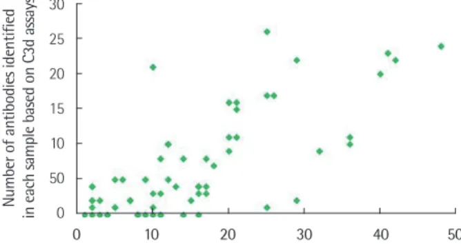
전체 검체 중 DSA 양성인 검체는 66예였고, 각 검체는 1개에서 48개의 항체를 가지고 있었고 중간값은 12.5개였다. 그 중 52예가 C3d 검사 양성이었고 1개에서 26개의 항체를 가지며 중간값은 4개 였다. SAB 검사와 C3d 검사에서 각각 996개와 442개의 항체가 양 성이었다. SAB 검사와 C3d 검사에서 확인된 항체수의 피어슨 상관 계수는 0.735로 양의 상관관계를 나타냈다(Fig. 1). 각 검사별 항체 수는 SAB 검사에서 항체수가 11-20개인 구간에서 24개(36.4%)로 가장 많았고, C3d 검사에서는 항체수가 2-5개인 구간에서 23개 (34.8%)로 가장 많았다.
2. SAB 검사와 C3d 검사의 MFI 값
SAB 검사의 항체는 MFI 값이 1,001≤MFI≤20,000인 구간에
802개(80.7%)가 속한 반면, C3d 검사는 MFI≤500인 구간과 1,001
≤MFI≤10,000인 구간에 299개(67.6%)의 항체가 속했다(Fig. 2).
3. SAB 검사와 C3d 검사의 비교
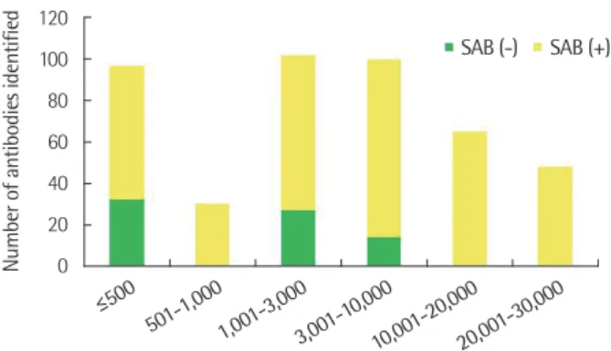
SAB 검사에서 양성인 항체 중에서 MFI≤3,000인 항체 440개 중 28개(6.4%)만이 C3d 검사 양성이었고, MFI≥3,001인 항체 556개 중 341개(61.3%)가 C3d 검사 양성이었다(Fig. 3). 반면, C3d 검사에 서 양성인 442개의 항체 중에서는 MFI≤500, 1,001≤MFI≤10,000 인 구간에서 각각 32개(7.2%), 41개(9.3%)를 제외하고는 모두 SAB 검사도 양성이었다(Fig. 4). 한편, SAB 검사에서 음성이었으나 C3d 검사 양성인 항체는 26종, 총 73개였다. 이들의 MFI 값은 98부터 9,710까지였으며 평균값은 2,062, 중간값은 1,817이었다. 반대로, SAB 양성이나 C3d 검사 음성인 항체는 총 92종, 627개로 63.1%를 차지했다. 이들의 MFI 값은 86부터 14,888까지였으며 평균값은 3,207, 중간값은 1,935였다. DSA만을 대상으로 했을 때 SAB 검사
Fig. 1. The number of antibodies identified by SAB assays correlated with those identified by C3d assays which are also SAB-positive sam- ples. Pearson’s r was 0.735.
Abbreviation: SAB, single antigen bead.
30 25 20 15 10 50 Number of antibodies identified in each sample based on C3d assays 0
Number of antibodies in each sample identified based on SAB assays 0 10 20 30 40 50
Fig. 2. Distribution pattern of MFI values of antibodies identified by SAB or C3d assays.
Abbreviations: MFI, median fluorescence intensity; SAB, single anti- gen bead.
MFI values of identified antibodies 350
300 250 200 150 100 50 0
Number of antibodies
SAB
≤500 501-1,0001,001-3,000 3,001-10,000
10,001-20,000 20,001-30,000
C3d
MFI values of antibodies identified based on SAB assays 350
300 250 200 150 100 50 Number of identified antibodies 0
C3d (-)
≤500 501-1,0001,001-3,000 3,001-10,000
10,001-20,000 20,001-30,000
C3d (+)
Fig. 3. Distribution of the numbers of C3d (-) and C3d (+) antibodies identified by SAB assays.
Abbreviations: MFI, median fluorescence intensity; SAB, single anti- gen bead.
양성이나 C3d 검사 음성인 항체는 총 42종, 103개로 56.9%를 차지 했다. 이들의 MFI 값은 206부터 8,505까지였으며 평균값은 2,488, 중간값은 1,591이었다.
고 찰
DSA의 임상적 중요성이 거론되면서 항체의 강도, 항체의 종류, 항원결정기, 대립유전자, 염기서열 분석 및 새로운 검사법과 검사 법 간 비교 등에 대한 많은 연구들이 이루어지고 있다[13, 16, 18- 21]. 그 중 총 IgG SAB 검사와 C1q, C3d 보체를 이용한 SAB 검사를 CDC PRA 검사법으로 동정된 항체와 비교한 한 연구에서는 IgG SAB 검사가 가장 높은 일치도를 보였다. 또한 IgG SAB 검사의 MFI 값이 10,000보다 클 경우에 C1q와 C3d 보체 검사의 정확도가 높은 것으로 나타났다[13]. HLA class I 항체를 대상으로 SAB 검사법과 C3d 검사법을 비교한 한 연구에 의하면 C3d 검사 양성 검체가 C3d 검사 음성 검체보다 SAB 검사의 최대 MFI 값이 큰 것으로 나 타났고 SAB 검사의 MFI 값은 C3d의 양성여부와 양의 상관관계를 보였다[19].
본 연구는 SAB 검사법과 C3d 검사법에서 HLA class II 항체를 비교한 것으로, 단일 기관에서 시행된 66예의 검체를 분석하였다.
그 결과 HLA class I 항체를 대상으로 한 앞선 연구와 동일한 양상 을 보였다. 이는 MFI 값이 C3d 검사 양성여부의 강력한 예측인자 라는 기존의 보고와 일치한다[20].
하지만 SAB 검사와 C3d의 양성여부 또는 MFI 값의 강도는 상관 관계를 보이지 않는 경우도 있었다. 특히 SAB 검사에서 양성이지 만 C3d 검사에서 음성인 항체가 63.1%였다. SAB 검사에서 DSA로 확인된 181개의 항체만을 대상으로 분석해봤을 때에도 동일하게 SAB 검사에서 양성이지만 C3d 검사에서 음성인 항체는 56.9%로
상당수가 존재했다. C3d 검사법은 면역반응에서 활성화되는 보체 를 이용한 검사법이므로 실제 보체의존성면역반응에 관여하는 HLA 항체만을 검출한다[23]. 따라서 이전에 SAB 검사에서 실제로 보체의존성면역반응에 관여하지 않지만 양성으로 검출되었던 항 체들이 음성으로 나왔을 것이라 예측해볼 수 있었다.
반대로 SAB 검사에서 음성이지만 C3d 검사에서 양성으로 검출 된 항체는 보체경로를 활성화하는 항체 자체의 특성 때문에 기존 방법으로 찾아내지 못했던 항체를 찾을 수 있어서 유용하다고 생 각하였으나 이에 대한 정확한 원인은 아직 알려지지 않았다. 흔히 면역검사법에서 위음성을 나타낼 수 있는 요인으로는 프로존 효 과(prozone effect) 등이 있으나 본 연구에서는 명확하게 프로존 효과를 보이는 것으로 생각되는 항체들이 관찰되지 않았다[24].
DSA가 환자의 치료와 임상 결과에 미치는 영향이 크기 때문에 이식 후의 거부반응이나 환자의 임상양상과의 연관성도 함께 연 구되어야 하지만 이번 연구에서는 이식 이후 환자의 임상양상을 포함하여 분석하지 못했다. 그러므로 C3d 검사 결과가 환자의 생 존율에 어떠한 영향을 미쳤는지는 확인하지 못하였다는 제한점을 가진다. 이에 대해 이전에 보고된 바에 따르면 MFI 값과 무관하게 C3d 양성 DSA가 존재할 경우 C3d 음성인 경우보다 장기 생존율 이 낮다는 연구결과가 있으며[16], DSA가 C1q와 C3d에 결합하는 경우 이식 후 예후가 좋지 않다는 보고도 있다[14]. 추가적으로, 항 체매개성 거부반응 진단 시 C4d 침착을 확인하기 위한 조직검사 는 침습적인 반면 DSA 검출은 말초혈액 채혈로 가능하기 때문에 훨씬 용이하다는 이점을 가진다. 또한 조직검사 결과가 항체매개 성 거부반응 진단기준에 만족하지 못하더라도 C3d에 결합하는 DSA가 존재할 때 조직 손상을 시사하는 미세혈관 염증 소견이 보 이는 경우도 있으므로[15], DSA의 중요성은 더욱 강조된다.
결론적으로 HLA class II 항체의 SAB 검사에서의 MFI 값은 C3d 검사 음성인 경우보다 양성인 경우에 더 큰 것으로 나타났다. 하지 만 C3d 검사의 양성여부와 SAB 검사에서의 MFI 값의 크기가 상관 관계를 보이지 않는 경우도 있었으므로 향후 이에 대한 검증이 필 요하며, 임상적 중요성에 대해서도 추가적인 연구가 필요할 것이다.
요 약
배경: 장기이식 전에 공여자 선택을 위해 사전에 공여자 HLA에 대 한 항체 유무를 확인하는 것이 중요하고, 이식 후의 급성 또는 만 성 거부반응을 예측하기 위해서도 HLA 항체에 대한 주기적인 추 적 검사가 필요하다. 본 연구에서는 보체 활성화를 유발하는 HLA 항체를 검출할 수 있는 Luminex 검사법 중 C3d 검사를 시행하여 기존의 SAB 검사법과 비교하였다.
방법: HLA 항체 검사를 위해 검사실로 의뢰된 검체 중 class II MFI values of antibodies identified based on C3d assays
120 100 80 60 40 20 0
Number of antibodies identified
SAB (-)
≤500 501-1,0001,001-3,000 3,001-10,000
10,001-20,000 20,001-30,000
SAB (+)
Fig. 4. Distribution of the numbers of SAB (-) and SAB (+) antibodies identified by C3d assays.
Abbreviations: MFI, median fluorescence intensity; SAB, single anti- gen bead.
SAB 검사(Immucor Transplant Diagnostics, USA)에서 양성인 혈 액 검체 중 최종적으로 DSA 양성인 66개의 검체를 대상으로 하였 다. 각 검체를 SAB 검사법과 C3d 검사법(Immucor Transplant Di- agnostics)으로 검사를 시행한 후 MATCH IT! Antibody v1.3.1.5 (Immucor Transplant Diagnostics) 프로그램으로 HLA 항체 종류, MFI 값, 양성여부를 파악한 후 두 검사의 상관관계를 분석하였다.
결과: SAB 검사의 항체수는 11-20개인 구간에서 24개(36.4%)로 가 장 많았고, C3d 검사는 항체수가 2-5개인 구간에서 23개(34.8%)로 가장 많았다. SAB 검사에서 양성인 항체 중에서 MFI≤3,000인 항 체 440개 중 28개(6.4%)만이 C3d 검사 양성이었고, MFI≥3,001인 항체 556개 중 341개(61.3%)가 C3d 검사 양성이었다. 반면, C3d 검 사에서 양성인 442개의 항체 중에서는 MFI≤500, 1,001≤MFI≤ 10,000인 구간에서 각각 32개(7.2%), 41개(9.3%)를 제외하고는 모 두 SAB 검사도 양성이었다. C3d 검사 양성 검체가 C3d 검사 음성 검체보다 SAB의 최대 MFI 값이 더 큰 것으로 나타났다.
결론: HLA class II 항체의 SAB 검사에서의 MFI 값은 C3d 검사 음 성인 경우보다 양성인 경우에 더 큰 것으로 나타났으며, C3d 검사 법의 임상적 중요성에 대해서는 추가적인 연구가 필요할 것이다.
이해관계
저자들은 본 연구와 관련하여 어떠한 이해관계도 없음을 밝힙니다.
REFERENCES
1. Tinckam KJ and Chandraker A. Mechanisms and role of HLA and non- HLA alloantibodies. Clin J Am Soc Nephrol 2006;1:404-14.
2. Valenzuela NM and Reed EF. Antibodies in transplantation: The ef- fects of HLA and non-HLA antibody binding and mechanisms of in- jury. Methods Mol Biol 2013;1034:41-70.
3. Loupy A, Suberbielle-Boissel C, Hill GS, Lefaucheur C, Anglicheau D, Zuber J, et al. Outcome of subclinical antibody-mediated rejection in kidney transplant recipients with preformed donor-specific antibod- ies. Am J Transplant 2009;9:2561-70.
4. Schönemann C, Groth J, Leverenz S, May G. HLA class I and class II antibodies: monitoring before and after kidney transplantation and their clinical relevance. Transplantation 1998;65:1519-23.
5. Jung S, Oh EJ, Yang CW, Ahn WS, Kim Y, Park YJ, et al. Comparative evaluation of ELISA and Luminex panel reactive antibody assays for HLA alloantibody screening. Korean J Lab Med 2009;29:473-80.
6. Joo DJ, Huh KH, Kim YS, Yoon SJ, Kim HJ, Sohn SS, et al. Predictive value of donor specific antibody measured by Luminex single antigen
assay for antibody mediated rejection after kidney transplantation. J Korean Soc Transplant 2011;25:169-75.
7. Aubert V, Venetz JP, Pantaleo G, Pascual M. Low levels of human leu- kocyte antigen donor-specific antibodies detected by solid phase as- say before transplantation are frequently clinically irrelevant. Hum Im- munol 2009;70:580-3.
8. Vlad G, Ho EK, Vasilescu ER, Colovai AI, Stokes MB, Markowitz GS, et al. Relevance of different antibody detection methods for the predic- tion of antibody-mediated rejection and deceased-donor kidney al- lograft survival. Hum Immunol 2009;70:589-94.
9. Zeevi A, Lunz J, Feingold B, Shullo M, Bermudez C, Teuteberg J, et al.
Persistent strong anti-HLA antibody at high titer is complement bind- ing and associated with increased risk of antibody-mediated rejection in heart transplant recipients. J Heart Lung Transplant 2013;32:98-105.
10. Loupy A, Lefaucheur C, Vernerey D, Prugger C, Duong van Huyen JP, Mooney N, et al. Complement-binding anti-HLA antibodies and kid- ney-allograft survival. N Engl J Med 2013;369:1215-26.
11. Llorente S, Boix F, Eguia J, López M, Bosch A, Martinez H, et al. C1q- fixing human leukocyte antigen assay in immunized renal patients:
correlation between Luminex SAB-C1q and SAB-IgG. Transplant Proc 2012;44:2535-7.
12. Kamburova EG, Wisse BW, Joosten I, Allebes WA, van der Meer A, Hilbrands LB, et al. Pretransplant C3d-fixing donor-specific anti-HLA antibodies are not associated with increased risk for kidney graft fail- ure. J Am Soc Nephrol 2018;29:2279-85.
13. Moreno Gonzales MA, Mitema DG, Smith BH, Schinstock CA, Stegall MD, Wakefield LL, et al. Comparison between total IgG, C1q, and C3d single antigen bead assays in detecting class I complement-binding anti-HLA antibodies. Transplant Proc 2017;49:2031-5.
14. Lee H, Han E, Choi AR, Ban TH, Chung BH, Yang CW, et al. Clinical im- pact of complement (C1q, C3d) binding de novo donor-specific HLA an- tibody in kidney transplant recipients. PLoS One 2018;13:e0207434.
15. Comoli P, Cioni M, Tagliamacco A, Quartuccio G, Innocente A, Fon- tana I, et al. Acquisition of C3d-binding activity by de novo donor-spe- cific HLA antibodies correlates with graft loss in nonsensitized pediat- ric kidney recipients. Am J Transplant 2016;16:2106-16.
16. Sicard A, Ducreux S, Rabeyrin M, Couzi L, McGregor B, Badet L, et al.
Detection of C3d-binding donor-specific anti-HLA antibodies at diag- nosis of humoral rejection predicts renal graft loss. J Am Soc Nephrol 2015;26:457-67.
17. Laboratory standardization of reporting HLA typing. http://www.ksdi- lm.org/rang_board/list.html?num=50&code=lib (Updated on Feb
2017)
18. Malheiro J, Santos S, Tafulo S, Dias L, Martins S, Fonseca I, et al. De- tection of complement-binding donor-specific antibodies, not IgG-an- tibody strength nor C4d status, at antibody-mediated rejection diagno- sis is an independent predictor of kidney graft failure. Transplantation 2018;102:1943-54.
19. Ko SY, Lee W, Jung CW, Cho Y. C3d assay in correlation with single an- tigen bead assay for human leukocyte antigen antibodies. Transplant Proc 2018;50:2354-8.
20. Claisse G, Absi L, Cognasse F, Alamartine E, Mariat C, Maillard N. Re- lationship between mean fluorescence intensity and C1q/C3d-fixing capacities of anti-HLA antibodies. Hum Immunol 2017;78:336-41.
21. Morales-Buenrostro LE, Terasaki PI, Marino-Vázquez LA, Lee JH, El- Awar N, Alberú J. “Natural” human leukocyte antigen antibodies
found in nonalloimmunized healthy males. Transplantation 2008;
86:1111-5.
22. El-Awar N, Lee JH, Tarsitani C, Terasaki PI. HLA Class I epitopes: rec- ognition of binding sites by mAbs or eluted alloantibody confirmed with single recombinant antigens. Hum Immunol 2007;68:170-80.
23. Pelletier RP, Balazs I, Adams P, Rajab A, DiPaola NR, Henry ML. Clini- cal utility of C3d binding donor-specific anti-human leukocyte antigen antibody detection by single antigen beads after kidney transplanta- tion-a retrospective study. Transpl Int 2018;31:424-35.
24. Schwaiger E, Wahrmann M, Bond G, Eskandary F, Böhmig GA. Com- plement component C3 activation: the leading cause of the prozone phenomenon affecting HLA antibody detection on single-antigen beads. Transplantation 2014; 97:1279-85.