Kor. J. Fertil. Steril., Vol. 31, No. 4, 2004, 12
배아줄기세포의 인슐린 분비세포로의 유도 분화에 대한 연구
성균관대학교 의과대학 삼성제일병원 생식생물학 및 불임연구실1, 산부인과2 미즈메디병원 의과학연구소3
성지혜1・임천규1・최혜원1・이형송1・신현상1・전진현1*・윤현수3・궁미경2
Induced Differentiation of Embryonic Stem Cells to Insulin Secreting Cells
Ji Hye Sung
1, Chun Kyu Lim
1, Hye Won Choi
1, Hyoung Song Lee
1, Hyeonsang Shin
1, Jin Hyun Jun
1*, Hyun Soo Yoon
3, Mi Kyoung Koong
21
Laboratory of Reproductive Biology & Infertility
2Department of Ob/Gyn Samsung Cheil Hospital, Sungkyunkwan University School of Medicine, Seoul, Korea
3
MizMedi Hospital, Medical Research Center, Seoul, Korea
Objective: Embryonic stem (ES) cells could be differentiated into the specific cell types by alternation of culture condition and modification of gene expression. This study was performed to evaluate the differentiation protocol for mouse and human ES cells to insulin secreting cells.
Methods: Undifferentiated mouse (JH-1) and human (Miz-hES1) ES cells were cultured on STO feeder layer, and embryoid bodies (EBs) were formed by suspension culture. For the differentiation, EBs were cultured by sequential system with three stage protocol. The differentiating ES cells were collected and marker gene expressions were analyzed by semi-quantitative RT-PCR in each stage. Amount of secreted insulin levels in culture media of human ES cells were measured by human insulin specific RIA kit.
Results: During the differentiation process of human ES cells, GATA-4, α-fetoprotein, glucose transporter-2 and Ngn-3 expression were increased whereas Oct-4 was decreased progressively. Insulin and albumin mRNAs were expressed from stage II in mouse ES cells and from stage III in human ES cells. We detected 3.0~7.9 µU/ml secretion of insulin from differentiated human ES cells by in vitro culture for 36 days.
Conclusion: The sequential culture system could induce the differentiation of mouse and human ES cells into insulin secreting cells. This is the first report of differentiation of human ES cells into insulin secreting cells by in vitro culture with serum and insulin free medium.
Key Words: Embryonic stem cell, Insulin secreting cell, Differentiation, Sequential system, Serum and insulin free medium
초기 포배기 배아의 미분화된 내세포괴에서 유 래한 배아줄기세포에 관한 연구는 Evans와 Kaufman
(1981)에 의해 생쥐 배아줄기세포주의 확립이 보고 되면서 많은 연구들이 시행되어 왔다.1 또한, 인간 주관책임자: 전진현, 우) 100-380 서울특별시 중구 묵정동 삼성제일병원 생식생물학 및 불임연구실
Tel: (02) 2000-7590, Fax: (02) 2265-5621, e-mail: [email protected]
*본 연구는 과학기술부 21세기 프론티어 연구개발 사업단인 세포응용연구사업단의 연구비 지원 (SC12022)으로 수행되었습니다.
에서의 체외수정 및 배아이식술 (in vitro fertilization and embryo transfer) 분야에 대한 많은 연구가 진행 됨에 따라 Thomson 등 (1998)에 의해 최초로 인간 의 배아줄기세포주 (human embryonic stem cell line)가 확립되었고, Shamblott 등 (1998)에 의해 인간 배아생 식세포주 (human embryonic germ cell line)의 확립이 보고되었다.2,3 그 후 국내 ・ 외의 여러 연구진에 의해 인간 배아줄기세포주가 확립되었음이 보고되었다.4,5
이러한 배아줄기세포로부터 유도 분화된 특정 세 포들은 질환 모델 동물에 이식되어 그 세포들의 생 체 내 생리학적 기능이 검증되었다. 그 예로, 척수 손상에 따른 운동장애의 개선, myelin 생성의 증가 및 증상 호전, 혈당량 조절을 통한 당뇨병의 개선, dopaminergic 신경세포의 이식을 통한 파킨슨병의 치 료 등이 보고됨으로써 이를 이용한 세포치료의 높 은 가능성을 시사해주고 있다.6~9
인간에서 나타나는 여러 가지 질병들 중에서 당뇨 병은 성인의 5~10%에서 나타나는 흔한 질환으로 그 빈도가 점차 증가하여 전 세계적으로 2000년에는 1300만 명이, 2005년에는 3000만 명이 이환될 것으로 추정되고 있다. 당뇨병은 두 가지 형태로 존재하는데 인슐린 의존형 (insulin-dependent type, IDDM, type 1) 과 인슐린 비의존형 (non-insulin-dependent type, NI- DDM, type 2)이 있다. 그 중에서 Type 1은 매일 인 슐린 주사를 맞아야 하고 많은 합병증, 즉, 신장장 애, 망막질환, 신경장애 등을 초래하게 되는데, 이는 췌장의 β-세포가 자가면역세포에 의해 파괴되기 때 문에 발생되는 것으로 알려지고 있다. 때로는 급성 합병증인 diabetic ketoacidosis, hypo- 또는 hyper- glycemia나, 만성 합병증인 nephropathy, neuropathy, retinopathy, cardiovascular disease 등으로 생명을 위협 받게 된다. 당뇨병은 철저한 혈당량 조절과 인슐린의 지속적인 투여가 필요한데, 이는 환자에게 큰 부담이 되며 때로는 조절이 불가능하여 췌장이식이나 췌장의 islet cell 이식을 시도하여야 한다. 그러나 이를 위해 서는 많은 췌장의 공여가 이루어져야 하고 면역거부반 응, 면역억제에 따르는 합병증, 수술의 위험 등을 수반 하게 된다. 따라서 줄기세포를 인슐린 분비세포로 분 화시켜 세포치료에 이용하면 많은 당뇨병 환자를 반 복적인 인슐린 주사로부터 해방시킬 수 있을 것이다.
Lumelsky 등 (2001)은 생쥐 배아줄기세포를 인슐
린 분비 췌도 및 췌도유사세포로 분화시키는데 성 공하였고, Assady 등 (2001)은 인간의 배아줄기세 포를 췌장 β-세포로 분화시켜 인슐린 분비 및 당 뇨병 증상 개선이 가능하다고 발표하였다.10,11 근래에 는 배아줄기세포를 인슐린 분비세포로 분화시키기 위한 분화 인자, 배양 조건, 생체 내 이식 등과 관 련된 연구들이 진행되어 그 효율성이 보고되고 있
다.12~17 이러한 연구들의 궁극적인 목표는 임상적으
로 이용 가능한 인슐린 분비세포로의 분화를 유도하 고, 이들을 환자에게 이식하는 세포치료를 통해 당뇨 병을 극복하는데 있다. 따라서, 본 연구에서는 생쥐 배아줄기세포를 이용하여 단계적 배양법의 효용성을 확인하고, 이러한 배양법을 발전시켜 혈청과 인슐린 이 존재하지 않는 배양액을 이용하여 인간 배아줄기 세포의 인슐린 분비세포로의 분화를 유도하였다.
연구 대상 및 방법
1. 단계적 배양법을 이용한 생쥐 및 인간 배아 줄기세포의 분화 유도
일차적으로 생쥐 배아줄기세포를 대상으로 단계적 배양법의 효용성을 확인하였다. 최근에 보고되었던 Lumelsky 등과 Moritoh 등의 생쥐 배아줄기세포의 인슐린 분비 세포로의 유도 분화 방법을 참고하여 다 음과 같이 실험을 진행하였다.10,16 계대배양 중인 생 쥐 배아줄기세포 (JH-1)를 수획하고, 부유 배양법을 이용하여 미분화 줄기세포에서 배아체 (embryoid body)의 형성을 유도하였다. 이를 insulin, transferrin, sodium selenate, fibronectin 등이 첨가된 ITSFn 배양 액에서 6일 동안 배양하고 (stage-I), nicotinamide, in- sulin, EGF, FGF 등이 첨가된 N2 배양액에서 8일 동 안 배양하였다 (stage-II). 마지막 단계에서는 N2 배 양액에 B27 supplement를 첨가하여 22일 동안 배양 하였다 (stage-III). 각각의 분화 단계에서 세포들을 수 획하여, Table 1에 나타낸 인슐린 분비세포로의 분화 관련 유전자에 대한 RT-PCR을 수행하여 분화 특이 성을 확인하였다.
2. 인슐린이 첨가되지 않은 배양액을 이용한 인 간 배아줄기세포의 분화 유도
위에서 기술한 단계적 배양법을 발전시켜 인슐린
이 존재하지 않는 배양액을 이용하여 인간 배아줄기 세포의 분화를 유도하였다. 계대배양 중인 인간 배아 줄기세포 (Miz-hESC1)를 수획하고, 부유 배양법을 이용하여 미분화 줄기세포에서 배아체 (embryoid
body)의 형성을 유도하였다. 이를 ITSFn 배양액에서 6일 동안 배양하고 (stage-I), N2 배양액 또는 insulin 대신에 IGF-I과 IGF-II를 첨가한 N2-insulin free 배 양액에서 각각 8일 동안 배양하였다 (stage-II). 마지막 단계에서는 N2, N2+B27 또는 N2-insulin free, N2+
B27-insulin free 등의 배양액에서 각각 22일 동안 배 양하였다 (stage-III). 각각의 분화 단계에서 세포들을 수확하여, Table 2에 나타낸 유전자들에 대한 RT-POR 을 수행하였다.
3. 유도 분화된 세포에서 RIA 방법으로 인슐린 분비량 측정
인간 배아줄기세포에서 최종적으로 유도 분화된 stage-III 세포들에서 실제적인 인슐린 분비 여부를 Table 1. List of marker genes for differentiation of mo-
use embryonic stem cells
Genes Sequences Product sizes Insulin 1 5'-ccagctataatcagagacca-3'
5'-gtgtagaagaagccacgct-3' 197 bp Insulin 2 5'-tccgctacaatcaaaaaccat-3'
5'-gctgggtagtggtgggtcta-3' 411 bp Glucagon 5'-actcacagggcacattcacc-3'
5'-ccagttgatgaagtccctgg-3' 353 bp Somatostatin 5'-tcgctgctgcctgaggacct-3' 5'-gccaagaagtacttggccagttc-3' 232 bp
IAPP 5'-actagctcagcacacaggat-3'
5'-agacaagagaggctgcaagt-3' 364 bp Pdx-1 5'-accatgaacagtgaggagca-3'
5'-tcctcttgttttcctcgggt-3' 451 bp Pax-4 5'-aaatggcgcaggcaagagaa-3'
5'-atgaggaggaagccacagga-3' 280 bp Isl-1 5'-agatatgggagacatgggcgat-3'
5'-acacagcggaaacactcgatg-3' 327 bp Ngn-3 5'-tggcactcagcaaacagcga-3'
5'-acccagagccagacaggtct-3' 444 bp Beta-2 5'-cttggccaagaactacatctgg-3'
5'-ggagtagggatgcaccgggaa-3' 222 bp Nkx2.2 5'-aaccgtgccacgcgctcaaa-3'
5'-agggcctaaggcctccagtct-3' 220 bp Glucose
transporter-2 5'-cggtgggacttgtgctgctgg-3'
5'-ctctgaagacgccaggaattccat-3' 416 bp Glucokinase 5'-tggatgacagagccaggatgg-3' 5'-acttctgagccttctggggtg-3' 208 bp Kir6.2 5'-ggctcctagtgacctgcacca-3'
5'-ccacagccacactgcgcttgcg-3' 317 bp PC2 5'-agagattccattgtgtggga-3'
5'-caaaatggacttggtgccca-3' 215 bp Oct-4 5'-accttccccatggc-3'
5'-acttgatcttttgcccttctg-3' 855 bp GAPDH 5'-accacagtccatgccatcac-3'
5'-tccaccaccctgttgctgta-3' 452 bp β-actin 5'-gtatgcctctggtcgtacca-3'
5'-cttctgcatcctgtcagcaa-3' 499 bp
Table 2. List of marker genes for differentiation of human embryonic stem cells
Genes Sequences Product
sizes Insulin 5'-gcctttgtgaaccaacacctg-3'
5'-gttgcagtagttctccagctg-3' 261 bp Pdx-1 5'-cccatggatgaagtctacc-3'
5'-gtcctcctcctttttccac-3' 262 bp α-fetoprotein 5'-tgaaaaccctcttgaatgcc-3' 5'-tcttgcttcatcgtttgcag-3' 492 bp Glucose
transporter-2 5'-aaccagcatttttcagacgg-3'
5'-agcactccagcaaagaggaa-3' 441 bp Sox-17 5'-agtgacgaccagagccagac-3'
5'-ccttagcccacaccatgaaa-3' 214 bp Ngn-3 5'-ccctctactccccagtctcc-3'
5'-ccttacccttagcacccaca-3' 176 bp GATA-4 5'-gacgggtcactatctgtgcaac-3'
5'-agacatcgcactgactgagaac-3' 475 bp Somatostatin 5'-cccagactccgtcagtttct-3' 5'-ccatagccgggtttgagtta-3' 205 bp
Glucagon 5'-ctcagtgatcctgatcagatgaacg-3' 5'-agtccctggcggcaagattatcaag-3' 370 bp Oct-4 5'-cgtgaagctggagaaggagaagctg-3'
5'-aagggccgcagcttacacatgttc-3' 244 bp GAPDH 5'-agccacatcgctcagacacc-3'
5'-gtactcagcgccagcatcg-3' 302 bp β-actin 5'-tggcaccacaccttctacaatgagc-3'
5'-gcacagcttctccttaatgtcacgc-3' 296 bp
확인하기 위해 인간의 인슐린에 특이적인 RIA kit (Linco Research Inc., USA)를 사용하였다. 유도 분화 된 세포를 0.1% bovine serum albumin (BSA)과 5 mM glucose 가 첨가된 Krebs buffer에서 각각 10분 동안 두 번 수세하고, glucose에 대한 반응을 확인하기 위 해 5 mM 또는 25 mM glucose가 포함된 Krebs buffer 에서 3시간 동안 배양한 후, 배양액을 수확하여 인슐 린 농도를 측정하였다. 본 연구에 사용한 RIA kit는 인간의 insulin에 특이적으로 반응하며, 세포를 배 양하지 않은 배양액에 첨가한 bovine insulin과는 반 응하지 않음 (< 0.1 µU/ml)을 확인하였다. Standard 와 quality control sample 그리고 시료에 대한 radio activity를 gamma counter를 이용하여 측정하고 au- tomated data reduction procedure를 사용하여, 시료
내의 human insulin을 µU/ml 단위로 계산하였다. 인 슐린 분비량에 대한 통계적인 분석은 Kruskal-Wallis ANOVA test와 Dunn’s multiple comparison test를 이 용하였다. 분석 후 p 값이 0.05보다 작은 경우를 통계 적으로 유의한 것으로 판정하였다.
결 과
1. 생쥐 배아줄기세포의 인슐린 분비세포로의 분화 유도
단계적 배양법을 이용하여 생쥐 배아줄기세포의 분화를 유도하였다. 분화과정에서 세포들의 형태적 인 변화를 관찰할 수 있었으며 (Figure 1 A-D), 인슐 린 분비세포로의 분화 관련 유전자들의 발현 양상 Figure 1. Morphological observation (A~D) and RT-PCR analysis (E) during the differentiation of mouse embryonic stem cells. A: EB stage, B: Stage-I (ITSFn for 6 days), C: Stage-II (N2 for 8 days), D: Stage-III (N2 for 22 days), E: Gel electrophoresis of RT-PCR products by sequential culture system. GAPDH and β-actin gene were used as internal controls.
A B C D
E
을 RT-PCR 방법으로 확인할 수 있었다 (Figure 1 E).
생쥐 배아줄기세포의 분화과정에서 미분화 관련 유 전자인 Oct-4 유전자 발현의 감소를 확인할 수 있었 으며, 인슐린 분비세포와 관련이 있는 유전자들의 발 현량이 분화가 진행됨에 따라 증가하는 것을 관찰할 수 있었다. 특히, insulin-1, Pdx-1, somatostatin과 PC2 유전자의 발현은 stage-III에서 현저하게 증가하였다.
2. 인간 배아줄기세포의 인슐린 분비세포로의 분 화 유도
단계적 배양법을 이용하여 인간 배아줄기세포의 분화를 유도하였으며, 분화과정에서 다양한 형태의 세포들을 관찰할 수 있었다 (Figure 2 A-D). 인슐린
분비세포로의 분화 과정에서 Oct-4와 같은 미분화 관련 유전자는 발현량이 점차적으로 감소되었으며, GATA-4, alpha fetoprotein, glucose transpo- rter-2, Ngn-3 등과 같은 내배엽성 분화 인자들의 발현이 점차적으 로 증가하였다. 또한, albumin과 insulin에 대한 mRNA 는 인간 배아줄기세포의 분화과정 stage-III에서 그 발현이 확인되었다 (Figure 2 E). 이러한 유전자들의 발현 양상을 semi-quantitative RT-PCR 방법을 이용 하여 정량적으로 분석하였다 (Figure 2 F).
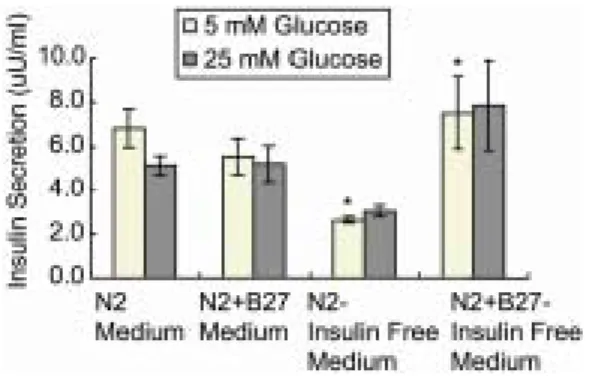
3. 인간 배아줄기세포에서 유도 분화된 세포에 서의 인슐린 분비량
최종적으로 분화시킨 인간 배아줄기세포 (stage-III) Figure 2. Morphological observation (A~D) and RT-PCR analysis (E, F) during the differentiation of human embryonic stem cells. A: EB stage, B: Stage-I (ITSFn for 6 days), C: Stage-II (N2 for 8 days), D: Stage-III (N2 for 22 days). E: Gel electrophoresis of RT-PCR products by sequential culture system. F: Schematic interpretation of the expr- ession pattern of marker genes. Data were calculated by the expression ratio of each group to undifferentiated human embryonic stem cells.
A B C D
E F
의 인슐린 분비량은 Figure 3에 나타난 바와 같이 3.0~7.9 µU/ml으로 측정되었다. 마지막 단계에서의 분화 조건에 따른 인슐린 분비량은 인슐린이 첨가 된 N2 배양액과 N2 + B27 배양액을 사용한 경우는 비슷하였지만, 인슐린을 첨가하지 않은 N2-insulin free 배양액에서는 5 mM glucose 조건에서 3.0 ± 0.2 µU/ml로 N2+B27-insulin free 배양액에서의 7.9 ± 2.0 µU/ml에 비해 통계적으로 유의하게 낮게 측정되었다 (p<0.05). 유도 분화된 세포들에서 고농도의 glucose에 의한 인슐린 분비량의 증가는 관찰할 수 없었다.
고 찰
본 연구에서는 지금까지 보고되었던 배아줄기세 포의 인슐린 분비세포로의 유도 분화 방법을 적용 하고 발전시켜, 인간 배아줄기세포를 혈청과 insulin 이 첨가되지 않은 배양액을 이용하여 인슐린 분비 세포로 유도 분화시킬 수 있었다. 이와 같은 단계적 배양법은 인슐린 분비세포뿐만 아니라 다른 종류의 특정 세포로의 유도 분화에도 이용이 가능하리라 생 각된다.
이러한 특정 세포로의 유도 분화는 궁극적으로 현 재의 치료 방법으로 치유가 어려운 난치병들에 대 한 세포치료를 목적으로 진행되고 있다. 본 연구에 서 유도 분화시킨 인슐린 분비세포를 이용한 당뇨 병 치료 가능성을 확인하기 위해 고농도의 glucose에 대한 반응을 살펴보았으나, 일반적인 췌장의 β-세 포에서 관찰되는 인슐린 분비의 증가를 확인하지 못하였다. 이는 최근에 보고된 Sipione 등 (2004)의
연구에서와 같이 배아줄기세포로부터 분화된 인슐 린 분비세포가 glucose에 반응하지 않음과 유사한 결과를 나타내었다.18 따라서, 실제적인 세포치료 에 이용하기 위해서는 보다 정교한 분화 유도 과 정을 통해 생체 내에서 glucose 농도에 반응하는 인슐린 분비세포로 분화시키는 것이 필수적이다.
그러나 생쥐 배아줄기세포를 이용한 연구에서 인 슐린 분비세포의 분화과정에서 중요한 역할을 수행 하는 Pax-4 유전자의 과발현을 유도하여 효과적으로 유도 분화가 가능함이 보고되었다.14 또한, Moritoh 등 (2003)도 유전자 도입방법을 이용한 Pdx-1의 일 시적인 발현 유도를 통해 여러 종류의 췌장세포로 분화시키는데 성공하였다.16 최근에는 Nkx6.1 prom- oter를 이용하여 세포를 선별하고 pancreatic rudim- ent (embryonic day 17.5)와의 공동배양을 통해 당뇨 동물 모델시스템에서 세포치료에 성공하였다는 보 고가 있었다.19 또한, 인간 배아줄기세포를 이용하여 본 연구와 유사한 단계적 배양법과 이로부터 얻어진 cell clusters를 분리한 후 배양하면 보다 효율적으로 인슐린 분비세포를 확보할 수 있음이 보고되었다.20
현재까지 여러 연구자들의 보고들을 종합해 보면 인간 배아줄기세포를 이용하여 췌장의 β-세포와 같 은 생리학적인 특성을 지니는 세포로의 분화에는 아 직까지 성공하지 못하였지만, 생쥐 배아줄기세포를 이용한 연구와 최근의 몇몇 연구들에서 배아줄기 세포를 이용한 당뇨병에 대한 세포치료 가능성은 계속해서 제시되고 있다. 향후 인간 배아줄기세포 로부터 유도 분화된 인슐린 분비세포에 대한 연구 가 지속적으로 수행된다면, 현대 의학으로 치유하기 어려운 당뇨병 환자에게 새로운 희망을 줄 수 있는 세포치료제 개발이 가능할 것으로 사료된다.
참 고 문 헌
1. Evans MJ, Kaufman M. Establishment in culture of pluripotential stem cells from mouse embryos. Nature 1981; 292: 151-6.
2. Thomson JA, Itskovitz-Eldor J, Shapiro SS, Waknitz MA, Swiergiel JJ, et al. Embyronic stem cell lines derived from human blastocysts. Science1998; 282:
1145-7.
Figure 3. Amount of secreted insulin from differenti- ated human embryonic stem cells (stage-III). *p<0.05.
3. Shamblott MJ, Axelman J, Wang S, Bugg EM, Li- ttlefield JW, Donovan PJ, et al. Human embryonic germ cell derivatives express a broad range of deve- lopmentally distinct markers and proliferate exten- sively in vitro. Proc Natl Acad Sci USA 2001; 98:
113-8.
4. Reubinoff BE, Pera MF, Fong CY, Trounson A, Bo- ngso A. Embryonic stem cell lines from human bla- stocysts: Somatic differentiation in vitro. Nat Biote- chnol 2000; 18: 399-404.
5. Park JH, Kim SJ, Oh EJ, Moon SY, Roh SI, Kim CG, et al. Establishment and maintenance of human em- bryonic stem cells on STO, a permanently growing cell line. Biol Reprod 2003; 69: 2007-14.
6. McDonald JW, Liu XZ, Qu Y, Liu S, Mickey SK, Tu- retsky D, Gottlieb DI, et al. Transplanted embryonic stem cells survive, differentiate and promote reco- very in injured rat spinal cord. Nat Med 1999; 5:
1410-2.
7. Brustle O, Jones KN, Learish RD, Karram K, Cho- udhary K, Wiestler OD, et al. Embryonic stem cell- derived glial precursors: A source of myelinating tr- ansplants. Science 1999; 285: 754-6.
8. Soria B, Roche E, Berná G, León-Quinto T, Reig JA, Martín F. Insulin-secreting cells derived from embr- yonic stem cells normalize glycaemia in streptozo- tocin-induced diabetic mice. Diabetes 2000; 49: 157- 62.
9. Deacon T, Dinsmore J, Costantini LC, Ratliff J, Isa- cson O. Blastula-stage stem cells can differentiate into dopaminergic and serotonergic neurons after tra- nsplantation. Exp Neurol 1998; 149: 28-41.
10. Lumelsky N, Blondel O, Laeng P, Velasco I, Ravin R, McKay R. Differentiation of embryonic stem cells to insulin-secreting structures similar to pancreatic islets.
Science 2001; 292: 1389-94.
11. Assady S, Maor G, Amit M, Itskovitz-Eldor J, Sko- recki K, Tzukerman M. Insulin production by human embryonic stem cells. Diabetes 2001; 50:
1691-7.
12. Hori Y, Rulifson IC, Tsai BC, Heit JJ, Cahoy JD, Kim SK. Growth inhibitors promote differentiation of in- sulin-producing tissue from embryonic stem cells.
Proc Natl Acad Sci USA 2002; 99: 16105-10.
13. Abraham EJ, Leech CA, Lin JC, Zulewski H, Habe- ner JF. Insulinotropic hormone glucagon-like pepti- de-1 differentiation of human pancreatic islet-derived progenitor cells into insulin-producing cells. Endocr- inology 2002; 143: 3152-61.
14. Blyszczuk P, Czyz J, Kania G, Wagner M, Roll U, St-Onge L, Wobus A. Expression of Pax4 in embr- yonic stem cells promotes differentiation of nestin- positive progenitor and insulin-producing cells. Proc Natl Acad Sci USA 2003; 100: 998-1003.
15. Houard N, Rousseau GG, Lemaigre FP. HNF-6- independent differentiation of mouse embryonic stem cells into insulin-producing cells. Diabetologia 2003;
46: 378-85.
16. Moritoh Y, Yamato E, Tasui Y, Miyazaki S, Miya- zaki J. Analysis of insulin-producing cells during in vitro differentiation from feeder-free embryonic stem cells. Diabetes 2003; 52: 1163-8.
17. Kim D, Gu Y, Ishii M, Fujimiya M, Qi M, Nakam- ura N, et al. In vivo functioning and transplantable mature pancreatic islet-like cell clusters differenti- ated from embryonic stem cell. Pancreas 2003; 27:
34-41.
18. Sipione S, Eshpeter A, Lyon JG, Korbutt GS, Blea- ckley RC. Insulin expressing cells from differentiated embryonic stem cells are not beta cells. Diabetologia 2004; 47: 499-508.
19. Leon-Quinto T, Jones J, Skoudy A, Burcin M, Soria B.
In vitro directed differentiation of mouse embryonic stem cells into insulin-producing cells. Diabetologia 2004; 47: 1442-51.
20. Segev H, Fishman B, Ziskind A, Shulman M, Itsko- vitz-Eldor J. Differentiation of human embryonic stem cells into insulin-producing clusters. Stem Cells 2004; 22: 265-74.