大 韓 不 妊 學 會 誌 : 第 32 卷 第 4 號 2005 Kor. J. Fertil. Steril., Vol. 32, No. 4, 2005, 12
Reliability of the Single Cell PCR analysis for Preimplantation Genetic Diagnosis of Single Gene Disorders
Hye Won Choi1, Hyoung-Song Lee1, Chun Kyu Lim1, Mi Kyoung Koong2, Inn Soo Kang2, Jin Hyun Jun1*
1Laboratory of Reproductive Biology & Infertility, 2Department of Obstetrics and Gynecology, Samsung Cheil Hospital, Sungkyunkwan University School of Medicine, Seoul, 100-380, Korea
단일 유전자 이상에 대한 착상전 유전진단을 위한 단일 세포 PCR 방법의 신뢰성
성균관대학교 의과대학 삼성제일병원 생식생물학 및 불임연구실
1, 산부인과
2최혜원
1·이형송
1·임천규
1·궁미경
2·강인수
2·전진현
1*연구목적: 단일 유전자 이상에 대한 착상전 유전진단을 성공적으로 시행하기 위해서는 효과적이고 신뢰도가 높은 PCR 방법의 확립이 중요하다. 본 연구에서는 alkaline lysis와 duplex nested PCR 방법을 단일 림프구와 할구의 유전자 분석에 적용하여 그 효용성을 확인하고자 하였다.
재료 및 방법: 단일 유전자의 이상이 확인된 Duchenne muscular dystrophy (DMD), ornithine transcarbamylase (OTC) 결핍증과 epidermolysis bullosa (EB) 가계의 대상자들에서 채취한 단일 림프구와 공여 받은 배아의 할구를 이용하여 각각 PCR, restriction fragment length polymorphism (RFLP)와 direct DNA sequencing 분석을 시행하였다. 이러한 분석에서 유전자 증폭률 (amplification rate)과 두개의 allele 중에서 하나의 allele이 증폭되지 않는 allele drop-out (ADO) 빈도에 대해 살펴보았다.
결 과: 단일 림프구와 할구를 이용한 PCR 방법의 유전자 증폭률은 DMD에서 91.1%와 81.8%, OTC 결핍증에서 96.0%와 78.1%, EB에서 91.3%와 90.0%를 각각 나타냈으며, ADO 빈도는 OTC 결핍증에서 13.3%, EB에서 16.8%로 관찰되었다.
결 론: 본 연구에서 적용한 alkaline lysis와 duplex nested PCR 방법은 단일 유전자에 대한 착상전 유 전진단에 성공적으로 적용할 수 있는 방법으로 생각되며, ADO 빈도를 최소화할 수 있는 효율적인 방법 의 개발에 대한 지속적인 연구가 필요하다.
Key Words: Preimplantation genetic diagnosis, Single cell PCR, Duchenne muscular dystrophy,
Epidermolysis bullosa, Ornithine transcarbamylase, Allele drop-outPreimplantation genetic diagnosis (PGD) for chromosomal abnormalities, X-linked diseases and single gene disorders has been successfully applied as an alternative to prenatal diagnosis of inherited diseases.1~4 The PGD can be used to differentiate
between unaffected and affected embryos before embryo transfer in human in vitro fertilization and embryo transfer (IVF-ET) program. The advan- tage of PGD is to avoid unethical terminations of affected pregnancies after prenatal diagnosis. The
주관책임자: 전진현, 우) 100-380 서울시 중구 묵정동 1-19, 삼성제일병원 생식생물학 Tel: (02) 2000-7590, Fax: (02) 2265-5621, e-mail: [email protected]
technique involves the blastomere biopsy and gene- tic diagnosis of single blastomeres derived from the cultured embryos by IVF.
Protocols for PGD of single gene disorders are based on the polymerase chain reaction (PCR) of single cells, which represent sensitive enough to detect single gene mutations by DNA amplifica- tion. Although the first PCR-based PGD case with healthy pregnancy was reported,1 several difficul- ties associated with single cell DNA amplification have become evident. It includes potential sample contamination, total amplification failure and allele drop-out (ADO). Occasionally, one of the alleles fails to amplify in PCR reaction to detectable levels when small amount of DNA samples such as single cell analysis. Therefore, a reliable PGD protocol must be established through the extensive pre- clinical tests before it can be applied to clinical trials.
For the diagnosis of single gene disorders in PGD, the target genes are amplified and the PCR products are subjected to further analyses for iden- tification of mutation sites.5 The PCR products from single blastomeres are analyzed by restriction fragment length polymorphism (RFLP) or direct DNA sequencing in the majority of PGD cases to detect the presence or absence of mutation such as small deletion, insertion or substitution.6~9 After the diagnosis of blastomeres, only unaffected embryos are transferred to the mother and consequently any resulting pregnancy is supposed to be unaffected.
In this study, we assessed the amplification and ADO rate of alkaline lysis and duplex nested PCR protocols using single lymphocytes and blastome- res in the pre-clinical diagnostic tests for Duchenne muscular dystrophy (DMD), ornithine transcarba- mylase (OTC) deficiency and epidermolysis bull- osa (EB). This PCR protocol with RFLP and direct DNA sequencing could provide a reasonable relia- bility for the single cell diagnosis of PGD in clini- cal trial.
MATERIALS AND METHODS
1. Duchenne muscular dystrophy (DMD) case
DMD (OMIM No. 310200) is an X-linked di- sease caused by mutations in the dystrophin gene located at Xp21.2 locus.10 It is a recessive inherited disorder and characterized as progressive muscle degeneration resulting in death during the second decade of life. Female partner was a heterozygous carrier of a deletion encompassing exon 45 of dystrophin gene. Her affected son had the mutated allele, which is the same deletion of exon 45 as his mother. For the diagnosis of DMD, the PCR pro- ducts of dystrophin and SRY gene were analyzed by 2% agarose gel electrophoresis.
2. Epidermolysis bullosa (EB) case
EB (OMIM No. 226730) is a group of inherited skin diseases characterized by blister formation of the skin and mucous membranes.11 The EB is caused by mutations in several kinds of genes.
Among the several genes, integrin beta 4 (ITGB4) located at chromosome 17 is a causative gene in this family. It is an autosomal recessive disease.
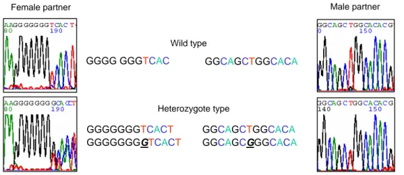
Unfortunately, each male and female partner in this family had a different mutation of ITGB4. Female partner was a heterozygous carrier of an insertion in the exon 7 of the ITGB4 gene (601CIns). Male partner was a heterozygous carrier a substitution in the exon 11 of the ITGB4 gene (1274 T>G, Q425P) (Figure 1). This couple previously had abnormal pregnancies resulting death of all two children.
For DNA sequencing of EB case, 20 ng of puri- fied PCR products were analyzed using fluorescent- labelled dideoxy terminators (Big Dye Terminator Cycle Sequencing Ready Reaction Kit; Applied Biosystems, USA), according to the manufacturer's protocol. The reaction conditions were as follows:
25 PCR cycles, a denaturation step of 10 sec at
96℃, annealing for 5 sec at 50℃ and extension for 4 min at 60℃. Sequencing products were then purified using AccuPrep bioneer purification kit (Bioneer, Korea) for unincorporated dye terminator removal. Then, purified product were resuspended to 15 µl of Hi-Di Formamide (Applied Biosys- tems, USA), heat-denatured at 90℃ for 4 min and run on ABI Prism 3100 Avant automated DNA sequencer (Applied Biosystems, USA). The data of DNA sequences were compared with the wild type controls using Seqscape Software (Applied Biosystems, USA) for mutation analysis.
3. Ornithine transcarbamylase (OTC) defici- ency case
OTC gene is located on the short arm of the X-chromosome with band Xp21.1.12 OTC defici- ency (OMIM No. 311250) is an X-linked semido- minant disorder and the most common inherited defect of the ureagenesis in hyperammonemia.13 After the neonatal death due to hyperammonemia and organ failure, molecular genetic analysis of the couple's OTC gene revealed that female partner had a single base substitution (R320X) in the exon 9 of OTC gene. The next pregnancy was termina- ted by therapeutic abortion after prenatal diagno-
sis of affected fetus. For the third pregnancy by PGD, the patient was referred to our hospital.
In case of OTC deficiency, after confirmation of PCR products for OTC gene by electrophoresis, 6 µl of the PCR products were digested with BclI restriction enzyme (New England Biolabs, Beverly, MA, USA) for 2 hours at 37℃. The digested PCR products were separated by electrophoresis on a 2% agarose gel and detected by ethidium bromide staining under UV transilluminator. By treatment with BclI, the 319 bp of 1st PCR and 218 bp of nested PCR products from affected allele were di- gested into 223 and 96 bp products, and into 178 and 40 bp products, respectively. However, the nor- mal allele was not digested by the BclI (Figure 2).
4. Isolation and preparation of single lym- phocyte
Single cell PCR was performed using peripheral single lymphocyte from heterozygous carriers of DMD, OTC and EB in the family, respectively.
Lymphocytes were isolated from peripheral blood collected from male and female carriers of each couple using Ficoll-Paque density gradient separa- tion (Amersham Pharmacia Biotech, Italy) accor- ding to the manufacturer's protocol. The cell layer GGGG GGGTCAC
GGGGGGGTCACT GGGGGGGGTCACT
GGCAGCTGGCACA
GGCAGCTGGCACA GGCAGCGGGCACA Wild type
Heterozygote type
Female partner Male partner
Figure 1. Identification of a mutation of a female partner (underlined G insertion) and a male partner (underlined G
substitution) in exon 7 and exon 11 of the ITGB4, respectively.
containing lymphocytes was removed and diluted with sterile phosphate-buffered saline to a suitable cell density for single cell isolation. Lymphocytes were then handled with a well-controlled fine heat- polished glass micropipette. The lymphocytes were selected and retrieved individually under visual control with a stereo microscope. Each single lym- phocyte was loaded into 0.2 ml thin wall PCR tubes containing 5 µl of lysis buffer (200 mmol/l KOH and 50 mmol/l dithiothreitol). The samples were stored at -70℃ before analysis.
5. Isolation of single blastomere from abnor- mally fertilized embryos
Blastomeres for this study were obtained from abnormally fertilized embryos donated by patients who were carried out IVF-ET program at Samsung Cheil Hospital. The embryos were incubated for 5 min in Ca2+/ Mg2+-free medium (EB-10TM, Vitro-
life Co. Sweden) before actual biopsy. The acid Tyrode's solution (ZD-10TM, Vitrolife Co. Sweden) was applied to make a hole in zona pellucida. Bio- psy was performed by gentle aspiration using a polished micropipette. Blastomere biopsy was per- formed using a single pipette with 30 µm of inner diameter. After the biopsy, each single blastomere was washed twice through two drops of HEPES- buffered medium, transferred into sterile 0.2 ml PCR tubes containing 5 µl of alkaline lysis buffer.
The samples were stored at -70℃ before analysis.
6. Alkaline lysis and duplex nested PCR
The stored samples with alkaline lysis buffer were incubated at 65℃ for 10 min. Then, the alka- line lysis buffer was neutralized by the addition of 5 µl of neutralization buffer (900 mmol/l Tris-HCl, 300 mmol/l KCl, 200 mmol/l HCl) before procee- ding to PCR.
The PCR strategy consisted of initial duplex PCR followed by nested PCR, specific for each interested regions involving mutations and SRY gene of Y-chromosome for sex determination. Pri- mers sequences for the target genes and their PCR conditions used in this study are summarized in Table 1. After lysis and neutralization, 200 mmol/l of each dNTP (Roche Diagnostic, Italy), 2 IU Supertherm DNA Polymerase (JMR HOLD INGS, UK), 3 µl of 10X PCR Buffer with 15 mmol/l MgCl2 and 10 pmol of each outer primer were added to each tube. The reaction was conducted with a 30 µl of total volume. The first round of PCR involved a 96℃ for 10 min as a means to reduce ADO, followed by a subsequent denatura- tion of 94℃ for 40 sec in 25 remaining cycles and followed by a final extension step of 10 min at 72℃. Primer sequences, annealing temperatures and expected product size of the analyzed genes is shown in Table 1. For the second round of PCR, 1 µl of the primary PCR products were added to another tube containing 2 µl of 10X PCR Buffer
Figure 2. Identification of the maternal mutation
(heterozygote type) in exon 9 of the OTC gene. After digesting the PCR product with BclI, the 319 or 218 bp PCR products of the affected mother were digested into 223 and 96 bp (lane 2) or 178 and 40 bp products (lane 5), respectively. The 40 bp product did not detect on the agarose gel electrophoresis. Lane 1 and 4, the first and nested PCR products of the affected mother, respectively;
lane 2 and 5, BclI-digested products from the first and nested PCR products of the affected mother, respectively;
lane 3 and 6, BclI-digested products from the first and
nested PCR products of the normal father, respectively.
(500 mmol/l KCl, 100 mmol/l Tris HCl, pH 8.3), 200 mmol/l of each dNTP (Roche Diagnostic, Ger- many), 2 IU SynergyN DNA Polymerase (Gene- craft, Germany) and 10 pmol of each inner primer.
The tube with a 20 µl of total volume were cycled as the first round reaction condition on a Gene- Amp PCR System 2700 (Applied Biosystems). To monitor successful amplification of single cell PCR, 5 µl of each PCR products was separated by elec- trophoresis for 20 min at 100 V on 2% agarose gel in 0.5X TBE buffer, and then visualized with ethi- dium bromide staining.
RESULTS
1. Duchenne muscular dystrophy (DMD) case
The efficiency and accuracy of the duplex nested PCR for DMD case were evaluated in pre-clinical experiments on 33 blastomeres and 86 single lym- phocytes from a normal male (30 lymphocytes) and a carrier female partner (56 lymphocytes) in the family. Amplification rates of single lympho- cytes and blastomeres were 91.1% (51/56) and 81.8% (27/33) for exon 45 of dystrophin gene, respectively. The SRY gene of Y-chromosome was
Table 1. Sequences of oligonucleotide primers and reaction conditions for duplex nested PCR
Name Sequences Annealing
temp. (℃)
Product sizes (bp) Duchenne muscular dystrophy (DMD)
5'-AAACATGGAACATCCTTGTGGGGAC-3'
Dystrophin exon 45 (outer)5'-CATTCCTATTAGATCTGTCGCCCTAC-3' 62 537
5'-AAAACTGGAGCTAACCGAGAG-3'
Dystrophin exon 45 (inner)5'-CTGTTTGCAGACCTCCTGCCAC-3' 64 370
Epidermolysis bullosa (EB)
5'-CGTCTTCCCCTGTGACACTC-3'
Integrin β4 exon 7 (outer)5'-CCTTGCCCAGAAGTGCTC-3' 61 274
5'-CTCTCTCTCCCTCCCACCTC-3'
Integrin β4 exon 7 (inner)5'-ACACAGCTGTCTGCAGGATG-3' 66 194
5'-TCGATGGCCCCCTGGTCCTT-3'
Integrin β4 exon 11 (outer)5'-CCTGGGTGCTTGGCTCAGCT-3' 61 274
5'-CCCTTGAGCACGTGGATG-3'
Integrin β4 exon 11 (inner)5'-GGTGCTTGGCTCAGCTCTC-3' 66 185
Ornithine transcarbamylase (OTC) deficiency
5'-CACTCTGCTCCTTTGTCTCT-3'
OTC exon 9 (outer)5'-GTTGGAACCACACAAAGAAC-3' 62 319
5'-GGCCATGTGTGTTTTTAGAT-3'
OTC exon 9 (inner)5'-GTCCACTTTCTGTTTTCTGC-3' 64 218
Sexing
5'-GAATATTCCCGCTCTCCGGA-3'
Sry5'-GCTGGTGCTCCATTCTTGAG-3' 64 472
amplified in 90.0% (27/30) of male lymphocytes.
2. Epidermolysis bullosa (EB) case
Duplex nested PCR and direct sequencing for the EB case were performed in pre-clinical experi- ments on 48 blastomeres and 173 single lympho- cytes from female and male partner with mutation of exon 7 and exon 11 of ITGB4 gene, respecti- vely. Amplification rates of single lymphocytes showed 95.3% (81/85) for the exon 7 and 96.6%
(85/88) for the exon 11. In the PCR of blastomeres, we observed 72.9% (35/48) for exon 7 and 83.3%
(40/48) for exon 11 of the ITGB4 gene PCR pro- ducts. In the direct DNA sequencing of PCR pro- ducts, the ADO rates were detected 19.8% (16/81) for exon 7 and 13.7% (7/51) for exon 11 of the ITGB4 gene.
3. Ornithine transcarbamylase (OTC) defici- ency case
In the OTC deficiency case, we analyzed 66 single lymphocytes from a carrier female (n=46) and a normal male partner (n=20) in the family by duplex nested PCR and RFLP analysis. Amplifi- cation rates of lymphocytes showed 91.3% (42/
46) for the exon 9 of OTC gene and 90.0% (18/20) for the SRY gene. In the RFLP analysis of PCR products with BclI, the 218 bp, PCR products of mutant allele were digested into 178 and 40 bp products (Figure 2), because BclI restriction site was created by this mutation (R320X) in the exon 9 of the OTC gene. As a result of RFLP analysis, ADO rate of single lymphocytes was detected 13.3% (4/30) for OTC gene.
DISCUSSION
The single cell PCR analysis for PGD has been slowly developed because the handling and analyz- ing single cell involve very difficult technical pro- cedures and pitfalls.5 Due to its sensitivity, PCR is
highly prone to have many sources of error. Espe- cially, ADO and amplification failure are the most frequent and significant problem in the single cell PCR. The ADO may lead to misdiagnosis of he- terozygous embryos, and is the most significant obstacle to the reliable diagnosis of dominant di- sorder in the single cell analysis. The exact cause of ADO remains elusive. However, as an origin of ADO, imperfect PCR conditions or incomplete cell lysis have been proposed. Incomplete cell lysis, anucleated blastomere and inadequate annealing temperature might be considered causes of ampli- fication failure. Therefore, the single cell PCR method has to be optimized to reduce the risk of ADO and amplification failure. Several strategies have been developed to decrease ADO, such as increasing the denaturation temperature and annea- ling time,14 using a more powerful lysis method,15 and applying the multiplex PCR and fluorescent PCR for mutations or linked markers.16,17
On the basis of our experience, the useful stra- tegy for PGD was to take nucleated lymphocytes and blastomeres, to use an alkaline lysis protocol, and to optimize the annealing temperature. In this study, the overall amplification rate using single lymphocytes PCR for the mutation site was 93.5%
(304/325), and ADOs were observed in 27 of 162 (16.7%). These data showed more than 80% accu- racy of single cell PCR analysis which might be acceptable for clinical trial of PGD for single gene disorders. It was reported that the alkaline lysis protocol showed significant lower ADO rates than those of proteinase K/SDS lysis protocol in single cell PCR analysis for PGD.15 Every researcher should make an effort to increase the amplification rate and to decrease the ADO rate for the pre- vention of misdiagnosis in single cell PCR-based PGD. Multiplex PCR of polymorphic markers re- duced overall ADO rate and more than 95% of multiplex PCR results were accepted to construct the informative genotypes for PGD of cystic fi-
brosis.17 Therefore, this polymorphic and multi- allelic marker analysis might be a reliable and applicable alternative for difficulties in mutation- directed PGD protocols such as repeated sequences and high content of G or C bases.
Our protocol provided 91.1% and 81.1% for DMD, 96.0% and 78.1% for OTC deficiency, and 91.3% and 90.0% for EB using single lympho- cytes and blastomeres, respectively. The ADO occurred 13.3% in OTC deficiency and 16.8% in EB. Based on these results, the alkaline lysis and duplex nested PCR method has proven to be a valuable method in single cell analysis for PGD.
This protocol has been successfully applied to clinical PGD cycles for couples at high risk for having children with single gene disorders. We could diagnose and transfer mutation free embryos into mother's uterus in all cycles. At the present time, two PGD babies from DMD and OTC de- ficiency case were born and they were confirmed to be genetically normal through post-natal genetic analysis.18,19 The alkaline lysis protocol of this study showed 100% reliability of single cell PCR analysis in PGD cycles for single gene disorders.
REFERENCES