34 www.kjtcvs.org
KJTCVSThe Korean Journal of Thoracic and Cardiovascular Surgery
CaseReport
https://doi.org/10.5090/kjtcs.2020.53.1.34 pISSN: 2233-601X eISSN: 2093-6516
Korean J Thorac Cardiovasc Surg. 2020;53(1):34-37
Cerebral Air Embolism and Cardiomyopathy Secondary to Large Bulla Rupture during a Pulmonary Function Test
Ha Lee, M.D., Hyun Soo Lee, M.D., Dulk Hwan Moon, M.D., M.Sc., Sungsoo Lee, M.D., Ph.D.
Department of Thoracic and Cardiovascular Surgery, Gangnam Severance Hospital, Yonsei University College of Medicine, Seoul, Korea
ARTICLE INFO Received July 29, 2019 Revised September 17, 2019 Accepted September 18, 2019 Corresponding author Sungsoo Lee
Tel 82-2-2019-3381 Fax 82-2-3461-8282 E-mail [email protected] ORCID
https://orcid.org/0000-0001-8998-9510
Cerebral air embolism combined with cardiomyopathy secondary to pulmonary baro- trauma is rare. Here, we report an unusual case of cerebral air embolism combined with transient cardiomyopathy secondary to large bulla rupture during a pulmonary function test after lung cancer surgery. The patient experienced loss of consciousness. Computed tomography and magnetic resonance imaging suggested a cerebral air embolism. Electro- cardiography showed ST-segment elevation and abnormally high plasma levels of cardiac enzymes. Echocardiography and coronary angiography suggested cardiomyopathy. The patient was discharged with no sequelae.
Keywords: Air embolism, Cardiomyopathies, Bullae, Barotrauma
Copyright©The Korean Society for Thoracic and Cardiovascular Surgery. 2020. All right reserved.
This is an Open Access article distributed under the terms of the Creative Commons Attribution Non-Commercial License (http://creativecommons.org/licenses/
by-nc/4.0) which permits unrestricted non-commercial use, distribution, and reproduction in any medium, provided the original work is properly cited.
Case report
A 63-year-old man with a history of hypertension and chronic kidney disease was admitted to the emergency de- partment for acute loss of consciousness after performing the Valsalva maneuver during a pulmonary function test (PFT). His medications included candesartan, hydrochlo- rothiazide, and aspirin. Three months ago, he had received neoadjuvant chemoradiation therapy for lung cancer (ade- nocarcinoma, cT3N2M0, stage IIIB) in the right upper lobe; after a successful response, he received right upper lobe lobectomy (ypT1aN0M0) 11 days before the event.
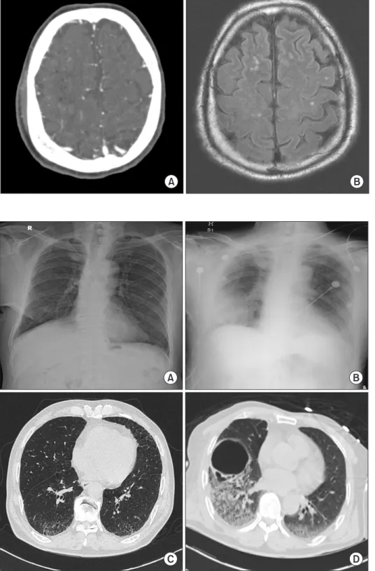
On examination, his level of consciousness was semi-co- ma with no response to pain; right-side deviation of both eyeballs was noted, and both pupils measured 5 mm with a sluggish response to light. Initially, he had a blood pressure of 100/52 mm Hg, a heart rate of 102 beats/min, and a tem- perature of 36.8°C, and he received rapid-sequence intuba- tion to protect his airway. Computed tomography (CT) scans of the brain revealed multifocal air density in the bi- lateral cerebral subarachnoid space and deep cerebral white matter. There was no evidence of brain hemorrhage. Mag- netic resonance imaging of the brain revealed diffuse lep- tomeningeal enhancement and diffuse enhancement in the perivascular space of the bilateral deep white matter. Both imaging studies suggested cerebral air embolism (Fig. 1).
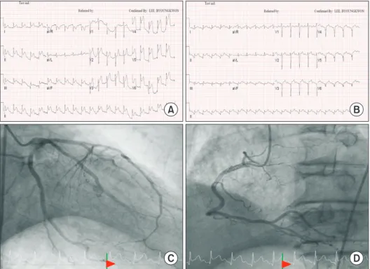
A chest radiograph showed a round area of consolidation in the right lower lobe lung field (Fig. 2B). Electrocardiog- raphy revealed sinus tachycardia at 130 beats/min, with ST-segment elevation of 2 to 3 mm in leads 2, 3, augment- ed vector foot, and V3 through V6 (Fig. 3A). Laboratory testing showed that his plasma levels of creatinine kinase (CK), creatinine kinase-muscle/brain (CK-MB), and tropo- nin I were 83 U/L, 1.59 μg/L, and 0.01 μg/L, respectively.
In a follow-up examination performed 3 hours after the event, electrocardiography showed sinus tachycardia at 117 beats/min and ST-segment elevation of 1 to 2 mm in leads 2 and V2 through V5 (Fig. 3B), and his plasma levels of CK, CK-MB, and troponin I were 156 U/L, 19.51 μg/L, and 4.70 μg/L, respectively. Transthoracic echocardiography (TTE) revealed a newly developed regional wall motion ab- normality, which was not compatible with the coronary territories. The ejection fraction decreased from 62% to 41%. Emergent coronary angiography revealed neither cor- onary artery disease nor acute myocardial infarction le- sions (Fig. 3C, D). In a follow-up examination 12 hours af- ter the event, the patient’s plasma levels of CK, CK-MB, and troponin I were 242 U/L, 31.21 μg/L, and 8.45 μg/L, respectively.
The patient was transferred to the intensive care unit.
His white blood cell count and hemoglobin, platelet, glu- cose, ammonia, sodium, potassium, calcium, and magne-
35
Ha Lee, et al. Cerebral Air Embolism and Cardiomyopathy due to Bulla Rupture
www.kjtcvs.org
KJTCVS
sium levels were all within the normal range. In a follow-up examination 6 hours after the event, his level of conscious- ness was confusion, both pupils were 3 mm with a prompt response to light, and one step obey to command was pos-
sible. Generalized tonic-clonic seizures took place 11 to 20 hours after the event, and the seizures were controlled by an intravenous infusion of levetiracetam. On the first day after the event, electrocardiography showed sinus tachycar-
A B
Fig. 1. (A) A computed tomography scan of the brain showed multifo- cal air density in the deep cerebral white matter, suggesting cerebral air embolism. (B) Magnetic reso- nance imaging of the brain showed diffuse enhancement in the peri- vascular space of the bilateral deep white matter in a T2 FLAIR image, suggesting acute cerebral infarc- tion.
A B
C D
Fig. 2. (A) A chest radiograph be- fore the pulmonary function test.
(B) An initial chest radiograph af- ter the patient lost consciousness shows a round consolidation, sug- gesting large bulla rupture. (C) A CT scan of the chest before right upper lobe lobectomy showed emphy- sematous lung, but no large bullae.
(D) A CT scan of the chest 2 days after cerebral air embolism showed large bullae. CT, computed tomog- raphy.
36
https://doi.org/10.5090/kjtcs.2020.53.1.34
www.kjtcvs.org
KJTCVS
dia at 107 beats/min with ST-segment elevation of 1 mm in leads V2 and V3, and his plasma levels of CK, CK-MB, and troponin I were 367 U/L, 13.51 μg/L, and 4.90 μg/L, re- spectively.
On the second day, the patient was alert, with chest radi- ography revealing a clear lung field. Mechanical ventilator weaning was performed. CT scans of the chest revealed a newly developed large bulla with a maximum diameter of 7 cm (Fig. 2D), and follow-up TTE showed an increased ejection fraction of 47%.
On the third day, the patient was transferred to the gen- eral ward. His plasma levels of CK, CK-MB, and troponin I were 1,642 U/L, 6.77 μg/L, and 1.12 μg/L, respectively. On the sixth day, electroencephalography revealed normal waveforms. On the seventh day, the patient was discharged without neurological symptoms or sequelae. Two weeks later, transesophageal echocardiography at the outpatient department revealed a grade I patent foramen ovale.
The patient provided written informed consent for the publication of clinical details and images.
Discussion
Cerebral air embolism secondary to pulmonary baro- trauma is rare, although a few case reports have described cerebral air embolism secondary to bulla rupture during air travel or diving [1,2]. In these reports, the clinical pre-
sentations and outcomes ranged from benign to fatal. Our patient experienced cerebral air embolism secondary to bulla rupture, as in previous reports, but during PFT; fur- thermore, he presented with acute loss of consciousness, followed by seizures. Nonetheless, our patient was dis- charged with no severe sequelae.
Risk factors for bulla formation and rupture in our pa- tient included emphysematous lung due a history of smok- ing, preoperative radiation therapy, fibrobullous changes in the ipsilateral residual lung after lobectomy, and a bleed- ing tendency due to aspirin. With regard to the first of those risk factors, Iwama et al. [3] showed a high frequency of emphysematous bullae in smokers, suggesting that smoking history is a risk factor for bulla formation. Our patient was an ex-smoker who quit 30 years ago and smoked 7.5 pack-years. Preoperative CT scans of the chest revealed diffuse pulmonary emphysema without large bul- lae. As a second risk factor, radiation-induced fibrosis may have occurred around the lung parenchyma during neoad- juvant chemoradiation therapy, making the bullae fragile to barotrauma, which led to rupture during the Valsalva maneuver. A total of 5,000 Gy of radiation was adminis- tered to the cancer mass in the right upper lobe and its margin. Intensity-modulated radiotherapy was used to minimize the dose to the surrounding normal tissue; how- ever, fibrotic changes in the other lobe of the lung, includ- ing the right middle lobe and right lower lobe, were inevi-
A B
C D
Fig. 3. (A) The initial electrocardi- ogram. (B) A 3-hour follow-up elec- trocardiogram. (C) Coronary angi- ography: left coronary artery. (D) Coronary angiography: right coro- nary artery. aVR, augmented vector right; aVL, augmented vector left;
aVF, augmented vector foot.
37
Ha Lee, et al. Cerebral Air Embolism and Cardiomyopathy due to Bulla Rupture
www.kjtcvs.org
KJTCVS
table. With regard to the third risk factor, Tanaka et al. [4]
reported fibrobullous changes in the lung parenchyma af- ter lobectomy. They showed that the initial changes ap- peared at an average of 2 years (range, 3 months to 6 years) after lobectomy, with an incidence rate of 3%. They consid- ered fibrobullous changes to be a late complication after lobectomy, but our patient developed fibrobullous changes only 3 weeks after lobectomy. This discrepancy may have been because our patient received radiation therapy preop- eratively, whereas their study excluded patients who re- ceived preoperative and postoperative radiation therapy.
Finally, the side effects of antiplatelet agents may result in bleeding around the bullae.
The direct cause of cerebral air embolism in this case was pulmonary barotrauma that occurred during PFT. The American Association for Respiratory Care (AARC) has suggested the following relative contraindications for spi- rometry: hemoptysis of unknown origin, unstable cardio- vascular status, a recent myocardial infarction or pulmo- nary embolus, and recent thoracic or abdominal surgery [5]. Although the recommendations of the AARC are based on minimal concrete evidence [6], our patient had recently undergone lobectomy. Moreover, we suggest careful con- sideration of PFT in patients with pulmonary bullae. Far- shchi Zarabi et al. [1] proposed 3 mechanisms of systemic air embolism secondary to bulla rupture: (1) direct emboli- zation of the gas bubbles into the pulmonary veins and from there into the systemic circulation; (2) gas emboliza- tion into the pulmonary arterial system with subsequent incomplete filtration of the gas bubbles by the pulmonary capillaries; and (3) paradoxical embolization through a functional right-to-left shunt, such as a patent foramen ovale. Our patient was susceptible to all these possibilities.
Our patient also received a coronary evaluation due to ST-segment elevation and abnormally high levels of cardiac enzymes. Echocardiography and coronary angiography suggested stress-induced cardiomyopathy (SCMP). Any of the following mechanisms may lead to SCMP: direct air embolization into the coronary arteries, direct pressure on the coronary arteries and myocardium due to increased thoracic pressure during the Valsalva maneuver, and large swings in blood pressure during the Valsalva maneuver causing stress on the myocardium. To the best of our knowledge, our patient is the first case in which cardiac function and the coronary arteries were evaluated during cerebral air embolism secondary to bulla rupture. We sug- gest evaluating electrocardiography findings and plasma levels of cardiac enzymes in patients with cerebral air em-
bolism due to bulla rupture, especially in those at high risk of cardiovascular disease.
In conclusion, we presented a rare case of cerebral air embolism during PFT in a patient with a large pulmonary bulla. Cardiomyopathy was accompanied by cerebral air embolism secondary to pulmonary barotrauma for un- known reasons. In patients with bullous lung disease, men- tal status and electrocardiography monitoring are neces- sary during PFT, and medical practitioners should observe the patient carefully after PFT is completed. Follow-up chest radiography must also be done in bullous lung pa- tients after PFT. Further studies are necessary to better ad- dress the relationship between cerebral air embolism sec- ondary to pulmonary barotrauma and cardiomyopathy.
Conflict of interest
No potential conflict of interest relevant to this article was reported.
ORCID
Ha Lee: https://orcid.org/0000-0003-3976-8554 Hyun Soo Lee: https://orcid.org/0000-0003-3684-6510 Duk Hwan Moon: https://orcid.org/0000-0003-1388-2471 Sungsoo Lee: https://orcid.org/0000-0001-8998-9510
References
1. Farshchi Zarabi S, Parotto M, Katznelson R, Downar J. Massive ischemic stroke due to pulmonary barotrauma and cerebral artery air embolism during commercial air travel. Am J Case Rep 2017;18:
660-4.
2. Gudmundsdottir JF, Geirsson A, Hannesson P, Gudbjartsson T. Major ischaemic stroke caused by an air embolism from a ruptured giant pulmonary bulla. BMJ Case Rep 2015;2015:bcr2014208159.
3. Iwama E, Okamoto I, Yabuuchi H, et al. Characteristics of smoking patients with lung cancer with emphysematous bullae. J Thorac On- col 2016;11:1586-90.
4. Tanaka H, Matsumura A, Ohta M, Ikeda N, Kitahara N, Iuchi K. Late sequelae of lobectomy for primary lung cancer: fibrobullous changes in ipsilateral residual lobes. Eur J Cardiothorac Surg 2007;32:859- 62.
5. Restrepo RD, Wettstein R, Wittnebel L, Tracy M. Incentive spirome- try: 2011. Respir Care 2011;56:1600-4.
6. Cooper BG. An update on contraindications for lung function testing.
Thorax 2011;66:714-23.