Mucormycosis is an invasive, fungoid disease caused by rhizopus, mucor or other fungoids be- longing to phycomycetes group. Although it mostly occurs in patients with diabetes through opportun- istic infections, patients with impaired immune functions or normal people may be affected by the disease in rare cases. Its mortality rate is high due to its rapid and foudroyant progression.
Mucormycosis can invade several organs, and can emerge in the form of a paranasal sinusitis in the area of otolaryngology as the patient inhales the mucor in the air. The mucor, attached to mucous membrane of paranasal sinus, primarily causes paranasal sinusitis and orbital inflammation. In addition, mucormycosis's high invasiveness with respect to blood vessels and tissues causes throm-
Case Reports
A Case Of Cavernous Sinus Syndrome and Mutifocal Cerebral Infarction Related To Mucormycosis Of Sphenoid Sinus
Seok Won Jeon, Chang Hoi Kim, Joo Yeon Kim, Jae Hwan Kwon
Department of Otorhinolaryngology-Head and neck surgery, College of Medicine, Kosin University, Busan, Korea
A 54-year-old man, suffering from severe headache and ophthalmoplegia after undergoing endoscopic sinus surgery was referred to a tertiary hospital. Computed tomography (CT) revealed soft tissue density lesions in the left sphenoid sinus. The internal carotid artery was shown to be occluded in brain magnetic resonance imaging (MRI) scans without any other cerebral lesion.
Endoscopic view of left nasal cavity shows whitish hyphae in the ethmoid and the sphenoid sinuses. We diagnosed him with cavernous sinus syndrome caused by mucormycosis and conducted endoscopic sinus surgery to remove remaining lesions and decompress orbit and optic nerves.
After the revision surgery the patient’s headache and ophthalmoplegia were improved. However, multifocal cerebral infarctions were newly discovered in a postoperative CT scan. We experienced a case of mucormycosis of sphenoid sinus resulting in occlusion of internal carotid artery and multifocal cerebral infarction, and report it with a brief review of these disease entities.
Key Words: Cavernous sinus, Cerebral infarction, Mucormycosis
Corresponding Author: Jae Hwan Kwon, Department of Otorhinolaryngology-Head and neck surgery, College of Medicine, Kosin University, 262, Gamcheon-ro, Seo-gu, Busan 49267, Korea Tel: +82-51-990-6470 Fax: +82-51-245-8539 E-mail: [email protected]
Received:
Revised:
Accepted:
Sep. 27, 2016 Feb. 02, 2017 Feb. 18, 2017
Articles published in Kosin Medical Journal are open-access, distributed under the terms of the Creative Commons Attribution Non-Commercial License (http://creativecommons.org/licenses/by-nc/4.0/) which permits unrestricted non-commercial use, distribution, and reproduction in any medium, provided the original work is properly cited.
bosis in the arteries and veins, and leads to the easy invasion of surrounding nerves; and partic- ularly, it can invade the optic nerve, internal car- otid artery, cavernous sinus, ocular motor nerve, and others, which are anatomically adjacent to the sphenoid sinus, thus causing severe complica- tions such as cavernous sinus syndrome and orbital apex syndrome. The authors have identified the rapidly-progressing loss of vision and successive occurrence of multiple cerebral artery occlusions in patients with cavernous sinus thrombosis caused by mucormycosis of the sphenoid sinus. While there have been reported overseas cases where mucormycosis caused cerebral artery occlusion or cerebral infarction,1 no cases have been reported in Korea. In that sense, this paper is intended to introduce such cases.
CASE
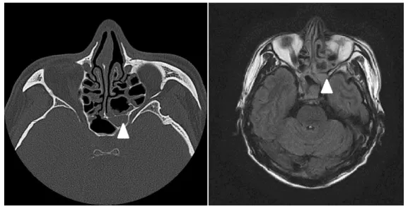
A 54-year-old man, suffering from severe head- ache that started 2 weeks previously, visited the department of neurology at another hospital, and then underwent brain magnetic resonance imag- ing (MRI) and computed tomography (CT). The imaging tests showed left sphenoid sinus lesion, but other abnormalities that suggested cerebral lesion were not found (Fig. 1).
It was concluded that the headache was a sec- ondary headache caused by paranasal sinusitis, and endoscopic sinus surgery was scheduled to be performed under general anesthesia at the ENT department of the same hospital. The blood tests of the patient prior to the surgery revealed that his blood sugar was somewhat high, but additional tests were not conducted because the patient said that he had no medical history related to diabetes.
According to operative findings, local fungal si- nusitis was suspected, and the antifungal agent (Ambisone® 50mg, IV QD) was administered, in
Fig. 1. Preoperative axial computed tomography (CT) and magnetic resonance images (MRI) show left sphenoid sinus lesion.
Arrow heads indicates left sphenoid sinus in both images.
consideration of the potential for postoperative invasiveness progression. In addition, vancomy- cin (Hanomycin® 500mg, IV BID) was also used because methicillin-resistant staphylococcus was found in the rhinorrhea.
Three days after the surgery, symptoms oc- curred, including left ocular motor dysfunction, decreased visual acuity, and ophthalmoplegia in the lower orbital area. Although a steroid (Solumedrol® 125mg, IV QD) was administered to address the neurologic symptoms in the left eye, such as decreased visual acuity and ocular motor dysfunction, these symptoms continued to worsen. As a result, the patient was transferred to this hospital.
At that time, headache was the severest symp- tom among others. It was accompanied by a dull pain that persisted and tightened around the left ocular area and the left parietal area. The visual acuity showed finger counting, but ocular move- ment showed complete ophthalmoplegia, which was considered to be related to the oculomotor nerve, trochlear nerve, and abducens nerve. In addition, the patient complained of oph- thalmoplegia in the left lower orbital area, which suggested that the maxillary branch of the trige- minal nerve was also invaded (Fig. 2A). The find- ings of the nasal endoscopy showed lesion and crusta, suspected as fungal hyphae, along the ex- ternal wall of the left ethmoidal sinus; and a small
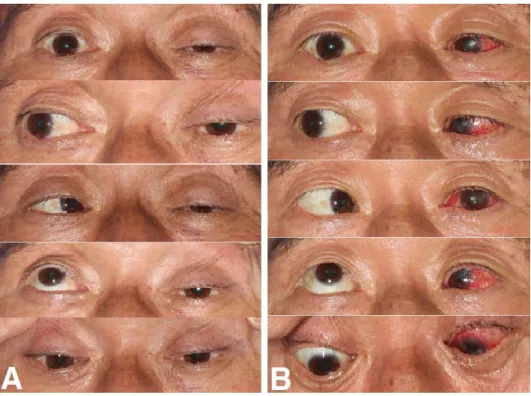
Fig. 2. These are postoperative extraocular movements of the patient.
A. Patient showed ptosis and ophthalmoplegia of his left eye in all directions after endoscopic sinus surgery.
B. 6 months after revision operation, patient’s ptosis and ophthalmoplegia had been improved partially when gazing medially.
amount of purulent discharge and granulation tis- sue were observed in the sphenoidal sinus. In the biopsy on the lesion site which the patient under- went at the outpatient department, the right-an- gled branched hyphae were observed. As a result, the patient was diagnosed with cavernous sinus syndrome caused by mucormycosis and mu- cormycosis (Fig. 3).
The blood test revealed that the WBC and CRP levels had increased to 10,000 /ul and 6.25 mg/dl respectively, and the urine test showed an in- crease in glucose to 3 +. According to additional tests, blood sugar and glycated hemoglobin after eight hour fasting were increased to 252 mg/dl and 10% respectively, indicating that uncontrolled diabetes had not been recognized for a long time.
Thus, joint treatment was immediately carried out with the department of endocrinology, and the sugar was controlled by focusing on insulin ther- apy rather than oral agents due to the risk of
nephrotoxicity and hepatotoxicity
To re-evaluate the patient's condition, imaging testing was performed. The paranasal sinus CT scan showed soft tissue density along with the site of previous surgery and heterogenous contrast enhancement around the orbital apex; the T1 and T2 imaging in the brain MRI revealed that in the pterygopalatine fossa and orbital apex, the lesion site was observed with low signal intensity, and the occlusion of the left internal carotid artery was confirmed (Fig. 4).
The antibiotic test was performed again by con- tinuously administering vancomycin (Vancocin®
1g, IV BID) and aseptically collecting rhinorrhea according to the findings of fungus identification of the previous hospital. As a result, methicillin-re- sistant staphylococcus was also identified. Although the antifungal agent (Fungizone® 50mg, IV QD)
Fig. 3. Both endoscopic and histologic findings revealed fungal infection after the first surgery in another hospital.
A. Endoscopic finding of left nasal cavity shows fungal hyphae and crust around posterior ethmoid sinus and sphenoid sinus before revision surgery.
arrowhead : left sphenoid sinus, arrow : lamina papyracea
B. Histopathologic scan shows non-septated, right angled branched hyphae (H&E stain,
and the high-dose steroid (Methysol® 125mg, IV BID) continued to be also administered, symptoms such as ophthalmoplegia, loss of vision, and head- ache were not improved. In particular, the patient showed a complete loss of vision as the decreased visual acuity continued to progress. On the fifth day of hospital visits, thus, he underwent the re- vision surgery to remove the residual lesion in the ethmoidal sinus and sphenoid sinus and decom- press orbital and optic nerves. First, the gran- ulation tissue and sphacelus were removed from the inner wall of the left orbit and the left sphenoid sinus. The optic nerve decompression was per- formed by removing and grinding the bone frag- ments of the superolateral sphenoid sinus and the external wall of ethmoidal sinus to the maximum along with the moving path of left optic nerve.
The absorbable material (Surgicel®) was applied to prevent bleeding, and another packing was not carried out (Fig. 5).
The brain MRI and the MR angiography, taken to monitor the patient's status on the first day of surgery, showed the occlusion of the left middle cerebral artery and left anterior cerebral artery as well as the occlusion of the internal carotid artery, already confirmed; and the cerebral in- farction in left middle cerebral artery. Although dysarthria or quadriplegia caused by cerebral ar- tery infarction was not found, the patient was transferred to the department of neurology to treat him and monitor his status (Fig. 6). The an- tithrombotic and antilipid agents (Plavix® 75mg, Lipitor® 80mg) were additionally administered along with antibiotics and antifungal agent, after he was transferred to the department of neurology.
According to the findings of the postoperative nasal endoscopy, mucor and hyphae were not ob- served; abnormalities were also not found in the site of orbital decompression; and the headache Fig. 4. Computed tomography (CT) and magnetic resonance images (MRI) were taken
before revision surgery.
A. CT scan shows heterogenous enhancing lesion involving orbital apex (black arrow).
B. White arrow indicates occlusion of left carotid artery in MRI.
disappeared, but the left ocular motor dysfunc- tion, the loss of vision, and the hypoesthesia in the left isthmus still persisted. Six months after the revision surgery, the loss of vision still per- sisted, but the ocular motor dysfunction and ptosis were partially recovered (Fig. 2B).
DISCUSSION
Mucormycosis is a rare disease that occurs
through opportunistic infections in patients with impaired immune function who mainly have dia- betes, hematologic malignancies, or suffer from chronic alcoholism; in particular, about 70% of mucormycosis patients are reported to have un- controlled diabetes.2,3 Mucormycosis divides into five major types: pulmonary type, dermal type, cerebral type, gastrointestinal type, and systemic disseminated type. And cerebral mucormycosis can be divided again into the rhino-orbito-cere- bral type with a bad prognosis and the rhi- Fig. 5. These pictures are presenting brief intraoperative findings during revision surgery.
A. Most of the area between lamina papyracea and sphenoid sinus was covered with granulomatous necrotic tissues.
asterisk : middle turbinate, arrow head : sphenoid sinus, arrow : lamina papyracea B. Magnified view of left sphenoid sinus
C. Black Circle indicates the site conducted partial optic nerve decompression.
D. Absorbable material (Surgicel®) was applied to prevent bleeding.
no-paranasal sinus type showing satisfactory progress.4 In particular, the rhino-orbito-cere- bral type exhibits rapid deterioration and high mortality. It occurs as a result of tissue invasion of the mucor aspirated through the nasal cavity.5 If it becomes morbid in the form of sphenoid sinus lesion, it may cause complications in the anatomi- cally adjacent cavernous sinus. Cavernous sinus syndrome collectively refers to neurological symptoms or thrombosis in lesions around cav- ernous sinus. Its most common cause is tumors (30%) and infection, deformity and trauma can al- so be the cause of the disease.6 It may cause neu- rological abnormalities by affecting the ocular branches of maxillary nerve and maxillary branches, and invades blood vessels and lymph nodes and, particularly, pierces the walls of the arteries and causes thrombosis, leading to cere- bral infarction. In addition, the intracranial in-
vasion may cause encephalitis, cerebral hemor- rhage and, in severe cases, death.7
The initial symptom of cavernous sinus syn- drome may be headache. Other symptoms include pains in the posterior ocular segment due to the invasion to ocular branches of trigeminal nerves,;
systemic symptoms occur such as fever or nausea, vomiting, ocular motor dysfunction, exoph- thalmos, and loss of vision.8
Mucormycosis of the sphenoid sinus, as in this present case, is not easy to access through the endoscopic examination because of the location of lesions, which can delay its diagnosis. In addi- tion, invasion to cavernous sinus may lead to fatal complications. In that sense, rapid diagnosis is a very important factor in the progress of treat- ment and the prognosis. Although imaging exami- Fig. 6. Brain magnetic resonance images (MRI) and MR angiography were taken after revision
surgery.
A. MR angiography cannot visualize left anterior cerebral artery, middle cerebral artery and ICA (white arrow).
B. Brain MRI shows infarction of left parietal lobe (white circle).
nations such as computed tomography (CT) and magnetic resonance images (MRI) can provide findings regarding the status of paranasal sinus and orbital apex, cavernous sinus, internal carotid artery or cerebral artery, its definite diagnosis is made through a biopsy.
To be specific, the definite diagnosis is made by identifying the non-septated, right-angled branched hyphae of irregular shape using the Grocott's Methenamine Silver-Periodic Acid Schiff or Hematoxylin & Eosin staining of the le- sion tissues.9
Prior to the treatment for mucormycosis, if the patient is suffering from lowered immune func- tion, it is essential to control underlying diseases.10 Drug therapy and surgical treatment may be considered for treatment. Drug therapy is carried out centering on antifungal agents, es- pecially Amphotericin B. High doses (1.0 to 1.5 mg/kg/day) are recommended to prevent compli- cations and their progress, and nephrotoxicity must be taken into consideration.11,12 Surgical treatment is based on aggressive and extensive resection of necrotic tissues, and all lesions should be removed until bleeding tissues are identified.
As the disease worsens over a few days or even a few hours, predicting its progress is difficult and complications can be fatal. Therefore, even total maxillectomy or eye enucleation should be ac- tively considered if the need arises according to the lesion's location or the surgeon's judgment.13
In this case, the revision surgery was performed for orbital decompression as a complete loss of vision was confirmed during the monitoring peri- od when the mucormycosis treatment was carried out accompanied by diabetes examination and control through joint treatment with the depart- ment of endocrinology. Other treatments can in- clude hyperbaric oxygen therapy and the admin- istration of a blood coagulation inhibitor.
However, despite the advancement of therapy methods, the mortality rate of rhino-orbito-cere- bral mucormycosis is still as high as 20 to 50 %.10,14
In this case, cavernous sinus syndrome occurred after the endoscopic sinus surgery for mucormy- cosis of the sphenoid sinus. It rapidly worsened within a few days after the first operation.
Currently, the patient still continues to show ocu- lar motor dysfunction, loss of vision and oph- thalmoplegia in the lower orbital area. However, it is believed that a relatively early diagnosis and a wide range of surgical interventions helped pre- vent a fatal outcome and ensure partial recovery of ocular movement. In general, the use of steroids in treatment for invasive mucormycosis is contra- indicated because it can accelerate the pro- gression of mucormycosis; however, in this case, steroids were unavoidably administered to de- compress optic nerves as the patient experienced rapid loss of vision.
The postoperative cerebral artery infarction is thought to have been caused by thrombosis due to mucormycosis invasion to the left internal car-
otid artery during the endoscopic sinus surgery or by the fungus itself that acted as an embolus in the middle cerebral artery. Therefore, antith- rombotic and antilipid agents are being administered to the patient who is under observation.
Several cases of orbital apex syndrome or cav- ernous sinus syndrome caused by cerebral mu- cormycosis have been reported. However, since no cerebral artery infarction caused by thrombo- sis accompanying the disease has been reported in Korea, this case is being introduced with a re- view of the literature.
REFERENCES
1. Thajeb P, Thajeb T, Dai D. Fatal strokes in patients with rhino-orbito-cerebral mucormycosis and associated vasculopathy. Scand J Infect Dis 2004;36:643–8.
2. Blitzer A, Lawson W, Meyers BR, Biller HF. Patient survivalfactors in paranasal sinus mucormycosis.
Laryngoscope 1980;90:635-8.
3. Patterson TF, Kirkpatrick WR, White M, Hiemenz JW,Wingard JR, Dupont B, et al. Invasive aspergillosis. Diseases pectrum, treatment practices, and outcomes. I3 Aspergillus Study Group.
Medicine 2000;79:250–60.
4. Vessely MB, Zitsch RP 3rd, Estrem SA, Renner G. Atypical presentations of mucormycosis in the head and neck. Otolaryngol Head Neck Surg 1996;115:573-7.
5. Bendet E, Talmi YP, Kronenberg J. Rhino-
orbito-cerebral mycormycosis. Otolaryngol Head Neck Surg 1996;114:830-2.
6. Keane JR. Cavernous sinus syndrome.Analysis of 151 cases. Arch Neurol 1996;53:967-71.
7. Onerci M, Gursel B, Hosal S, Gulekon N, Gokoz A. Rhinocerebral mucormycosis with extension to the cavernous sinus. Rhinology 1991;29:321-4.
8. Jin YW, Cho JS, Kim KH, Cha CI.
Rhinocerebralmucormycosis with selective cranial nerve palsy. Korean J Otolaryngol-Head Neck Surg 2001;44:674-7.
9. Bendet E, Talmi YP, Kronenberg J. Rhino-orbito- cerebral mycormycosis. Otolaryngol Head Neck Surg 1996;114:830-2.
10. Talmi YP, Goldschmied-Reouven A, Bakon M, Barshack I, Wolf M,Horowitz Z, et al. Rhino-orbital and rhino-orbito-cerebral mucormycosis. Head Neck Surg 2002;127:22–31.
11. Baumann A, Zimmerli S, Hausler R, Caversaccio M. Invasive sphe¬noidalaspergillosis: successful treatment with sphenoidotomy and voriconazole.
ORL J OtorhinolaryngolRelat Spec 2007;69:121-6.
12. Sugar AM. Mucormycosis.Clin Infect Dis 1992;14:s126-9.
13. Viterbo S, Fasolis M, Garzino-Demo P, Griffa A, Boffano P, Iaquinta C, et al. Management and outcomes of three cases of rhinocerebralmucormycosis.
Oral Surg Oral Med Oral Pathol Oral RadiolEndod 2011;112:e69-74.
14. Mohamed MS, Abdel-Motaleb HY, Mobarak FA.
Management of rhino-orbital mucormycosis.
Saudi Med J. 2015;36:865-8.