Comparison of Dental Ages Estimated according to Oral Habit for the Patients with Temporomandibular Disorders
Won-Seop Lee, D.D.S., Ki-Suk Kim, D.D.S.,M.S.D.,Ph.D.
Department of Oral Medicine, School of Dentistry, Dankook University
This study was performed to investigate the dental ages of the patients with temporomandibular disorders(TMD), using a method of age estimation by dental attrition, and to compare the differences of dental ages between control and experimental groups. In addition, they were compared according to oral habit.
Clinical evaluations for the tooth attrition were to examine the study models of 65 TMD patients (mean age of 26.6 years, aged 18 to 40) had visited to the Department of Oral Medicine and Orofacial Pain Clinic, Dental Hospital, Dankook University as a experimental group and 22 volunteers (mean age of 25.4 years, aged 23 to 36) without TMD as a control group. Age differences were calculated subtracting real age from estimated age. Age differences between experimental and control groups were compared statistically between groups classified according to oral habit.
As a results, dental age was increased significantly in TMD patients compared with normal subjects. Oral parafunctions which increased dental age were bruxism, clenching and heavy chewing in order.
Conclusively, it should be consider that there can be much discrepancy between real age and age estimated by dental attrition in patients with TMD. In addition, it is suggested that dentists should try to protect the tooth from attrition by various parafunction on treating the patients with TMD.
Key words: Temporomandibular disorder, Oral habit, Bruxism, Dental attrition, Age estimation
1)I. INTRODUCTION
Several predisposing factors are responsible for the development of the TMD.1) There is general agreement that oral parafunction is occlusal activity
Corresponding author : Ki-Suk Kim
Dept. of Oral Medicine, School of Dentistry, Dankook University Sinbu-dong san 7-1, Chunan, Choongnam 330-716 Korea
Tele : 82-41-550-1914 Fax : 82-41-556-9665 Email : [email protected] Received: 2007-08-18
Accepted: 2007-11-28
* The present research was conducted by the research fund of Dankook University in 2006.
outside the realm of masticatory function(i.e, chewing, speaking, swallowing) and includes bruxism(grinding or clenching of teeth), diet habits (chewing gum or solid food), unilateral chewing, thumb-sucking, finger-sucking, nail biting, lip biting, and abnormal jaw posturing. These activities have been related to problems of the masticatory system, such as muscular pain, degenerative joint conditions, headache, and occlusal traumatism. Like the muscles and joints, the dentition can show signs and symptoms of functional disorders. These are normally associated with breakdown created by heavy occlusal forces to the teeth and their supportive structures. By far the most sign associated with functional disturbances of the dentition is tooth wear. This is observed as shiny
flat areas of the teeth that do not match the natural occlusal form of the tooth. If an abrasive diet is not a factor, such wear will usually be due to nonfunctional activities. Examination of wear patterns often reveals that bruxism occurs in eccentric mandibular positions, beyond the normal functional range and in contrast to the normal physiologic wear of aging seen in the functional range. Tooth wear stems predominantly from parafunctional activity.2)
Dental attrition is considered the most visible sign of functional wear. Parafunctional habits of bruxism and clenching are subjects of concern for dental professionals, and it is frequent for dentists to draw strong clinical associations of TMD with bruxism. It is clear that parafunctional activity is a mainly contributing factor for development of TMD as well as tooth wear.3,4)
The purposes of this study were to compare the differences of dental ages of TMD patients with that of normal subjects, using a method of age estimation by dental attrition, and to investigate a relation of tooth attrition and oral habit in TMD patients.
II. MATERIAL AND METHODS 1. Subjects
1) Experimental group
Sixty five patients were selected for this study.
Their mean age was 26.62±5.90 years. These subjects were referred to the Department of Oral Medicine and Orofacial Pain Clinic, Dental Hospital, Dankook University for TMD treatment and exclusion criteria are as follows:
1) previous occlusal equilibration therapy; 2) trauma, surgery, or systemic joint, muscle, or skin diseases influencing the symptoms; 3) pathologic conditions in temporomandibular joint(TMJ), facial skeleton, or teeth.
2) Control group
Twenty two normal volunteers were included.
Their mean age was 25.37±2.89 years. None of
them had any symptoms and signs of TMD and were screened using a history taking and a clinical examination.
2. Methods
1) Oral habit examination
Intraoral and radiographic examination were performed for all subjects. Oral habits such as bruxism, clenching, unilateral chewing, heavy chewing, gum chewing, and nail biting were investigated by questionaires and interview. Based on the above method, in addition to oral habits which the patient subjectively recognized, we also examined those recognized by the third-party individuals, such as nocturnal bruxism.
2) Evaluation of attrition and determination of age differences
A set of maxillary and mandibular study casts were obtained from each subjects. Upper and lower permanent teeth except third molars were examined to determine the degree of tooth wear.
The degree of incisal and occlusal wear was evaluated with naked eye or magnifying glass based on the Takei’s5) method and estimated age related with dental attrition was determined.
Age difference was determined by calculating to the following formula: Age Difference=Estimated Age-Real Age.
3) Age estimation from dental attrition and state of dental treatment(by Tetsuji Takei)
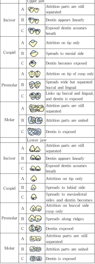
As shown in Fig. 1 and 2, the type of attrition was examined and then evaluated using a 5-grade category classification system, in which such unevaluable areas as prosthesis, missing tooth, etc.
were also included. Based on the Calculating Table for Age Estimation(Fig. 3), each of 28 teeth was assigned with a grading point and dental age was estimated using the formula.
Upper jaw
Incisor
A Attrition parts are still separated
B Dentin appears linearly C Exposed dentin assumes
breath
Cuspid
A Attrition on tip only
B Spreads to mesial side C Dentin becomes exposed
Premolar
A Attrition on tip of cusp only B Spreads wide but separated
buccal and lingual
C Links up buccal and lingual, and dentin is exposed
Molar
A Attrition parts are still separated
B Attrition parts are united
C Dentin is exposed
Lower jaw
Incisor
A Attrition parts are still separated
B Dentin appears linearly C Exposed dentin assumes
breath
Cuspid
A Attrition on tip only B Spreads to labial side C Spreads to mesiodistal
sides and dentin becomes
Premolar
A Attrition on buccal side cusp only
B Spreads along ridges
C Dentin exposed
Molar
A Attrition parts are still separated
B Attrition parts are united
C Dentin is exposed
Fig. 1. Ranking standards of attrition degree
Category
No. Classification
1 Attrition degree A
2 Attrition degree B
3 Attrition degree C
4 Caries, Filling, Crown
5 Stump of tooth, Missing, Pontic, Denture Fig. 2. Category classification
Category No.
1 2 3 4 5
#17 -1.24 -0.76 1.17 1.24 1.38
#16 -1.12 0.23 2.10 -0.33 0.74
#15 -0.52 -0.49 1.88 0.59 0.25
#14 -0.44 -0.91 1.89 0.55 0.40
#13 -0.54 -0.46 -0.34 1.51 1.30
#12 0.53 -0.69 2.40 -1.18 0.44
#11 -1.66 -1.58 -0.43 1.20 2.71
#21 1.01 -0.06 2.69 -0.93 -1.84
#22 -0.55 0.56 -0.34 0.36 -0.05
#23 -0.95 -0.71 -0.64 1.41 3.11
#24 -0.58 0.04 1.19 -0.66 0.76
#25 -1.05 0.13 -0.07 0.83 0.67
#26 -0.42 0.14 1.02 -1.23 1.08
#27 -2.47 -0.54 2.70 -0.30 3.31
#37 -0.74 -0.15 1.95 0.73 0.38
#36 -0.08 0.66 -1.36 0.02 -0.01
#35 -0.45 0.39 2.88 -0.89 0.64
#34 -1.54 -0.42 1.08 1.42 2.58
#33 -1.48 -0.34 0.16 2.27 2.30
#32 -1.03 0.18 0.28 -1.76 2.54
#31 -2.33 -0.34 0.11 2.94 5.51
#41 0.61 0.22 0.27 1.48 -3.45
#42 0.03 -0.36 1.27 0.50 -1.05
#43 0.25 -0.45 0.29 -1.62 1.10
#44 0.34 -1.14 1.33 0.57 -0.77
#45 -1.48 -0.65 -0.94 1.38 1.54
#46 -0.97 2.50 1.72 -1.21 0.04
#47 -0.11 0.24 1.49 -1.50 0.94
Mean 38.78
Estimated Age = 38.78 + ∑[value(#17)~value(#47)]
Fig. 3. The Calculating Table for Age Estimation
3. Statistical analysis
Unpaired t-test were used to compare the age differences of estimated age and real age of TMD patients and normal subjects and one-way ANOVA and multiple comparison t-test were used to compare the age differences of groups classified according to oral habits.
Ⅲ. RESULTS
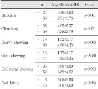
Table 1 shows age differences between the estimated age and real ages in the TMD patients and normal controls. While the estimated age of the TMD patients was 4.78±4.39 years older than the real age, the age difference of the controls was 1.26±3.25 years. There was significant age difference between the TMD patients and the controls(p=0.001).
When all the subjects including the TMD patients and controls were divided according to type of oral habits, mean age difference related to presence or absence of each habit was seen in Table 2 and Fig. 1. There was significant higher increase of age difference in the bruxers as compared with non-bruxers, indicating that the age estimated by the tooth attrition in the bruxers was older than in the non-bruxers(p=0.001). The subjects with the habit of heavy chewing also exhibited significant age difference compared with those without the habit(p=0.009), but the estimated age was older in the those who didn't have the
Control group Experimental group t-test
n 22 65
Δage
(Mean±SD) 1.26±3.25 4.78±4.39 p=0.001 Δage : estimated age-real age
Table 1. Mean and standard deviations of differences between estimated and real ages in and experimental groups and the result of t-test.
n Δage(Mean±SD) t-test
Bruxism + 24
- 63
6.46±4.93
2.91±3.76 p=0.001
Clenching + 28
- 59
4.95±5.37
3.38±3.79 p=0.121 Heavy chewing + 18
- 69
1.52±3.77
4.50±4.35 p=0.009 Gum chewing + 13
- 74
1.77±4.17
4.25±4.35 p=0.059 Unilateral chewing + 35
- 52
3.89±4.09
3.89±4.62 p=0.993 Nail biting + 6
- 16
5.05±2.96
3.68±4.60 p=0.305 Δage : estimated age-real age
Table 2. Mean and standard deviations of differences between estimated and real ages in all subjects classified according to type of oral habit and the results of t-test.
n Δage(Mean±SD) t-test
Bruxism + 8
- 14
1.80±3.24
0.95±3.33 p=0.570 Clenching + 9
- 13
2.25±2.97
0.57±3.37 p=0.243 Heavy chewing + 8
- 14
0.60±2.91
1.63±3.37 p=0.488 Gum chewing + 2
- 20
4.48±0.89
0.93±3.23 p=0.145 Unilateral chewing + 8
- 14
1.47±3.62
1.13±3.16 p=0.826 Nail biting + 6
- 16
3.85±1.11
0.28±3.27 p=0.018 Δage : estimated age-real age
Table 3. Mean and standard deviations of differences between estimated and real ages in control group classified according to type of oral habit and the results of t-test.
habit. There was no significant difference incomparisons between presence and absence of the other habits.
Table 3 and Fig. 2 represent the age difference related with presence or absence of the oral habits in the normal controls and Table 4 and Fig. 3 in the TMD patients. In comparison of the normal controls, significant age difference was found only in a group with nail biting habit(p=0.018). The subjects with nail biting habit showed 3.85±1.11
n Δage(Mean±SD) t-test
Bruxism + 19
- 49
8.79±3.87
3.46±3.71 p=0.000
Clenching + 19
- 46
6.23±5.83
4.17±3.54 p=0.087 Heavy chewing + 10
- 55
2.26±4.35
5.23±4.27 p=0.047 Gum chewing + 11
- 54
1.28±4.36
5.49±4.07 p=0.003 Unilateral chewing + 27
- 38
4.61±4.00
4.90±4.69 p=0.799 Nail biting + 7
- 58
6.06±3.71
4.62±4.46 p=0.415 Δage : estimated age-real age
Table 4. Mean and standard deviations of differences between estimated and real ages in experimental group classified according to type of oral habit and the results of t-test.
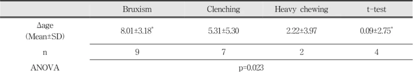
Bruxism Clenching Heavy chewing t-test
Δage
(Mean±SD) 8.01±3.18* 5.31±5.30 2.22±3.97 0.09±2.75*
n 9 7 2 4
ANOVA p=0.023
Δage : estimated age-real age
* means that there is a very significant difference between Bruxism and Gum chewing(p=0.004)
Table 5. Mean and standard deviations of differences between estimated and real ages in experimental group with single oral habit and the results of one-way ANOVA and multiple comparison t-test.
years of age difference whereas those without the habit revealed 0.28±3.27 year of age difference (Table 3).
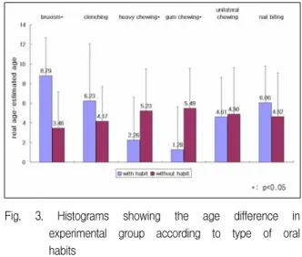
In regard with the TMD patients, significant age differences were observed related with bruxism, heavy chewing and gum chewing. The patients with bruxism showed that their age estimated by the tooth attrition was 8.79±3.87 years older than the real age while those without bruxism showed 3.46±3.71 years of age difference(p=0.000). However the habit of heavy chewing and gum chewing exhibited that the age difference was greater than those who didn't have the habits(p=0.047, 0.003, respectively, Table 4).
Table 5 represents comparison of age differences among the various oral habits in the TMD patients with only single habit. Bruxism
Fig. 1. Histograms showing the age difference in all subjects according to type of oral habits
Fig. 2. Histograms showing the age difference in control group according to type of oral habits
resulted in the most age difference (8.01±3.18 years), and followed by clenching (5.31±5.30 years), heavy chewing (2.22±3.97 years) and gum chewing (0.09±2.75 years) in order. The comparison between the single habit groups, significant difference was found between the bruxsim and gum chewing groups.(p=0.004).
Ⅳ. DISCUSSION
Oral habit that can be detrimental to oral health include a number of behaviors, such as bruxism, clenching, unilateral chewing, heavy (solid food) chewing, unusual postural habits, occupational- related activities, nail biting or gum chewing, etc.6) Such parafunctional behaviors are common and usually do not harm the stomatognathic apparatus.7) However, when the activity exceeds the individual's physiological tolerance, the system begins to alter and breakdown may occur.
The initial breakdown is seen in the tissue with the lowest structural tolerance. In other words, the effects of diurnal or nocturnal parafunctional activities can cause damage to dentition, musculature, or joints.8) Most functional activities occur close to the maximal intercuspal position and distributing forces over a maximum number of teeth, whereas parafunctional activities occur in eccentric positions and apply forces to a fewer
Fig. 3. Histograms showing the age difference in experimental group according to type of oral habits
number of teeth and often in an unstable joint position.8) Also, protective reflexes that control functional activities are less responsive or absent during parafunctional activities.9,10) Therefore, the likelihood of damage to the teeth and the TMJ is increased. Additionally, because these parafun- ctional behaviors differ from normal function in that they are often subconscious, it is inevitable that abnormal force from parafunctional activities promote the functional load of teeth.
Like the muscles and joints, the dentition can show sign and symptoms of functional disorders, by far the most common functional disturbance is tooth wear. Generally, occlusal and incisal attrition can occur during normal deglutition and mastication.11-14) As priorly noted, however, severe tooth attrition can occur in patients who have persisted the abnormal tooth function to such an extent as to exceed the physiological resistance, this can be verified by merely observing the location of most wear facets. Wear facets are simply identified as noncontiguous anatomy on occlusal or incisal tooth surfaces. It can therefore be assumed that tooth attrition can occur with a high probability in patients who have the oral habit to such an extent as to develop the TMD.
There was many studies performed on relation dental attrition and TMD, but it was remain controversial. That is, negative correlation was
found between dental attrition and the sign of TMD,15-19) whereas, severe dental attrition was found to have a very positive correlation to TMD symptoms.20-22) Also, Fujita23) reported that notable dental attrition was observed in the patients with unilateral chewing and bruxism compared to patients without these habits. It can therefore be assumed that there be a considerable difference in the effect of oral habits, the causative factor for TMD, on attrition. A complete understanding of this difference is essential for dentist to prevent additional tooth wear as well as to treat TMD.
In the present study using age estimation, estimated age was significantly higher in patients with TMD than normal controls as shown in Table 1. This result suggests that tooth attrition should be increased significantly in patients with TMD having oral habits. This result is also in agreement with the report of Lee et al.24) that attrition of teeth was significantly increased in patients with TMD showing higher frequency of oral parafunctional habit. However, this study shows the different effect of oral habits on tooth attrition (Table 5), this can be the clue for previous controversial reports about the relation between TMD symptoms and tooth attrition.
In this study, the estimated age was the highest in patients with bruxism, this indicates that bruxism had a most significant effect on the tooth attrition. As represented in Table 2 and 4, including consideration of the result in Table 5, the contradictory outcomes in heavy chewing or gum chewing can therefore be explained as the most significant effect of bruxism on attrition, as previously described. Among oral habits of the control group, nail biting had a significant effect on attrition. Presumably, this might be due to a limited number of subjects.
Bruxism, defined as “the parafunctional grinding of teeth”,25) may include clenching, grinding and tapping of the teeth as well as the chewing of pencils, pips, cheeks and lips. If left untreated, bruxism can lead to attrition of tooth surface, loss of vertical dimension of occlusion, increased muscle
tonus, and adaptive changes in the temporo- mandibular joint.26) There is reason to believe that bruxism is the commonest and most important of the functional disturbances.27) Molina28) reported that TMD and bruxing patients may present many other additional oral parafunctional habit which may combine to increase masticatory muscle activity, thus leading to TMD signs and symptoms.
Therefore, it is concluded that bruxism is not only a contributing factor for TMD but also the most powerful factor for tooth attrition.
A barrier to previous objective evaluation of occlusal features as factors in TMD has been difficulty in constructing mathematical models and considering functional wear according to aging. This study sets age estimation which quantify the severity of dental attrition. The present study examined particularly patients in the 20s and 30s who commonly develop TMD, for which Takei’s method of age estimation based on tooth attrition.
Currently, this can be an essential method for estimating dental age after 20s because tooth age must be estimated based on the physiologic change of tooth after the growth and development of tooth were completed.29) Based on these results of dental age estimation, an approximately 3.5-years difference between TMD patients and normal controls(Table 1), and an approximately 5.3-years difference between those with and without bruxism(Table 4) can be reference values for the age estimation based on attrition in patients with TMD.
There was significant increase of estimated age in experimental bruxism group as seen in table 4, but was not increase of estimated age in control bruxism group as seen in table 3. It can be assumed, although the sample size was not enough to suggest a conclusion, that TMD have been occurred in patients with severe bruxism to the extent of being attrited, not in patients with mild bruxism.
The attrition was comparatively and widely recognized in the patients with the specific habits in comparison with patients without the habits in this
study. The results of this study indicated that some predisposing factors strongly influence on tooth wear as well as development of TMD. It is evident that various oral habits give a loading to the tooth and TMJ to a greater extent in patients with TMD based on these results, dentist must therefore consider concomitantly performing the procedure to remove these multifactorial causes in treating patients with TMD. It was thought that although several significant correlations exist between occlusal factors and signs and symptoms of TMD in the material presented, the occlusal factor as a etiologic factor of TMD was not investigated in this study. Further studies are required to be performed in large population including older age than this study and to extend various factors for TMD and attrition.
Ⅴ. CONCLUSION
Dental attrition was increased significantly in TMD patients compared with normal subjects.
Parafunctional activities which influence on tooth wear were bruxism, clenching and heavy chewing in order.
In conclusion, specific consideration should be required on estimating age for TMD patients.
Approximately 3.5 years in patients with TMD, approximately 5.3 years particularly in those with bruxism, must be subtracted in estimating dental age in the 20s and 30s. In addition, it is suggested that dentist consider various procedure reducing loading force to the tooth and TMJ in treating TMD patients.
REFERENCES
1. Meng HP, Dibbets JM, van der Weele LT, Boering G.
Symptoms of temporomandibular joint dysfunction and predisposing factors. J Prosthet Dent 1987;57:
215-222.
2. Molina OF, dos Santos J, Nelson SJ, Nowlin T. A clinical study of specific sings and symptoms of CMD in bruxers classified by the degree of severity.
J Cranio 1999;17:268-279.
3. Seligman DA, Pullinger AG. Analysis of occlusal variables, dental attrition, and age for distinguishing healthy controls from female patients with intracapsular temporomandibular disoredrs. J Prosthet Dent 2000;83:76-82.
4. Gavish A, Halachmi M, Winocur E, Gazit E. Oral habits and their associatio with signs and symptoms of temporomandibualr disorders in adolescent girls. J Oral Rehabil 2000;27:22-32.
5. Takie T. Age estimation from dental attrition and state of dental treatment(by application of the theory of quantification Type I). J Nihon university school of dentistry 1984;26:119-132.
6. Okeson JP. Management of Temporomandibular Disorders and Occlusion. 3rd ed., St Louis, 1993, Mosby Year Book.
7. Rugh JD, Ohrbach R. Occlusal parafunction. In A textbook of Occlusion, Chicago, 1988, Quintessence Publishing Co., pp.249-261.
8. Seligman DA, Pullinger AG, Solberg WK. The prevalence of dental attrition and its association with factors of age, gender, occlusion, and TMJ symptomatology. J Dent Res 1988;67:1323-1333.
9. Ash MM, Ramfjord SP. Occlusion. 4th ed., Philadelphia, 1995, WB Saunders, pp.500.
10. Guyton AC. Textbook of medical physiology. 8th ed., Philadelphia, 1991, WB Saunders, pp.1013.
11. Lehman ML, Meyers ML. Relationship of dental caries and stress:concentrations in teeth as revealed by photoelastic tests. J Dent Res 1966;45:1706-1714.
12. Shore NA. Temporomandibular joint dysfunction and occlusal equilibration. 2nd ed., Philadelphia, 1976, Lippincott, pp.11.
13. Straub WJ. Malfunctions of the tongue. Am J Ortho 1960;40:404-420
14. Kydd WL. Maximum forces exerted on the dentition by the perioral and lingual musculature. JADA 1957;55:646-651.
15. Droukas B, Lindee C, Carlsson GE. Occlusion and mandibular dysfunction: A clinical study of patients referred for functional disturbances of the masticatory system. J Prosthet Dent 1985;53:402-406.
16. Reuling N. Comparative study of clinical examination, occlusal analysis and new radiological imaging procedure in patients with functional disorders. J Oral Rehabil 1987;14:165-174.
17. Pullinger AG, Seligman DA. TMJ osteoarthrosis:A differentiation of diagnostic subgroups by symptom history and demographics. J Craniomandib Disord
1987;1:251-256.
18. Seligman DA, Pullinger AG. TMJ derangement and osteoarthrosis subgroups differentiated according to active range of mandibular opening. J Craniomandib Disord 1988;2:33-40.
19. Roberts CA, Tallents RH, Katzberg RW, Sanche- zwoodworth RE, Espeland MA, Handelman SL.
Comparison of internal derangements of the TMJ with occlusal findings. J Oral Surg Oral Med Oral Pathol 1987;63:645-650.
20. Lieberman MA, Gazit E, Fuchs C, Lilos P. Mandibular dysfunction in 10-18 years old school children as related to morphological malocclusion. J Oral Rehabil 1985;12:209-214.
21. Gazit E, Lieberman MA, Eini R, Hirsch N, Fuchs C, Lilos p. Prevalence of mandibular dysfunction in 10-18 years old Israeli school children. J Oral Rehabil 1984;11:307-317.
국문요약
측두하악장애환자의 구강습관에 따른 치아감정연령의 비교
단국대학교 치과대학 구강내과학 교실 이원섭․김기석
본 연구에서는 치아교모도에 의한 연령감정법을 이용하여 측두하악장애환자의 치아연령을 일반 정상인과 비교하고, 측두 하악장애환자의 구강습관이 치아교모에 미치는 영향을 조사하고자 하였다.
단국대학교 치과병원 구강내과에 측두하악장애를 주소로 내원한 환자 65명을 실험군으로 하고, 측두하악장애증상이 없는 정상인 22명을 대조군으로 하였다. 문진 및 설문조사를 통해 대상자의 구강습관을 조사하였으며, 임상 및 방사선검사후 치아 교모도를 조사하여 치아연령을 산출하였다.
실험군과 대조군의 치아교모도의 차이 및 각각의 구강습관이 치아교모에 미치는 정도를 분석하기 위해 t-test 및 one-way ANOVA, multiple comparison t-test를 이용하였다.
연구 결과 대조군에 비해 실험군의 치아교모도가 유의성 있게 증가했으며, 조사된 구강습관중 이갈이, 이악물기, 식사습관 (단단하고 질긴 음식)순으로 치아교모도에 영향을 주었다.
따라서, 측두하악장애환자의 연령감정시 증가된 치아교모도에 의해 나타날 수 있는 실제 연령과의 차이를 고려해야 한다.
특히, 20-30대의 연령감정시에 측두하악장애환자의 경우 약 3.5세, 이중 특히 이갈이의 습관이 있는 경우에는 약 5.3세의 감정연령의 증가가 있을 수 있음을 감안해야 한다. 또한, 측두하악장애환자는 일반 정상인보다 치아 교모가 심한 경향이 있 고 이는 치아와 턱관절에 더 많은 하중이 가해지고 있음을 의미하므로 측두하악장애환자의 치료시에는 반드시 이러한 요인 을 제거하는 치료술식을 병행해야 할 것으로 사료된다.
주제어: 측두하악장애, 구강습관, 이갈이, 치아교모도, 연령감정
22. Molina O, Santoz J, Mazzetoo M et al. Oral jaw behaviors in TMD and bruxism: a comparison study by severity of bruxism. J Craniomandibular Pract 2001;19:114-122.
23. Yamakage S. Read and classify wear facet-Guideline for clinicians, 2001, Quintessence., pp.2001-2029.
24. Lee SY, Kim KS. Study on the dental attrition of the patients with craniomandibualr disorders using age estimation. Korean J Oral Med 1995;20:421-427.
25. Glossary of prostohdontic terms. J Prosthet Dent 1987;58:723.
26. Dawson PE. Evaulation, diagnosis, and treatment of occlusal problem. Baltimore, 1989, Mosby, pp.457-458.
27. Drum W. Klassifikation von Parafunktionen. Dtsch Zahnarztl Zschr. 1962;17:411-415.
28. Molina O, Santos J, Mazzetto M, Nelson S, Nowlin T, Mainieri ET. Oral jaw behaviors in TMD and bruxism: a comparison study by xeverity of bruxism.
J Craniomandibular Pract 2001;19:114-122.
29. 김종열. 법치의학. 서울, 2005, 지성출판사, pp.317-332.