1341
Copyrights © 2021 The Korean Society of Radiology
Non-Enhancing Intradural
Extramedullary Ependymoma:
A Case Report
조영증강이 되지 않는 경막내 수외 뇌실막세포종:
증례 보고
Jaemin Kim, MD1 , Hyunjung Kim, MD1* , Hyeongju Kwon, MD2
Departments of 1Radiology and 2Pathology, Yonsei University Wonju College of Medicine, Wonju, Korea
Spinal ependymomas are generally located in the intramedullary compartment in adults. In- tradural extramedullary spinal ependymomas are extremely rare. Spinal ependymomas show various contrast enhancements on MRI. In this study, we report a rare case of a 52-year-old female who had a pathologically confirmed intradural extramedullary ependymoma that showed no en- hancement on MRI.
Index terms Spinal Cord Neoplasms, Intradural-Extramedullary; Ependymoma;
Magnetic Resonance Imaging
INTRODUCTION
Spinal ependymoma is generally found in the intramedullary location in adults, ac- counting for 60% of the total intramedullary lesions (1). Intradural extramedullary ep- endymoma is relatively rare (2-4). Most spinal ependymomas have well-defined and in- tense contrast enhancement after the injection of contrast medium (5-7). This case report focuses on the unusual MR finding of intradural extramedullary ependymoma at conus medullaris without contrast enhancement.
CASE REPORT
A 52-year-old female presented at the hospital in March 2020 with low back pain with direct tenderness and both leg weakness. Neurologic examination revealed bilateral paraparesis grade IV. The results of Deep tendon reflexes and Patrick straight leg raise tests were normal.
Degenerative spondylosis was confirmed through lateral thoraco-lumbar (TL) spine
Received August 10, 2020 Revised November 14, 2020 Accepted December 31, 2020
*Corresponding author Hyunjung Kim, MD Department of Radiology, Yonsei University Wonju College of Medicine, 20 Ilsan-ro, Wonju 26426, Korea.
Tel 82-33-741-1474 Fax 82-33-732-8281 E-mail radkhj@yonsei.ac.kr This is an Open Access article distributed under the terms of the Creative Commons Attribu- tion Non-Commercial License (https://creativecommons.org/
licenses/by-nc/4.0) which permits unrestricted non-commercial use, distribution, and reproduc- tion in any medium, provided the original work is properly cited.
ORCID iDs Jaemin Kim https://
orcid.org/0000-0001-9959-8978 Hyunjung Kim
https://
orcid.org/0000-0001-6522-9883 Hyeongju Kwon
https://
orcid.org/0000-0003-1615-7152
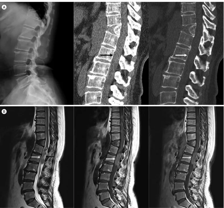
radiograph, and wedge-shaped T11 vertebral body was incidentally found (Fig. 1A). TL spine CT revealed an intradural iso-dense mass at the T12–L1 level without calcification. The inci- dentally founded wedge-shaped T11 vertebral body was a butterfly vertebra, normal varia- tion (Fig. 1A). The results of spinal MRI demonstrated an intradural lesion, posteriorly located and compressing the conus medullaris anteriorly. There was an enlargement of cerebrospinal fluid (CSF) space adjacent to the mass. The intradural extramedullary mass was approximate- ly 3.9 cm in length and was located at the T12–L1 level near conus medullaris.
A
B
Fig. 1. A 52-year-old female with spinal intradural extramedullary ependymoma.
A. Radiograph shows degenerative spondylosis and incidentally found butterfly vertebra at T11 (left). Sagittal CT scans with a soft tissue win- dow (middle) and bone window (right) show the intradural isodense mass (arrows) at the T12–L1 level. The mass has no internal calcification.
B. Sagittal T2-weighted (left), T1-weighted (middle), and postcontrast T1-weighted MRI (right) show the intradural extramedullary tumor at T12–L1 (white arrows). The central portion of the mass shows high signal intensity on the T2-weighted image (black arrow).
https://doi.org/10.3348/jksr.2020.0147 1343 The mass showed a slightly high signal intensity on both T1 and T2-weighted images. The central region of the mass had a higher signal intensity on T2-weighted image than in the pe- ripheral region. After gadolinium injection, the lesion showed no contrast enhancement (Fig. 1B, C). The preoperative diagnosis was schwannoma. The possibility of hematoma and myxopapillary ependymoma was not excluded.
T12–L1 total laminectomy and dural incision were performed under general anesthesia.
The white round well-defined mass located in the intradural space was found intraoperative- ly. A strand of the nerve was attached to the mass, and it was well separated. Post-operative complications were not developed.
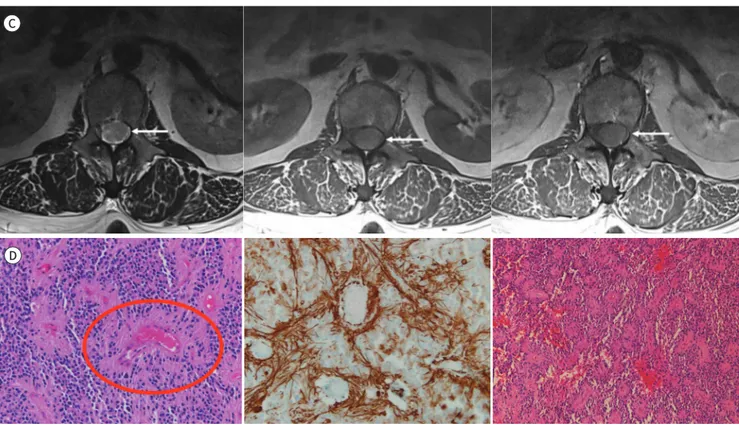
The results of histologic examination showed perivascular pseudorosettes. Immunohisto- chemical staining was performed and positive for glial fibrillary acid protein (GFAP). Addition- ally, partial hemorrhage was observed (Fig. 1D). The final histologic diagnosis was a World Health Organization grade II ependymoma.
DISCUSSION
Intradural extramedullary ependymoma is much rarer than intramedullary ependymoma.
The mechanism underlying the occurrence of intradural extramedullary ependymoma re- mains unknown (1). Overall, two hypotheses for the ectopic ependymoma have been pro- C
D
Fig. 1. A 52-year-old female with spinal intradural extramedullary ependymoma.
C. Axial T2-weighted (left), T1-weighted (middle), and postcontrast T1- weighted MRI (right). The mass (arrows) shows no contrast enhancement.
D. Histologic feature of ependymoma. Ependymal pseudorosettes (red circle) are composed of tumor cells arranged around blood vessels (hematoxylin and eosin stain, × 200) (left). Pseudorosettes are positive for glial fibrillary acidic protein (GFAP, × 200) (middle). Ependymoma shows partial internal hemorrhage (hematoxylin and eosin stain, × 100) (right).
posed (8, 9). To date, only nine cases of intradural extramedullary ependymoma have been reported, and all of them had intense contrast enhancement. All patients, including the one reported in the present study, were female. Accordingly, the hypothesis has been formulated that hormonal mechanism may have contributed to the development of the tumor, which seems reasonable in our case (9). Additionally, previous studies have also discussed the pos- sibility that the location of these exceptional tumors is caused by a heterotopic glial cell in the intradural extramedullary space (8, 9).
Previously reported intradural extramedullary ependymomas were usually iso-to-hypoin- tense on T1-weighted image and hyperintense on T2-weighted image, with a homogeneous enhancement after gadolinium injection (3, 4). However, none of the reported cases showed image findings similar to those observed in our patient. Intramedullary ependymoma can be accompanied by cystic changes and may not be enhanced after the contrast medium inject- ed (5-7). However, in our case, it was an intradural extramedullary solid tumor without cystic change, and there was no contrast enhancement. Non-enhancing ependymoma without cys- tic change was reported among pediatric brain tumors (5). Only three cases of intramedul- lary ependymoma without contrast enhancement were reported in the literature (10). In ad- dition, on contrast-enhanced T1-weigthed image, the signal intensity of this tumor was slightly higher than that of CSF and muscle. We considered the reason for hyperintensity on T1-weighted image as partial hemorrhage.
In conclusion, ependymoma should be considered when we encounter intradural extra- medullary spinal neoplasm, even if it is not common. Furthermore, it is important to know that intradural extramedullary ependymoma may not be enhanced after the injection of contrast medium.
Author Contributions
Conceptualization, K.H.; data curation, K.H., K.J.; formal analysis, K.H., K.J.; funding acquisition, K.H.; investigation, K.H., K.J.; methodology, K.H., K.J.; project administration, K.H.; resources, K.H., K.J.; software, K.H., K.J.; supervision, K.H.; validation, K.H.; visualization, all authors; writing—origi- nal draft, K.H., K.J.; and writing—review & editing, K.H., K.J.
Conflicts of Interest
The authors have no potential conflicts of interest to disclose.
Funding None
REFERENCES
1. Graça J, Gültasli N, D’Haene N, Brotchi J, Salmon I, Balériaux D. Cystic extramedullary ependymoma. AJNR Am J Neuroradiol 2006;27:818-821
2. Koeller KK, Rosenblum RS, Morrison AL. Neoplasms of the spinal cord and filum terminale: radiologic- pathologic correlation. Radiographics 2000;20:1721-1749
3. Robles SG, Saldaña C, Boto GR, Martinez A, Zamarron AP, Jorquera M, et al. Intradural extramedullary spi- nal ependymoma: a benign pathology? Spine (Phila Pa 1976) 2005;30:E251-E254
4. Moriwaki T, Iwatsuki K, Ohnishi Y, Umegaki M, Ishihara M, Yoshimine T. Intradural extramedullary spinal ep- endymoma: a case report of malignant transformation occurring. Asian Spine J 2013;7:139-142
5. Yuh EL, Barkovich AJ, Gupta N. Imaging of ependymomas: MRI and CT. Childs Nerv Syst 2009;25:1203-1213 6. Fanous AA, Jost GF, Schmidt MH. A nonenhancing World Health Organization Grade II intramedullary spinal
https://doi.org/10.3348/jksr.2020.0147 1345
ependymoma in the conus: case illustration and review of imaging characteristics. Global Spine J 2012;
2:57-64
7. Shin YJ, Lee E, Lee JW, Kang Y, Hyun SJ, Kim KJ, et al. Various MRI findings of spinal epenymoma. J Korean Soc Radiol 2017;76:411-419
8. Cooper I, Craig WM, Kernohan JW. Tumors of the spinal cord; primary extramedullary gliomas. Surg Gyne- col Obstet 1951;92:183-190
9. Duffau H, Gazzaz M, Kujas M, Fohanno D. Primary intradural extramedullary ependymoma: case report and review of the literature. Spine (Phila Pa 1976) 2000;25:1993-1995
10. Choi JY, Chang KH, Yu IK, Kim KH, Kwon BJ, Han MH, et al. Intracranial and spinal ependymomas: review of MR images in 61 patients. Korean J Radiol 2002;3:219-228
조영증강이 되지 않는 경막내 수외 뇌실막세포종: 증례 보고
김재민1 · 김현중1* · 권형주2
척수 뇌실막세포종은 일반적으로 성인에서 척수 내에 위치한다. 경막내 수외 척수 뇌실막세 포종은 극히 드물다. 대부분의 척수 뇌실막세포종은 MRI에서 다양한 조영증강을 보인다. 저 자는 MRI에서 조영증강을 보이지 않으며, 병리학적으로 확인된 52세 여자 환자의 경막내 수 외 뇌실막세포종을 경험하여 이에 대해 보고하고자 한다.
연세대학교 원주의과대학 1영상의학교실, 2병리학교실