Epidemiology and Factors Related to Clinical Severity of Acute Gastroenteritis in Hospitalized Children after the Introduction of Rotavirus Vaccination
We aimed to investigate epidemiology and host- and pathogen-related factors associated with clinical severity of acute gastroenteritis (AGE) in children after rotavirus vaccination introduction. Factors assessed included age, co-infection with more than 2 viruses, and virus-toxigenic Clostridium difficile co-detection. Fecal samples and clinical information, including modified Vesikari scores, were collected from hospitalized children with AGE. The presence of enteric viruses and bacteria, including toxigenic C. difficile, was detected by polymerase chain reaction (PCR). Among the 415 children included, virus was detected in stool of 282 (68.0%) children. Co-infection with more than 2 viruses and toxigenic C.
difficile were found in 24 (8.5%) and 26 (9.2%) children with viral AGE, respectively.
Norovirus (n = 130) infection, including norovirus-associated co-infection, was the most frequent infection, especially in children aged < 24 months (P < 0.001). In the severity- related analysis, age < 24 months was associated with greater diarrheal severity (P < 0.001) and modified Vesikari score (P = 0.001), after adjustment for other severity- related factors including rotavirus status. Although the age at infection with rotavirus was higher than that for other viruses (P = 0.001), rotavirus detection was the most significant risk factor for all severity parameters, including modified Vesikari score (P < 0.001). Viral co-infection and toxigenic C. difficile co-detection were not associated with any severity- related parameter. This information will be helpful in the management of childhood AGE in this era of rotavirus vaccination and availability of molecular diagnostic tests, which often lead to the simultaneous detection of multiple pathogens.
Keywords: Acute Gastroenteritis; Clinical Severity; Age; Rotavirus; Norovirus;
Co-Infection; Clostridium difficile; Children Ahlee Kim,1 Ju Young Chang,1,2
Sue Shin,3,4 Hana Yi,5 Jin Soo Moon,1 Jae Sung Ko,1 and Sohee Oh6
1Department of Pediatrics, Seoul National University College of Medicine, Seoul, Korea; 2Department of Pediatrics, Seoul Metropolitan Government-Seoul National University Boramae Medical Center, Seoul, Korea; 3Department of Laboratory Medicine, Seoul National University College of Medicine, Seoul, Korea; 4Department of Laboratory Medicine, Seoul Metropolitan Government-Seoul National University Boramae Medical Center, Seoul, Korea; 5School of Biosystem and Biomedical Science, Korea University College of Health Science, Seoul, Korea;
6Department of Medical Statistics, Seoul
Metropolitan Government-Seoul National University Boramae Medical Center, Seoul, Korea
Received: 2 September 2016 Accepted: 19 November 2016 Address for Correspondence:
Ju Young Chang, MD
Department of Pediatrics, Seoul National University College of Medicine, Seoul Metropolitan Government-Seoul National University Boramae Medical Center, 20 Boramae-ro 5-gil, Dongjak-gu, Seoul 07061, Korea
E-mail: [email protected]
Funding: This work supported by a grant from a Basic Science Research Program through the National Research Foundation of Korea (NRF), funded by the Ministry of Science, ICT & Future Planning (NRF-2012R1A1A3010058).
https://doi.org/10.3346/jkms.2017.32.3.465 • J Korean Med Sci 2017; 32: 465-474
INTRODUCTION
Introduction of the rotavirus vaccine significantly decreased the incidence of acute gastroenteritis (AGE) in children (1,2). Nev- ertheless, among children, AGE remains a major cause of hos- pitalization. Recently, evidence-based guidelines for the man- agement of AGE in children have been published in Europe (3).
According to this report, the major risk factors affecting gastro- enteritis severity are both pathogen- and host-related. Among the pathogens, rotavirus has been reported to be the main cause of persistent or severe diarrhea in Europe (4,5). Among the host- related factors, age, underlying chronic disease, and immune deficiencies have been reported to be associated with specific diarrheal pathogens and the severity of diarrhea (6,7).
However, previous studies on the severity of viral gastroen- teritis were conducted mostly before the introduction of rotavi-
rus vaccination; hence, rotavirus infection was most prevalent and mostly occurred in those less than 24 months of age. The clinical epidemiology of viral gastroenteritis, including preva- lence and vulnerable age, has been changing since the intro- duction of rotavirus vaccination (1,2,8). This might affect both clinical severity and the factors related to clinical severity of vi- ral gastroenteritis. In addition, the association between age and severity of diarrhea was not consistent among previous studies performed before the introduction of rotavirus vaccination. An age of less than 6 months was associated with less severe diar- rhea in a large-scale prospective study in Europe (9), but in stud- ies in developing countries, infants less than 6 months of age tended towards persistent or severe diarrhea (6). It has been thought that the high incidence of dehydration in infants less than 6 months is related to a high exposure to rotavirus.
Among pathogen-related factors that affect the clinical sever-
ity of gastroenteritis, the influence of co-infections with differ- ent viruses also varies among studies (10,11). Toxigenic Clos- tridium difficile infection, which is known to be mostly asymp- tomatic in infants and young toddlers, could also affect the se- verity of viral gastroenteritis as C. difficile co-infections in viral gastroenteritis have been reported to undergo a more severe clinical course in a few case reports (12,13). Currently, with the increasing use of diagnostic multiplex polymerase chain reac- tion (PCR) testing for viruses and enteric bacteria, multiple patho- gens have often been simultaneously detected in the feces of children with AGE. Hence, the assessment of clinical severity of virus-virus or virus-C. difficile co-infection is important for the determination of management for viral AGE in children.
This study aimed to investigate the epidemiology and host- and pathogen-related factors, including host age, co-infection with multiple viruses, and toxigenic C. difficile co-detection, as- sociated with the clinical severity of gastroenteritis in hospital- ized Korean children in the era of rotavirus vaccination and mo- lecular diagnosis of multiple pathogens. To assess the clinical severity of gastroenteritis, a modified Vesikari scoring system was used according to recent European guidelines (3,14).
MATERIALS AND METHODS Patients and clinical information
This study was conducted in the pediatric ward of the Seoul Metropolitan Government-Seoul National University Boramae Medical Center. From January 2012 to December 2013, i.e., 5–6 years after the introduction of rotavirus vaccination in Korea, stool samples were collected within 3 days of admission from children under 16 years of age who were hospitalized with a clinical suspicion of AGE. The patients were admitted via the emergency department or outpatient clinics. Children with known immunosuppressive underlying chronic illnesses, in- cluding inflammatory bowel diseases or malnutrition, were ex- cluded. Informed consent was obtained from the guardians of the children at enrolment. At discharge, subjects were further excluded based on the following criteria: failure to obtain per- mission, insufficient amount of fecal samples to complete the tests, and having a final diagnosis of an acute or chronic illness that mimics AGE, such as sepsis, cyclic vomiting syndrome, vi- ral infection with mucocutaneous manifestations, allergic or eosinophilic enterocolitis, or inflammatory bowel disease.
Clinical information including the age of onset, date of hos- pitalization, anthropometry (including weight and height/length), fever, duration and maximum number of episodes of diarrhea before and during hospitalization, duration and maximum num- ber of vomiting episodes before and during hospitalization, re- spiratory symptoms (including cough and rhinorrhea), and med- ications (including antibiotics taken in the 4 weeks before ad- mission and during hospitalization) were collected. From these
records, weight-for-height Z-scores (WHZ) and modified Vesi- kari scores were calculated (3). Laboratory examinations, in- cluding complete blood cell count and albumin, blood urea ni- trogen, and calcium level measurement, were performed on all of the children in the hospital laboratory. A stool test for white blood cells, hemoglobin, Rotavirus antigen (Bioline rotavirus®; SD standard diagnostics, Youngin, Korea), Salmonella species (spp.), Shigella spp., and Vibrio spp. were also conducted for all included children. The C. difficile toxin immunoassay (Tox A/B QUIK CHEK®; TechLab, Blacksburg, VA, USA) was performed for the selected children: those with a history of taking antibiot- ics within 2 weeks prior to the onset of diarrhea. For children who tested positive for rotavirus stool antigen, a vaccination history was obtained from the individual vaccination cards and the Korea Centers for Disease Control and Prevention website (http://is.cdc.go.kr), which records histories as part of the na- tional vaccination program.
Stool examination for viruses
Stool samples were collected in sterile tubes, transferred to the laboratory, and stored at −70°C until further processing. Stool samples were diluted to 10% (w/v) with phosphate-buffered saline and centrifuged. Viral double stranded RNA and DNA were extracted from the supernatant using the QIAamp Min- iElute Virus Spin Kit (Qiagen, Hilden, Germany). PCR was per- formed to detect adenovirus (15) and reverse transcription PCR (RT-PCR) was performed to detect rotavirus (8), norovirus (16), and astrovirus (17) as described previously (18). Multiplex PCR using a Seeplex Diarrhea-V ACE detection kit (Seegene Inc., Seoul, Korea) was also performed to detect astrovirus, group A rotavirus, enteric adenovirus, and norovirus, according to the manufacturer’s instructions (19). Amplification products were examined by electrophoresis on 1.5% agarose gels and docu- mented with the Bio-Rad Gel Doc 1000 Documentation System (BioRad, Hercules, CA, USA). The samples were scored as posi- tive for rotavirus if the rotavirus antigen test and/or PCR reac- tions were positive. The samples were scored as positive for nor- ovirus, adenovirus, or astrovirus if the PCR reactions were positive.
Stool examination for bacteria including toxigenic C. difficile
For bacterial DNA extraction, a portion of the 10% faucal slush was incubated for 10 minutes at 95°C and then centrifuged. Bac- terial DNA was extracted from the supernatant using a QIAamp DNA Mini Kit (Qiagen). Multiplex PCR using the Seeplex Diar- rhea-B1 and -B2 ACE detection kits (Seegene Inc.) was performed on stool samples from included children in accordance with the manufacturer’s instructions to detect Vibrio spp., C. difficile tox- in B, Salmonella spp., Shigella spp., Campylobacter spp., Clos- tridium perfringens, Yersinia enterocolitica, Escherichia coli O157, E. coli H7, VTEC, and Aeromonas spp. (20). Samples were scored
as positive for toxigenic C. difficile if either C. difficile toxin im- munoassay or the PCR reaction was positive.
Clinical severity analysis
In clinical severity related analyses of AGE, children with the following conditions were excluded: children with presence of enteric bacterial pathogen other than C. difficile, gross frequent hematochezia or the presence of tenesmus without toxigenic C.
difficile being detected, diagnosis of co-morbid acute lower re- spiratory infection, documented respiratory virus or enterovi- rus co-infection, and suspicion of co-morbid focal or systemic bacterial infection requiring ongoing antibiotic use.
Statistical analysis
We examined the data with the Kolmogorov-Smirnov test for normality. For age-related analyses, four age groups, < 6 months, 6–23 months, 24–59 months, and ≥ 60 months, or 2 age groups,
< 24 months, and ≥ 24 months were used. Comparisons of cat- egorical data were evaluated using the χ2 test. Continuous vari- ables were summarized using the median and interquartile range (IQR). Comparisons of continuous data were evaluated using the Mann-Whitney test or the Kruskal-Wallis test. Data were analyzed using ordered logit models to determine the cumula- tive odds ratio (c-OR) for clinical severity-related factors. Poten- tial clinical severity-related factors including age, virus status, viral co-infection status, C. difficile status, presence of upper re- spiratory tract infection-related symptoms, and previous use of antibiotics for more than 1 day within 2 weeks prior to the onset of diarrhea were chosen as the explanatory variables for ordinal logistic regression. All analyses were performed using SPSS ver- sion 20.0 (IBM Corp., Chicago, IL, USA).
Ethics statement
The study protocol was approved by the Institutional Review Board of Seoul Metropolitan Government-Seoul National Uni- versity Boramae Medical Center (IRB No. 02-2011-11, 06-2012-
151, 16-2015-78). Informed consent was confirmed by the IRB.
RESULTS
Clinical characteristics of subjects
During the study period, a total of 1,023 children (2012: n = 496, 2013: n = 527) were diagnosed with AGE according to electron- ic medical records. Of those, 415 children (40.6%) met the in- clusion criteria and were enrolled in this study. The median age of these 415 children was 27.2 (IQR 13.8–56.0) months and there were 244 males (58.8%). Of the 415 children, 319 (76.9%) were under 5 years of age and 190 (45.8%) were under 2 years of age (Table 1). There were no significant differences between the pa- tients admitted in 2012 (n = 179, 43.1%) and 2013 (n = 236, 56.9%) in terms of demographic and anthropometric characteristics.
Enteric pathogen distribution
A total of 355 (80.7%) children tested positive for enteric patho- gens. Viruses, toxigenic C. difficile, and other enteric bacteria were detected in 282 (68.0%), 38 (9.2%), and 80 (19.3%) children, respectively (Table 1). The age distribution was not significantly different between those pathogen positive vs. negative, howev- er, it was significantly different between those with virus posi- tive vs. negative status (P = 0.018). Toxigenic C. difficile infec- tion was significantly more common in children less than 24 months of age (P < 0.001). In contrast, enteric bacterial infec- tion other than toxigenic C. difficile was significantly more fre- quent in children older than 24 months (P = 0.016).
Virus distribution
Of the 282 children (68.0%) who tested positive for viral infec- tion by PCR, norovirus was the most commonly detected (n = 130), followed by rotavirus (n = 103), adenovirus (n = 56), and astro- virus (n = 20) (Table 1). The age distributions of the children differed significantly according to virus type (P = 0.008): noro- virus was found mostly in those aged 19.0 (IQR 12.0–40.1) months, Table 1. Distribution of enteric pathogens according to the age group in 415 children with AGE
Subjects/pathogens No. (%) Age, mon, median (IQR)
Age group, mon, No. (%)
P* P†
< 6 6–23 24–59 ≥ 60
Total 415 (100.0) 27.2 (13.8–56.3) 19 (100.0) 171 (100.0) 129 (100.0) 96 (100.0) - -
Pathogen positive 335 (80.7) 27.4 (14.2–55.4) 12 (63.2) 140 (81.9) 108 (83.7) 75 (78.1) 0.166 0.732
Virus positive 282 (68.0) 25.2 (13.8–48.3) 10 (52.6) 126 (73.7) 91 (70.5) 55 (57.3) 0.018 0.146
Norovirus 130 (31.3) 19.0 (11.8–40.9) 4 (21.1) 73 (42.7) 30 (23.3) 23 (24.0) 0.001 < 0.001
Rotavirus 103 (24.8) 30.5 (20.3–57.3) 4 (21.1) 30 (17.5) 44 (34.1) 25 (26.0) 0.012 0.003
Adenovirus 56 (13.5) 19.7 (12.0–40.1) 3 (15.8) 31 (18.1) 16 (12.4) 6 (6.3) 0.009 0.016
Astrovirus 20 (4.8) 26.2 (14.5–55.9) 0 (0.0) 10 (5.8) 7 (5.4) 3 (3.1) 0.679 0.698
Virus coinfection 24 (5.8) 17.7 (12.5–38.6) 1 (5.3) 15 (8.8) 6 (4.7) 2 (2.1) 0.033 0.034
Clostridium difficile 38 (9.2) 13.4 (9.9–21.5) 1 (5.3) 29 (17.0) 5 (3.9) 3 (3.1) < 0.001 < 0.001
Other enteric bacteria 80 (19.3) 38.2 (15.2–79.4) 2 (10.5) 25 (14.6) 26 (20.2) 27 (28.1) 0.012 0.016
The values were calculated using the χ2 test.
AGE = acute gastroenteritis, IQR = interquartile range.
*Among the 4 age groups; †Between children aged less than 24 months and those older than 24 months.
rotavirus in those aged 30.5 (IQR 20.3–57.3) months, adenovi- rus in those aged 19.7 (IQR 12.0–40.0) months, and astrovirus in those aged 26.2 (IQR 14.5–55.9) months. Rotavirus infection was significantly more frequent in children more than 24 months of age (P < 0.001) and showed a preference for older age groups compared with other enteric viruses (P = 0.001). Among chil-
Fig. 1. Monthly distribution of enteric viruses in 415 children.
0 5 10 15 20 25 30 35 40 45 50
Jan Feb Mar Apr May Jun Jul Aug Sep Oct Nov Dec
Number of patients
Month
rotavirus norovirus adenovirus astrovirus virus negative
Number of patients
Month
Jan Feb Mar Apr May Jun Jul Aug Sep Oct Nov Dec 5045
4035 3025 2015 105 0
0 5 10 15 20 25 30 35 40 45 50
Jan Feb Mar Apr May Jun Jul Aug Sep Oct Nov Dec
Number of patients
Month
rotavirusRotavirus norovirusNorovirus Adenovirusadenovirus astrovirusAstrovirus virus negativeVirus negative
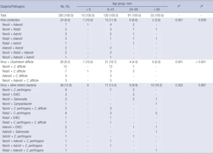
Table 2. Distribution enteric pathogens involved in coinfections according to the age group in 282 children with viral AGE
Subjects/Pathogens No. (%) Age group, mon
P* P†
< 6 6–23 24–59 ≥ 60
Total 282 (100.0) 10 (100.0) 126 (100.0) 91 (100.0) 55 (100.0) - -
Viral coinfection 24 (8.5) 1 (10.0) 15 (11.9) 6 (6.6) 2 (3.6) 0.261 0.059
NoroV + AdenoV 7 1 4 2 -
NoroV + RotaV 5 - 3 1 1
NoroV + AstroV 3 - 2 1 -
RotaV + AdenoV 3 - 1 1 1
RotaV + AstroV 1 - - 1 -
AdenoV + AstroV 2 - 2 - -
NoroV + RotaV + AdenoV 2 - 2 - -
NoroV + AdenoV + AstroV 1 - 1 - -
Virus + Clostridium difficile 26 (9.2) 1 (10.0) 21 (16.7) 4 (4.4) 0 (0.0) 0.001 < 0.001
NoroV + C. difficile 13 - 12 1 -
RotaV + C. difficile 7 1 3 3 -
AdenoV + C. difficile 3 - 3 - -
NoroV + AdenoV + C. difficile 3 - 3 - -
Virus + other enteric bacteria 36 (12.8) 0 17 (13.5) 9 (9.9) 10 (18.2) 0.302 0.897
NoroV + C. perfringens 9 - 7 2 -
NoroV + EHEC 3 - - 3 -
NoroV + Salmonella 3 - - 2 1
NoroV + Campylobacter 1 - - - 1
NoroV + C. perfringens + C. difficile 3 - 3 - -
RotaV + C. perfringens 8 - 3 - 5
RotaV + EHEC 1 - 1 - -
RotaV + C. perfringens + C. difficile 1 - 1 - -
AdenoV + EHEC 2 - - 1 1
AdenoV + Salmonella 1 - - - 1
AstroV + C. perfringens 1 - - 1 -
NoroV + AdenoV + C. perfringens 1 - 1 - -
NoroV + AstroV + C. perfringens 1 - 1 - -
RotaV + AdenoV + C. perfringens 1 - - - 1
The values were calculated using the χ2 test.
AGE = acute gastroenteritis, NoroV = Norovirus, AdenoV = Adenovirus, RotaV = Rotavirus, AstroV = Astrovirus, EHEC = enterohemorrhagic Escherichia coli.
*Among the 4 age groups; †Between children less than 24 months and those older than 24 months.
dren with rotavirus infection, 14 (13.6%) had been vaccinated with more than 2 doses of rotavirus vaccines. The age of vacci- nated children was significantly younger than that of unvacci- nated children (22.9 [IQR 15.7–29.7] months vs. 34.0 [IQR 21.9–
63.1] months, P = 0.010).
Seasonal differences were also observed among the viruses (Fig. 1). Mixed viral infections were found in 8.5% of children with AGE (Table 2). Norovirus co-infection (n = 18) occurred most frequently, followed by adenovirus and rotavirus co-in- fection. Median age of virus–virus co-infection was 17.7 (IQR 12.5–38.6) months: viral co-infection tended to be more frequent in children less than 24 months of age, although this was not statistically significant (P = 0.059).
Co-infection of C. difficile and other bacteria in viral AGE Co-infection by toxigenic C. difficile and other enteric bacteria in viral AGE was present in 26 (9.2%) and 36 (12.8%) children, respectively (Table 2). Median age of virus-toxigenic C. difficile co-infection was 12.7 (IQR 10.2–20.6) months, which was young-
er than that of virus–virus co-infection, albeit not statistically significant. Toxigenic C. difficile co-infection with viruses was significantly more frequent in those less than 24 months of age (P < 0.001). Toxigenic C. difficile co-infection with viruses was most frequently associated with norovirus infection (n = 19), followed by rotavirus infection. However, there was no signifi- cant association between presence of toxigenic C. difficile and norovirus detection.
The prevalence of co-infection with other enteric bacteria and viruses was 12.8%, which was slightly higher than the rate of virus–virus co-infection or virus-toxigenic C. difficile co-in- fection. Norovirus (n = 21) was the most frequent virus involved in this type of co-infection. The median age of children with vi- rus–other enteric bacterial co-infection was 28.4 (IQR 11.4–64.4) months, which was significantly higher than those with virus- C. difficile co-infection (P = 0.010).
Association of age and clinical severity of AGE
Among 415 children, 313 children were included in the severity analysis, after excluding 102. The exclusion criteria were as fol- lows: presence of enteropathogens other than C. difficile on mul-
tiplex PCR or stool culture (n = 80), gross frequent hematoche- zia or presence of tenesmus without toxigenic C. difficile detec- tion (n = 4), or presence of co-diagnosis of an acute lower respi- ratory infection, documentation of a respiratory virus or entero- virus co-infection or other suspicious focal or systemic bacterial co-infections with previous and/or ongoing antibiotic use (n = 18).
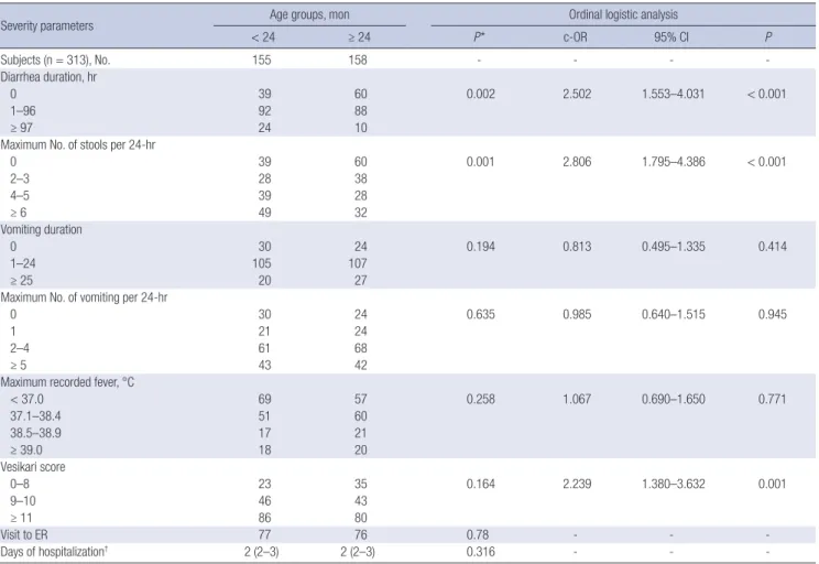
In the ordinal logistic regression, age less than 24 months was significantly associated with greater diarrheal severity in terms of duration and frequency and a higher modified Vesikari score than age more than 24 months, after adjustment for other po- tential severity-related factors (Table 3). Age less than 6 months was significantly associated with less vomiting severity in terms of duration (cumulative odds ratio [c-OR], 0.16; 95% confidence interval [CI], 0.06–0.49; P = 0.001) and frequency (c-OR, 0.29;
95% CI, 0.09–0.90; P = 0.032) compared to other age groups, af- ter adjustment for other potential severity-related factors.
Association of viral pathogen and clinical severity of AGE In the severity analyses of 313 children, virus detection was sig- nificantly associated with vomiting severity in terms of duration and frequency (c-OR, 2.5; 95% CI, 1.4–4.4; P = 0.002; c-OR, 1.8;
Table 3. Clinical severity of AGE according to the age group in 313 children
Severity parameters Age groups, mon Ordinal logistic analysis
< 24 ≥ 24 P* c-OR 95% CI P
Subjects (n = 313), No. 155 158 - - - -
Diarrhea duration, hr
0 39 60 0.002 2.502 1.553–4.031 < 0.001
1–96 92 88
≥ 97 24 10
Maximum No. of stools per 24-hr
0 39 60 0.001 2.806 1.795–4.386 < 0.001
2–3 28 38
4–5 39 28
≥ 6 49 32
Vomiting duration
0 30 24 0.194 0.813 0.495–1.335 0.414
1–24 105 107
≥ 25 20 27
Maximum No. of vomiting per 24-hr
0 30 24 0.635 0.985 0.640–1.515 0.945
1 21 24
2–4 61 68
≥ 5 43 42
Maximum recorded fever, °C
< 37.0 69 57 0.258 1.067 0.690–1.650 0.771
37.1–38.4 51 60
38.5–38.9 17 21
≥ 39.0 18 20
Vesikari score
0–8 23 35 0.164 2.239 1.380–3.632 0.001
9–10 46 43
≥ 11 86 80
Visit to ER 77 76 0.78 - - -
Days of hospitalization† 2 (2–3) 2 (2–3) 0.316 - - -
AGE = acute gastroenteritis, c-OR = cumulative odds ratio, CI = confidence interval, ER = emergency room.
*P values were calculated using the χ2 test. †The values are presented as median (interquartile range).
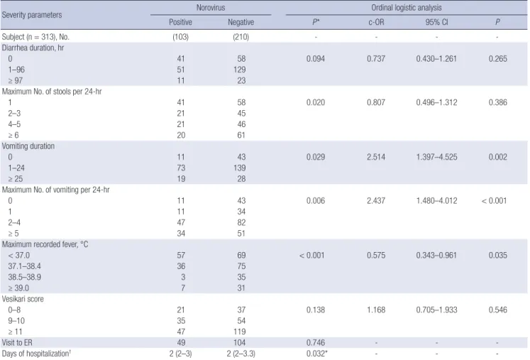
95% CI, 1.1–2.9; P = 0.028) and with the modified Vesikari score (c-OR, 2.3; 95% CI, 1.4–3.8; P = 0.001). Rotavirus infection (in- cluding 13 vaccinated cases) was significantly associated with severity of diarrhea, vomiting, fever, and overall clinical severity (Table 4). Detection of norovirus was associated with vomiting severity, and less severe fever (Table 5). However, viral co-infec- tion (n = 20) was not significantly associated with any of the clin- ical severity-related parameters.
Association of C. difficile co-infection and the clinical severity of viral AGE
The severity assessment for toxigenic C. difficile co-detection was performed with a subpopulation of children who tested pos- itive for viral infections (n = 237). C. difficile co-infection (n = 22) was not significantly associated with any clinical severity-relat- ed parameters after adjustment for other potential severity-re- lated factors. In this subgroup analysis, younger age, infection with rotavirus, and infection with norovirus were severity-relat- ed factors associated with AGE, as they were in the previous anal- ysis of all 313 children.
DISCUSSION
During the study period, norovirus infection was the leading cause of AGE in hospitalized children, exceeding rotavirus in- fection. This has been observed in several recent studies from various countries after implementing rotavirus vaccination (1, 21-23). The prevalence of adenovirus was also relatively high compared with that reported by previous Korean studies from the pre-vaccine era, which enrolled hospitalized children (16, 24,25). In some recent multi-center Asian studies conducted in inpatient or outpatient settings, an increased prevalence of ad- enovirus or enteric adenovirus infection was not demonstrated (26,27). However, recent studies from Korea (16.6% of children with documented viral AGE) and Brazil (12.5% of AGE) conduct- ed with inpatient and outpatient children reported a prevalence similar to that found in the present study (19.9% of children with documented viral AGE and 13.5% of children with AGE). These findings suggest the increasing importance of other viruses in cases of severe childhood AGE after the introduction of rotavi- rus vaccination, at least in some regions (21,28).
The prevalence of mixed viral infections (5.8% of AGE) was Table 4. Clinical severity of AGE according to the rotavirus infection status in 313 children
Severity parameters Rotavirus Ordinal logistic analysis
Positive Negative P* c-OR 95% CI P
Subject (n = 313), No. 90 223 - - - -
Diarrhea duration, hr
0 13 86 0.051 1.959 1.133–3.387 0.016
1–96 73 107
≥ 97 4 30
Maximum No. of stools per 24-hr
1 13 86 0.002 2.582 1.545–4.317 < 0.001
2–3 27 39
4–5 20 47
≥ 6 30 51
Vomiting duration
0 5 49 0.048 2.221 1.224–4.030 0.009
1–24 73 139
≥ 25 12 35
Maximum no. of vomiting per 24-hr
0 5 49 0.004 2.447 1.463–4.094 0.001
1 12 33
2–4 47 82
≥ 5 26 59
Maximum recorded fever, °C
< 37.0 13 113 < 0.001 3.216 1.896–5.455 < 0.001
37.1–38.4 38 73
38.5–38.9 23 15
≥ 39.0 16 22
Vesikari score
0–8 3 55 < 0.001 6.962 3.655–13.262 < 0.001
9–10 16 73
≥ 11 71 95
Visit to ER 51 102 0.080 - - -
Days of hospitalization† 3 (2–4) 2 (2–3) 0.001* - - -
AGE = acute gastroenteritis, c-OR = cumulative odds ratio, CI = confidence interval, ER = emergency room.
*P values were calculated using the χ2 test. †The values are presented as median (interquartile range).
similar to that noted by previous studies from various countries, which reported a prevalence of 3%–6% in hospitalized children with AGE. However, the prevalence of co-infections varied across studies (18,24,26,29,30) and seemed to be higher (up to 15%) in recent studies conducted in outpatient settings (27,31). The dom- inant types of co-infection were quite different even among stud- ies, partly because of variations in the study inclusion criteria, such as those related to age, hospitalization, season, or year, and variations in the diagnostic methods used to detect enteric vi- ruses. Mostly, rotavirus co-infection was the most frequent type in studies conducted in the pre-vaccine period (29,30,32,33).
However, norovirus co-infection has become most prevalent in a few studies conducted in the era of rotavirus vaccination (21, 27,31), similar to the findings of this study. Few large-scale stud- ies have examined the co-detection rate of toxigenic C. difficile and enteric virus in childhood viral gastroenteritis. In a recent single center European study with a modest number of fecal samples, a prevalence of 14.4% was reported (10). The overall prevalence of toxigenic C. difficile (including both mono-infec- tion and co-infection) was approximately 5%–20% in hospital- ized children with diarrhea, no different from the prevalence in
hospitalized children without diarrhea (34-37). The diversity in prevalence may be largely due to the different age distribution of included children, which could be deduced from the results of this study: there was a 17.0% prevalence of toxigenic C. diffi- cile in children aged 6–23 months in contrast to 4.4% in those 24–59 months (P < 0.001). Toxigenic C. difficile co-infection was also most frequently associated with norovirus infection in this study population, which is in contrast to the results of previous studies conducted in the pre-vaccine era. In those previous stud- ies, rotavirus-toxigenic C. difficile co-infection was the most fre- quent virus-toxigenic C. difficile co-infection (10,36). However, the association between the norovirus and toxigenic C. difficile was not statistically significant.
To our knowledge, the association that we found between younger age (less than 24 months) and the clinical severity of diarrhea after adjustment for rotavirus infection may be one of the first observed in a non-developing country. Previous Euro- pean studies showed that rotavirus infection is associated with severe clinical manifestations of AGE (4,5). However, these stud- ies were performed in the pre-vaccine era in which most chil- dren with rotavirus infections were less than 24 months of age.
Table 5. Clinical severity of AGE according to the norovirus infection status in 313 children
Severity parameters Norovirus Ordinal logistic analysis
Positive Negative P* c-OR 95% CI P
Subject (n = 313), No. (103) (210) - - - -
Diarrhea duration, hr
0 41 58 0.094 0.737 0.430–1.261 0.265
1–96 51 129
≥ 97 11 23
Maximum No. of stools per 24-hr
1 41 58 0.020 0.807 0.496–1.312 0.386
2–3 21 45
4–5 21 46
≥ 6 20 61
Vomiting duration
0 11 43 0.029 2.514 1.397–4.525 0.002
1–24 73 139
≥ 25 19 28
Maximum No. of vomiting per 24-hr
0 11 43 0.006 2.437 1.480–4.012 < 0.001
1 11 34
2–4 47 82
≥ 5 34 51
Maximum recorded fever, °C
< 37.0 57 69 < 0.001 0.575 0.343–0.961 0.035
37.1–38.4 36 75
38.5–38.9 3 35
≥ 39.0 7 31
Vesikari score
0–8 21 37 0.138 1.168 0.705–1.933 0.546
9–10 35 54
≥ 11 47 119
Visit to ER 49 104 0.746 - - -
Days of hospitalization† 2 (2–3) 2 (2–3.3) 0.032* - - -
AGE = acute gastroenteritis, c-OR = cumulative odds ratio, CI = confidence interval, ER = emergency room.
*P values were calculated using the χ2 test. †The values are presented as median (interquartile range).
Hence, age-related severity may, at least partly, be attributed to rotavirus-related severity (9). In contrast, in this study, the me- dian age of children with rotavirus infection was 31 months. In spite of the higher age of rotavirus-infected children, we found that an age of less than 24 months was a significant severity-re- lated host factor. Additionally, we found that rotavirus infection (in mostly unvaccinated children in this study) was the primary pathogenic cause of severe clinical gastroenteritis. Because this study was performed between 2012 and 2013, which corresponds to 5–6 years after the introduction of the rotavirus vaccine in Ko- rea (the rate of rotavirus vaccination more than 2 times was re- ported to be 65.6% in Seoul, and nationwide rate 52.4% in 2013 by Korean National Immunization Survey; http://www.cdc.go.
kr), the age of children vulnerable to rotavirus infection seems to have transiently increased over the study period (8).
In this study, children less than 6 months of age, as well as children aged between 6 and 23 months, had more severe diar- rhea than the older age groups. These findings are contrary to previous European studies but similar to results from previous studies from developing countries (6,9). Although other host- related factors, such as breastfeeding, were not investigated in this study, WHZ and macronutrient status (including albumin, blood urea nitrogen, and serum calcium) were examined to ex- clude malnutrition. Therefore, malnutrition-associated immune deficiencies were not likely to have caused the age-related dif- ference in diarrheal severity. Given that an age of less than 24 months was significantly associated with toxigenic C. difficile detection and also tended to be associated with a higher inci- dence of viral co-infection, the greater diarrheal severity in these age groups might, at least in part, be related to the relative insta- bility of the intestinal microbiota in these age groups in com- parison with those in older age groups. In other words, intesti- nal mucosal immunity might play a role in this age-related di- arrheal severity.
It is controversial whether gastroenteritis cases with co-infec- tion with multiple viruses are more severe than gastroenteritis cases with mono-infection (10,11,30). A previous study showed greater severity in co-infection cases, but did not separate viral co-infections from bacterial co-infections (10). In our study, bac- terial co-infection was excluded in the severity-related analyses.
In this condition, viral co-infection was not associated with great- er clinical severity, which was compatible with the results of some previous studies (11,29,30). Because rotavirus infection cases showed the most severe clinical manifestations, and nor- ovirus infection was associated with greater vomiting severity, one could expect more severe clinical symptoms in co-infec- tion cases with rotavirus and norovirus than in mono-infection cases, if both viruses play roles as infecting agents. Although evaluation of the severity of gastroenteritis resulting from this co-infection was limited, as only 7 of our patients were co-in- fected with rotavirus and norovirus, these patients did not show
any greater clinical severity compared to mono-infection cases with rotavirus or norovirus or co-infection cases with other vi- ruses. Further studies that include a sufficient number of rota- virus and norovirus co-infection cases may be needed to deter- mine the clinical significance of this co-infection and to deter- mine whether this co-infection doubles the clinical manifesta- tions or whether 1 virus functions as the main pathogen and the other as a bystander.
Regarding the clinical severity of co-infection with C. difficile, 2 contradictory results were also found in the literature. In one study, viral co-infection with C. difficile was clinically indistin- guishable from C. difficile mono-infection, although the bacte- rial burden of C. difficile was significantly higher in viral co-in- fection cases than in cases without viral co-infection (38). How- ever, in another study, the severity of viral gastroenteritis in chil- dren co-infected with C. difficile was significantly greater than viral or bacterial mono-infection (10). In our study, clinical se- verity of viral AGE was not affected by presence of toxigenic C.
difficile: Vomiting severity and the modified Vesikari score, but not diarrheal severity, were significantly different between vi- rus-C. difficile co-infection and C. difficile mono-infection cas- es. Because these associations were also found in children with viral gastroenteritis without C. difficile, it seems that co-infec- tion with C. difficile may not further contribute to severity of childhood gastroenteritis in most cases. This is in line with the previous opinion that detection of C. difficile toxin cannot be assumed to be the causative agent for diarrhea in most children between 1–3 years of age (39,40). Thus, in most such cases, if toxigenic C. difficile is detected unexpectedly in typical viral gastroenteritis cases, it may not be necessary to treat C. difficile.
A limitation of this study is that this was a single-center study that enrolled approximately half of the children hospitalized with gastroenteritis during the study period. However, a con- siderable number of epidemiologic findings share similar trends with the results of previous multicenter or national studies from other countries, as mentioned previously. Moreover, the num- ber of samples examined is not that small, compared with pre- vious epidemiologic studies. Additionally, the patients included in the severity analysis were strictly chosen to exclude potential confounders that may affect the clinical severity of gastroenteri- tis such as presence of co-morbid acute illnesses.
In summary, this study captured the changing prevalence and age-related distribution of viruses and their co-infections after implementing rotavirus vaccination in Seoul: norovirus infection, including norovirus-associated co-infection, was the leading cause of AGE in hospitalized children, especially in those less than 24 months of age. Age-dependent clinical severity was also observed: age less than 24 months was associated with high- grade diarrheal severity and overall severity of gastroenteritis independent of rotavirus status. Co-infection with multiple vi- ruses may not affect clinical severity, but this needs to be inves-
tigated further with a sufficient number of children co-infected with rotavirus and norovirus. Mostly, the clinical severity of vi- ral gastroenteritis may not be affected by the co-existence of toxigenic C. difficile. This study provides information on the changing epidemiology and clinical severity-related factors of childhood gastroenteritis. It will aid in the management of child- hood AGE in this era of rotavirus vaccination and molecular di- agnostic techniques to detect multiple pathogens.
ACKNOWLEDGMENT
We thank Kyung Mi Choi (Seoul Metropolitan Government Seoul National University Boramae Medical Center) for the expert technical assistance.
DISCLOSURE
The authors have no potential conflicts of interest to disclose.
AUTHOR CONTRIBUTION
Conceptualization: Chang JY, Yi H. Data curation: Kim A, Chang JY, Shin S, Oh S. Formal analysis: Kim A, Chang JY, Shin S, Oh S.
Investigation: Kim A, Chang JY, Shin S.Writing - original draft:
Kim A, Chang JY. Writing - review & editing: Moon JS, Ko JS.
ORCID
Ahlee Kim http://orcid.org/0000-0002-0803-0402 Ju Young Chang http://orcid.org/0000-0002-4760-6777 Sue Shin http://orcid.org/0000-0003-4791-8671 Hana Yi http://orcid.org/0000-0002-4910-122X Jin Soo Moon http://orcid.org/0000-0001-9760-297X Jae Sung Ko http://orcid.org/0000-0002-3064-2974 Sohee Oh http://orcid.org/0000-0002-3010-448X REFERENCES
1. Wikswo ME, Desai R, Edwards KM, Staat MA, Szilagyi PG, Weinberg GA, Curns AT, Lopman B, Vinjé J, Parashar UD, et al. Clinical profile of children with norovirus disease in rotavirus vaccine era. Emerg Infect Dis 2013; 19:
1691-3.
2. Choi UY, Lee SY, Ma SH, Jang YT, Kim JY, Kim HM, Kim JH, Kim DS, Kim YS, Kang JH. Epidemiological changes in rotavirus gastroenteritis in chil- dren under 5 years of age after the introduction of rotavirus vaccines in Korea. Eur J Pediatr 2013; 172: 947-52.
3. Guarino A, Ashkenazi S, Gendrel D, Lo Vecchio A, Shamir R, Szajewska H; European Society for Pediatric Gastroenterology, Hepatology, and Nu- trition; European Society for Pediatric Infectious Diseases. European So- ciety for Pediatric Gastroenterology, Hepatology, and Nutrition/Europe- an Society for Pediatric Infectious Diseases evidence-based guidelines for the management of acute gastroenteritis in children in Europe: up-
date 2014. J Pediatr Gastroenterol Nutr 2014; 59: 132-52.
4. Friesema IH, de Boer RF, Duizer E, Kortbeek LM, Notermans DW, Nor- bruis OF, Bezemer DD, van Heerbeek H, van Andel RN, van Enk JG, et al.
Etiology of acute gastroenteritis in children requiring hospitalization in the Netherlands. Eur J Clin Microbiol Infect Dis 2012; 31: 405-15.
5. Gimenez-Sanchez F, Delgado-Rubio A, Martinon-Torres F, Bernaola-Iturbe E; Rotascore Research Group. Multicenter prospective study analysing the role of rotavirus on acute gastroenteritis in Spain. Acta Paediatr 2010;
99: 738-42.
6. Mathew A, Rao PS, Sowmyanarayanan TV, Kang G. Severity of rotavirus gastroenteritis in an Indian population: report from a 3 year surveillance study. Vaccine 2014; 32 Suppl 1: A45-8.
7. Strand TA, Sharma PR, Gjessing HK, Ulak M, Chandyo RK, Adhikari RK, Sommerfelt H. Risk factors for extended duration of acute diarrhea in young children. PLoS One 2012; 7: e36436.
8. Shim JO, Chang JY, Shin S, Moon JS, Ko JS. Changing distribution of age, clinical severity, and genotypes of rotavirus gastroenteritis in hospitalized children after the introduction of vaccination: a single center study in Seoul between 2011 and 2014. BMC Infect Dis 2016; 16: 287.
9. Albano F, Bruzzese E, Bella A, Cascio A, Titone L, Arista S, Izzi G, Virdis R, Pecco P, Principi N, et al. Rotavirus and not age determines gastroenteri- tis severity in children: a hospital-based study. Eur J Pediatr 2007; 166:
241-7.
10. Valentini D, Vittucci AC, Grandin A, Tozzi AE, Russo C, Onori M, Menichel- la D, Bartuli A, Villani A. Coinfection in acute gastroenteritis predicts a more severe clinical course in children. Eur J Clin Microbiol Infect Dis 2013; 32: 909-15.
11. Matthijnssens J, Zeller M, Heylen E, De Coster S, Vercauteren J, Braeck- man T, Van Herck K, Meyer N, Pirçon JY, Soriano-Gabarro M, et al. Higher proportion of G2P[4] rotaviruses in vaccinated hospitalized cases com- pared with unvaccinated hospitalized cases, despite high vaccine effec- tiveness against heterotypic G2P[4] rotaviruses. Clin Microbiol Infect 2014;
20: O702-10.
12. Bignardi GE, Staples K, Majmudar N. A case of norovirus and Clostridi- um difficile infection: casual or causal relationship? J Hosp Infect 2007;
67: 198-200.
13. Lukkarinen H, Eerola E, Ruohola A, Vainionpää R, Jalava J, Kotila S, Ruus- kanen O. Clostridium difficile ribotype 027-associated disease in children with norovirus infection. Pediatr Infect Dis J 2009; 28: 847-8.
14. Ruuska T, Vesikari T. Rotavirus disease in Finnish children: use of numeri- cal scores for clinical severity of diarrhoeal episodes. Scand J Infect Dis 1990; 22: 259-67.
15. Allard A, Girones R, Juto P, Wadell G. Polymerase chain reaction for de- tection of adenoviruses in stool samples. J Clin Microbiol 1990; 28: 2659- 67.
16. Koh H, Baek SY, Shin JI, Chung KS, Jee YM. Coinfection of viral agents in Korean children with acute watery diarrhea. J Korean Med Sci 2008; 23:
937-40.
17. Belliot G, Laveran H, Monroe SS. Detection and genetic differentiation of human astroviruses: phylogenetic grouping varies by coding region. Arch Virol 1997; 142: 1323-34.
18. Yang HR, Jee YM, Ko JS, Seo JK. Detection and genotyping of viruses de- tected in children with benign afebrile seizures associated with acute gas- troenteritis. Korean J Pediatr Gastroenterol Nutr 2009; 12: 183-93.
19. Higgins RR, Beniprashad M, Cardona M, Masney S, Low DE, Gubbay JB.
Evaluation and verification of the Seeplex Diarrhea-V ACE assay for si- multaneous detection of adenovirus, rotavirus, and norovirus genogroups I and II in clinical stool specimens. J Clin Microbiol 2011; 49: 3154-62.
20. Onori M, Coltella L, Mancinelli L, Argentieri M, Menichella D, Villani A, Grandin A, Valentini D, Raponi M, Russo C. Evaluation of a multiplex PCR assay for simultaneous detection of bacterial and viral enteropathogens in stool samples of paediatric patients. Diagn Microbiol Infect Dis 2014;
79: 149-54.
21. Jin HI, Lee YM, Choi YJ, Jeong SJ. Recent viral pathogen in acute gastroen- teritis: a retrospective study at a tertiary hospital for 1 year. Korean J Pedi- atr 2016; 59: 120-5.
22. Doll MK, Gagneur A, Tapiéro B, Charest H, Gonzales M, Buckeridge DL, Quach C. Temporal changes in pediatric gastroenteritis after rotavirus vaccination in Quebec. Pediatr Infect Dis J 2016; 35: 555-60.
23. Lopman BA, Steele D, Kirkwood CD, Parashar UD. The vast and varied global burden of norovirus: prospects for prevention and control. PLoS Med 2016; 13: e1001999.
24. Chung JY, Huh K, Kim SW, Shin BM, Han TH, Lee JI, Song MO. Molecular epidemiology of human astrovirus infection in hospitalized children with acute gastroenteritis. Korean J Pediatr Gastroenterol Nutr 2006; 9: 139- 46.
25. Lee JI, Lee GC, Chung JY, Han TH, Lee YK, Kim MS, Lee CH. Detection and molecular characterization of adenoviruses in Korean children hos- pitalized with acute gastroenteritis. Microbiol Immunol 2012; 56: 523-8.
26. Chen CJ, Wu FT, Huang YC, Chang WC, Wu HS, Wu CY, Lin JS, Huang FC, Hsiung CA. Clinical and epidemiologic features of severe viral gastroen- teritis in children: a 3-year surveillance, multicentered study in Taiwan with partial rotavirus immunization. Medicine (Baltimore) 2015; 94: e1372.
27. Thongprachum A, Takanashi S, Kalesaran AF, Okitsu S, Mizuguchi M, Hay- akawa S, Ushijima H. Four-year study of viruses that cause diarrhea in Japanese pediatric outpatients. J Med Virol 2015; 87: 1141-8.
28. Reis TA, Assis AS, do Valle DA, Barletta VH, de Carvalho IP, Rose TL, Portes SA, Leite JP, da Rosa e Silva ML. The role of human adenoviruses type 41 in acute diarrheal disease in Minas Gerais after rotavirus vaccination. Braz J Microbiol 2016; 47: 243-50.
29. Tran A, Talmud D, Lejeune B, Jovenin N, Renois F, Payan C, Leveque N, Andreoletti L. Prevalence of rotavirus, adenovirus, norovirus, and astrovi- rus infections and coinfections among hospitalized children in northern
France. J Clin Microbiol 2010; 48: 1943-6.
30. Román E, Wilhelmi I, Colomina J, Villar J, Cilleruelo ML, Nebreda V, Del Alamo M, Sánchez-Fauquier A. Acute viral gastroenteritis: proportion and clinical relevance of multiple infections in Spanish children. J Med Microbiol 2003; 52: 435-40.
31. Lu L, Jia R, Zhong H, Xu M, Su L, Cao L, Dong Z, Dong N, Xu J. Molecular characterization and multiple infections of rotavirus, norovirus, sapovi- rus, astrovirus and adenovirus in outpatients with sporadic gastroenteri- tis in Shanghai, China, 2010-2011. Arch Virol 2015; 160: 1229-38.
32. Marie-Cardine A, Gourlain K, Mouterde O, Castignolles N, Hellot MF, Mal- let E, Buffet-Janvresse C. Epidemiology of acute viral gastroenteritis in chil- dren hospitalized in Rouen, France. Clin Infect Dis 2002; 34: 1170-8.
33. González GG, Liprandi F, Ludert JE. Molecular epidemiology of enteric viruses in children with sporadic gastroenteritis in Valencia, Venezuela. J Med Virol 2011; 83: 1972-82.
34. Cerquetti M, Luzzi I, Caprioli A, Sebastianelli A, Mastrantonio P. Role of Clostridium difficile in childhood diarrhea. Pediatr Infect Dis J 1995; 14:
598-603.
35. Mårdh PA, Helin I, Colleen I, Oberg M, Holst E. Clostridium difficile toxin in faecal specimens of healthy children and children with diarrhoea. Acta Paediatr Scand 1982; 71: 275-8.
36. de Graaf H, Pai S, Burns DA, Karas JA, Enoch DA, Faust SN. Co-infection as a confounder for the role of Clostridium difficile infection in children with diarrhoea: a summary of the literature. Eur J Clin Microbiol Infect Dis 2015; 34: 1281-7.
37. Rexach CE, Tang-Feldman YJ, Cantrell MC, Cohen SH. Epidemiologic surveillance of Clostridium difficile diarrhea in a freestanding pediatric hospital and a pediatric hospital at a university medical center. Diagn Microbiol Infect Dis 2006; 56: 109-14.
38. El Feghaly RE, Stauber JL, Tarr PI, Haslam DB. Viral co-infections are com- mon and are associated with higher bacterial burden in children with Clos- tridium difficile infection. J Pediatr Gastroenterol Nutr 2013; 57: 813-6.
39. Schutze GE, Willoughby RE; Committee on Infectious Diseases; Ameri- can Academy of Pediatrics. Clostridium difficile infection in infants and children. Pediatrics 2013; 131: 196-200.
40. Borali E, De Giacomo C. Clostridium difficile infection in children: a re- view. J Pediatr Gastroenterol Nutr 2016; 63: e130-40.