INTRODUCTION
Torus hyperplasia is a very rare condition also known as focal pyloric hypertrophy (1). This lesion is caused by a cir- cular muscle hypertrophy affecting the lesser curvature near the pylorus. Pathogenesis of the lesion is unclear, some con- sidering it as a persistence of infantile pyloric hypertrophy to adulthood, others noting that it develops within a few weeks in adult (1, 2). Some have speculated that the lesion results from chronic gastritis or from repeated spastic con- tractions of the area secondary to visceral-visceral reflexes that could be established as a result of local irritation stimulating afferent fibers and starting a reflex arc involving efferents that locally innervate the circular pyloric musculature (1, 3). In summary, focal hypertrophic stenosis of the pyloric antrum in adult is classified as primary or secondary types; 1) prima- ry type being an idiopathic torus hyperplasia uncertain of origin, and 2) secondary type from a result of muscular thick- ening due to inflammation, ulceration, or carcinoma (4).
After the first report on 1946, torus hyperplasia has been reported as a rare disease (5). Although there are some typi- cal findings (i.e., recognizable radiologically by narrowing and enlongation of the pyloric cancal and endoscopically by appearances resembling those of the cervix so called charac- teristic pyloric ‘‘cervix sign’’) in this benign disease (6, 7), it is difficult to confirm without resection. Moreover, it is hard
to predict the prevalence of the disease because of its’ rarity (3). Herein, we report a 56-yr-old man who was diagnosed as torus hyperplasia with appendiceal mucocele after visiting our hospital because of abdominal discomfort. To the best of our knowledge, this is the first case of torus hyperplasia with appendiceal mucocele which is found incidentally.
CASE REPORT
A 56-yr-old man with a history of hypertension presented with abdominal discomfort for one month. He reported no bowel habit change, nausea, vomiting, or fever, but compla- ined of anal pain. He had a family history since his father died of advanced gastric cancer. On arrival, his blood pressure was 128/87 mmHg, heart rate 80 beats/min, respiratory rate was 20/min, and body temperature was 36.0℃. The physical examination revealed no abnormalities except rectal exami- nation. His abdomen was soft and non-tender with normal bowel sound. On rectal examination, a fissure was noticed on 6 o’clock direction.
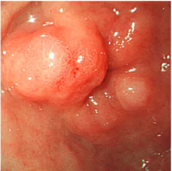
There was no remarkable finding on laboratory examina- tions on arrival. Upper gastrointestinal endoscopy showed a 1.0 cm sized irregular submucosal lesion proximal to the pylorus to the distal antrum on the lesser curvature. Central erosion was noticed on the surface of the lesion due to previ-
152
Chi-Hun Kim1, Hye Seung Han2, Sun-Young Lee1, Byung Kook Kim1, In-Kyung Sung1, Moo Kyung Seong3 and Kyung Yung Lee3
Departments of Internal Medicine1, Pathology2, and Surgery3, Konkuk University School of Medicine, Seoul, Korea
Address for correspondence Sun-Young Lee, M.D.
Department of Internal Medicine, Konkuk University School of Medicine, 4-12 Hwayang-dong, Gwangjin-gu, Seoul 143-729, Korea Tel : +82-2-2030-7747, Fax : +82-2-2030-7748 E-mail : sunyoung@kuh.ac.kr
J Korean Med Sci 2010; 25: 152-4 ISSN 1011-8934
DOI: 10.3346/jkms.2010.25.1.152
Torus Hyperplasia of the Pyloric Antrum
Primary or idiopathic hypertrophy of the pyloric muscle in adult, so called torus hyper- plasia, is an infrequent but an established entity. It is caused by a circular muscle hypertrophy affecting the lesser curvature near the pylorus. Since most of the lesions are difficult to differentiate from tumor, distal gastrectomy is usually preformed to rule out most causes of pyloric lesions including neoplastic ones through a patho- logical study. A 56-yr-old man with a family history of gastric cancer presented with abdominal discomfort of 1 month duration. Upper gastrointestinal endoscopy showed a 1.0 cm sized irregular submucosal lesion proximal to the pylorus to the distal antrum on the lesser curvature. On colonoscopy examination, a 1.5 cm sized protruding mass was noticed on the appendiceal orifice. Gastrectomy and cecectomy were done, and histological section revealed marked hypertrophy of the distal circular pyloric musculature and an appendiceal mucocele. To the best of our knowledge, this is the first case of torus hyperplasia with appendiceal mucocele which is found incidentally.
Key Words : Hyperplasia; Antrum, Pyloric; Appendix; Mucocele
ⓒ 2010 The Korean Academy of Medical Sciences.
This is an Open Access article distributed under the terms of the Creative Commons Attribution Non-Commercial License (http://creativecommons.org/licenses/by-nc/3.0) which permits unrestricted non-commercial use, distribution, and reproduction in any medium, provided the original work is properly cited.
Received : 24 October 2007 Accepted : 18 July 2008
Torus Hyperplasia of the Pyloric Antrum 153
ous biopsy (Fig. 1). Cushion sign revealed negative finding, and biopsy revealed no significant abnormality. On colono- scopy examination which was performed on the same day with upper gastrointestinal endoscopy, a 1.5 cm sized pro- truding mass was noticed on the appendiceal orifice. This lesion revealed positive tent sign suggesting a cystic change (Fig. 2).
Gastrectomy and cecectomy were underwent after abdom- inal computed tomography. Histological section revealed marked hypertrophy of the distal circular pyloric muscula- ture without an evidence of neoplasia (Fig. 3). Finally, gas- tric lesion was diagnosed as torus hyperplasia. Appendiceal mass was diagnosed as mucinous cystadenoma after the sur-
gical resection (Fig. 4). The patient tolerated well and was discharged without any complication. He is being followed up in the outpatient clinic without any symptom.
DISCUSSION
The present case revealed marked hypertrophy of the dis- tal circular pyloric musculature on histological finding, and was diagnosed as torus hyperplasia after the resection. It was consistent with adult idiopathic hypertrophic pyloric steno- sis, a benign disease resulting from hypertrophy of the circu- lar fibers of the pyloric canal of unknown etiology. Since the
Fig. 1. Upper gastrointestinal endoscopic finding. A 10 mm sized hemispheric shaped elevated lesion with central erosion on the lesser curvature side of the distal antrum near the pylorus.
Fig. 2. Colonoscopic finding. It shows a large round mass on the cecal end with positive tent sign.
Fig. 3. Microscopic finding. In low power view, this mass-like lesion is consists of a localized area of hypertrophic circular muscle (H&
E stain, ×12.5).
Fig. 4. Gross finding. Abundant mucoid material is present within the lumen of the appendix.
154 C.-H. Kim, H.S. Han, S.-Y. Lee, et al.
patient had no predisposing factors for the development of secondary pyloric stenosis, we considered this lesion as con- genital in origin. Although the endoscopic finding revealed typical cervix sign, we were not able to differentiate from true neoplastic lesion before the resection.
Despite the recent progress in radiology and endoscopy, it is very hard to define hypertrophic stenosis in adults (8). On endoscopic finding, torus hyperplasia reveals as submucosal tumor since it arise from circular muscle consisting the pyloric canal which makes it hard to diagnose through endoscopic biopsy (6, 8). In addition, it is usually misdiagnosed as car- cinoma of the antrum (6). Although recent progress of endo- scopic ultrasonography and endoscopic resection had been made, it is hard to diagnose only with the aid of endoscopic procedures.
Most of these lesions are found incidentally like our case because it cause minimal clinical signs or is asymptomatic.
In the absence of the symptoms, no clinical treatment is re- quired, and resection is advocated only when stenosis gives rise to symptoms or when a malignancy is suspicious (6, 8).
Although endoscopic dilatation or laparascopic hypertrophy have been reported as alternative treatments (9, 10), distal gastrectomy with gastroduodenostomy is still the main ther- apy for final diagnosis and treatment. Gastrectomy was per- formed in our patient because of several reasons. First, the antral lesion was hard to differentiate from true neoplastic lesion. Second, the patient had a fear of gastric cancer since his father died of this malignancy. Third, elective operation for appendiceal mucocele was planned for suspicious malig- nant potential.
In summary, we have experienced a rare disease, tours hyper- plasia which was difficult to differentiate from gastric cancer
mimicking submucosal tumor. Our experience including typical cervix sign on endoscopic finding would help other endoscopists for their diagnosis.
REFERENCES
1. Aron J, Newman A, Heaton J. Torus hyperplasia of the pyloric antrum presenting as a gastric pseudotumor. Gastroenterology 1973; 64:
634-6.
2. Graadt van Roggen JF, M van Krieken JH. Adult hypertrophic pyl- roic stenosis: case report and review. J Clin Pathol 1998; 51: 479-80.
3. Quigley RL, Pruitt SK, Pappas TN, Akwari O. Primary hypertrophic pyloric stenosis in the adult. Arch Surg 1990; 125: 1219-21.
4. Wellmann KF, Kagan A, Fang H. Hypertorphic pyloric stenosis in adults. Gasteroenterology 1964; 46: 601-8.
5. Andersen K, Gammerlgaard A, Licht E de F. Hypertrophy of the pylorus in adults. Acta Radiol 1946; 27: 552.
6. Ikenaga T, Honmyo U, Takano S, Murakami A, Harada K, Mizumo- to S, Yoshinaka I, Hirata T, Maeda M, Kiyohara H. Primary hyper- trophic pyloric stenosis in the adult. J Gastroenterol Hepatol 1992;
7: 524-6.
7. Dye TE, Vidals VG, Lockhart CE, Snider WR. Adult hypertrophic pyloric stenosis. Am Surg 1979; 45: 478-84.
8. Hellan M, Lee T, Lerner T. Diagnosis and therapy of primary hyper- trophic stenosis in adults: case report and review of literature. J Gas- trointest Surg 2006; 10: 265-9.
9. Franco L, Dryden N. Gastric-outlet obstruction. N Engl J Med 2007;
356: 942.
10. Danikas D, Peter Geis W, Ginalis E, Gorcey S, Stratoulias C. Laparas- copic pyloroplasty in idiopathic hypertrophic pyloric stenosis in an adult. JSLS 2000; 4: 173-5.