Effect of Circuit Training on Aging-related Hormones in Obese Middle-aged Women
Won-Mok Son1, Yi-Sub Kwak2, Gun-Do Kim3, Min-Sung Ha1, Song-Young Park4 and Gi-Dong Sung1*
1Department of Physical Education, Pusan National University, Busan 46241, Korea
2Department of Physical Education, Dong-Eui University, Busan 47340, Korea
3Department of Microbiology, Pukyong National University, Busan 48513, Korea
4School of Health and Kinesiology, University of Nebraska-omaha, Omaha, NE, USA
Received May 12, 2017 /Revised June 27, 2017 /Accepted June 28, 2017
Due to aging, the physical fitness of middle-aged women decreases after menopause. This results in increased body fat and reduced lean body mass, both of which can lead to obesity. This phenomenon is accompanied by changes in hormone secretion in the body. The purpose of this study was to ana- lyze the effects of circuit training on aging-related hormones in obese middle-aged women. The sub- jects were 20 obese middle-aged women. The subjects were divided into two groups: a circuit training group (n=10) and nonexercise control group (n=10). Growth hormone (GH), insulin-like growth fac- tor-1 (IGF-1), estrogen, and DHEA-S were measured before and after the circuit training program. The circuit training group performed circuit training for 12 weeks, three times per week. Exercise intensity was increased gradually from 60% to 80% of heart rate reserve every 6 weeks. In the circuit training group, at the end of the 12-week training program, the IGF-1 levels were significantly increased com- pared to pre-exercise levels, and they were higher than those of the control group. GH levels were also significantly increased in the circuit training group, but the differences were not statistically significant. DHEA-S was significantly increased in the circuit training group, but the difference was not statistically significant. The results showed that circuit training improved aging-related hormones levels in obese middle-aged women.
Key words : DHEA-S, estrogen, GH, IGF-1, obese
*Corresponding author
*Tel : +82-51-510-1627, Fax : +82-51-515-1991
*E-mail : [email protected]
This is an Open-Access article distributed under the terms of the Creative Commons Attribution Non-Commercial License (http://creativecommons.org/licenses/by-nc/3.0) which permits unrestricted non-commercial use, distribution, and reproduction in any medium, provided the original work is properly cited.
Journal of Life Science 2017 Vol. 27. No. 9. 1047~1051 DOI : https://doi.org/10.5352/JLS.2017.27.9.1047
Introduction
The human body has many physiological control mecha- nisms that contribute to the maintenance of homeostasis.
However, due to aging, the physical fitness of middle-aged women decreases after menopause. This results in increased body fat and reduced lean body mass, both of which could lead to obesity. This phenomenon is accompanied by changes in hormone secretion in the body.
Aging decreases the secretion of growth hormone (GH) and insulin-like growth factor 1 (IGF-1), which could lead to metabolic disorders [12]. There are reports of increased body fat resulting from GH deficiency [24]. Changes in body composition and function from aging leads to decreased con-
centrations of IGF-1, and the reduction is associated with cardiovascular diseases [9].
Moreover, aging causes changes in the female endocrine system. After menopause, the concentration of estrogen de- creases significantly, and the reduction is reported to in- crease the incidence of metabolic diseases [14].
DHEA-S, a steroid hormone secreted from the adrenal glands in its ester form [5], circulates in the blood in high concentrations during puberty but in significantly lower concentrations in older populations. Given that reduced DHEA-S levels could lead to degenerative diseases, diabetes, and atherosclerosis, it is an important index of aging [4, 27].
As such, the aging process is accompanied by various symptoms, such as decreased hormone secretion. However, increase in physical activities is known to regulate the secre- tion of aging-related hormones, which prevent and delay ag- ing [7].
Previous studies reported that regular exercise stimulates the secretion of GH/IGF-1 [18, 25] while increasing the levels of estrogen [20] and DHEA-S [19].
Circuit training is a form of exercise that combines multi-
Table 1. Participant characteristics
EX (n=10) CON (n=10)
Age (yrs) Height (cm) Weight (kg)
%Body fat (%)
43.11±2.89 161.31±5.03 70.89±5.46 34.60±2.37
42.67±2.87 163.36±4.35 70.02±9.94 33.91±3.05
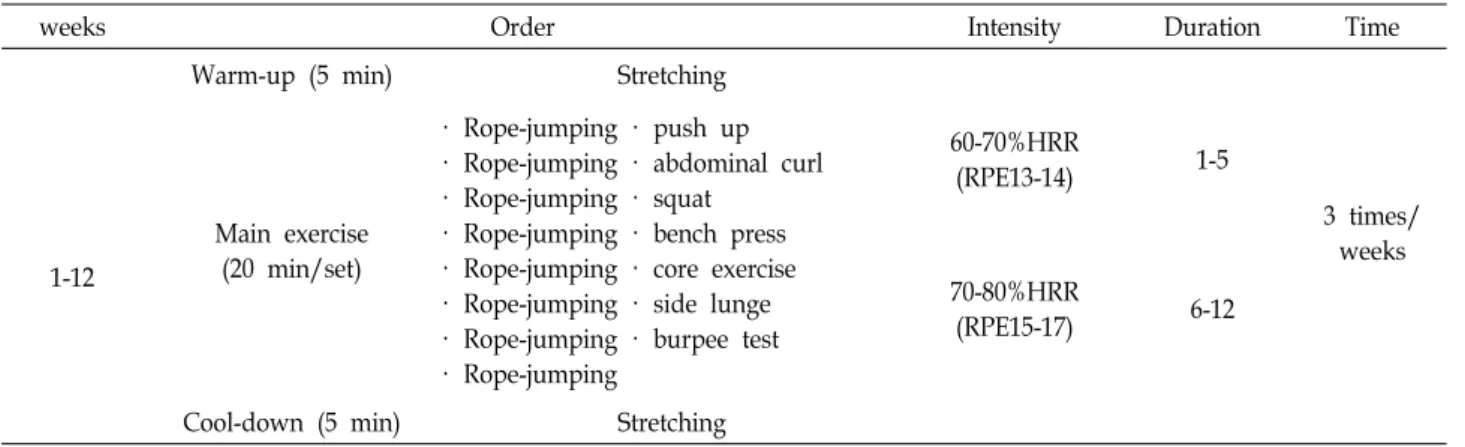
Table 2 Circuit training protocol
weeks Order Intensity Duration Time
1-12
Warm-up (5 min) Stretching
Main exercise (20 min/set)
· Rope-jumping · push up
· Rope-jumping · abdominal curl
· Rope-jumping · squat
· Rope-jumping · bench press
· Rope-jumping · core exercise
· Rope-jumping · side lunge
· Rope-jumping · burpee test
· Rope-jumping
60-70%HRR
(RPE13-14) 1-5
3 times/
weeks 70-80%HRR
(RPE15-17) 6-12
Cool-down (5 min) Stretching
joint resistance training and calisthenic workouts. Reducing the resting time between each set and each workout is an important factor in increasing and maintaining a higher heart rate and blood pressure [8].
Circuit training not only enhances muscular strength but also improves the overall cardiorespiratory function [17].
Therefore, this study aims to identify the effects of circuit training on aging-related hormones in middle-aged women, among whom the obesity rate has been recently increasing.
Materials and Methods
Participant characteristics
This study involved 20 obese middle-aged women. The subjects were divided into 2 equal groups: to ensure biologic homogeneity: the circuit training group (EX, n=10) and non-circuit training control group (CON, n=10). All subjects provided written informed consent prior to participation.
and ethical approval was granted by our Institutional Human Research Committee (PNU IRB/2016_45_HR).
Body composition
Body composition was measured prior to the experiment by using Inbody X-scan plus (Jawon medical, Korea).
Circuit Training
The subjects subsequently engaged in preliminary ex-
ercise for one week to adapt to the circuiting training pro- gram application. The circuiting training consisted of 5 min warm-up, 20 min of main exercise, and 5 min cool down, for a total duration of 30 min (Table 2).
Aging related hormone
Serum estrogen concentrations were measured in dupli- cate by radioimmunoassay (Diagnostic Systems Laborato- ries, Webster, TX, USA). GH was measured using the Beckman Access Ultra sensitive human GH assay, a para- magnetic particle, chemiluminescent immuno assay (Beck- man Coulter, Chaska, MN). IGF-I concentrations were meas- ured by means of the Ray−Bio Human IGF-I ELISA KIT (Norc ross GA. USA). A monoclonal antibody specific to the appropriate human sequence of IGF-1 had been pre-coated in a 96-well microtitre plate. IGF-I was separated from bind- ing proteins. DHEA-S was measured in serum using a com- mercial single antibody radioimmunoassay (RIA) for human DHEA-S (Siemen’s Coat-a-Count DHEA-Sulphate RIA kit, Siemens Medical Solutions USA, Inc., Los Angeles, CA).
Data analysis
All data are presented as mean ± SD, and all statistical analyses were completed using SPSS version 23.0 for Windows (SPSS Inc., Chicago, IL, USA). All statistical tests used an alpha level set at p<0.05. Changes from baseline to the end of the intervention were determined using the paired t-test and the independent t-test.
Results
All the participants characteristics and percent body fat were measured before the start and at the end of the 12-week circuiting training. There were no significant differences in
Table 3. Changes of aging related hormone within each group
Group Pre Post T-value
IGF-1
EX (n=10) CON (n=10)
T-value
160.43±26.68 166.20±32.25
182.85±21.53 164.43±40.05
2.885*
0.341 2.596*
GH
EX (n=10) CON (n=10)
T-value
2.60±2.06 2.80±1.85
3.65±3.06 2.34±1.55
2.510*
1.113 1.470
DHEA-S
EX (n=10) CON (n=10)
T-value
130.64±31.23 126.14±19.59
143.93±28.00 125.68±28.21
2.583*
0.058 1.591
Estrogen
EX (n=10) CON (n=10)
T-value
471.06±180.38 420.29±150.08
510.63±142.37 447.61±208.73
1.483 0.710 0.727 Values are M±SD * p<0.05
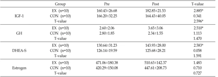
baseline demographic characteristics between the study groups. Tables 3 show that the baseline values between the two groups were comparable for aging related hormone activity. Circuiting training resulted IGF-1 in the circuiting training group had significantly increased (p<0.05) and cir- cuiting training group was higher than that of control group.
GH, DHEA-S in the circuiting training group significantly increased (p<0.05).
Discussion
Synthesis and secretion of GH increase after the prenatal stage of life and decrease with age [22]. Exercise stimulates the natural secretion of GH [13], and IGF-1, which is regu- lated by GH, is known to contribute to GH-mediated growth stimulation [15].
After endurance exercise, the GH level increased sig- nificantly in obese women [6]. Similarly, 12 weeks of circuit training also led to a significant increase in GH levels in middle-aged obese women [2]. From this result, it can be concluded that regular physical activity activates GH secre- tion in subjects with obesity [16].
Increased physical activity improves insulin function and consequently reduces insulin resistance. Reduced insulin levels activate and significantly increase IGF-1 [10], and high-impact exercise leads to greater increases in IGF-1 than low-impact workouts do [28]. In this study, the levels of GH and IGF-1 were higher (p<0.05) in the circuit-training group than in the control group. Appropriate exercise increases GH secretion, which leads to higher IGF-1 concentrations [1].
Circuit training causes growth and hypertrophy of skel-
etal muscles, increasing the use of fat as the energy source.
Hence, circuit training is an effective way of preventing obe- sity in middle-aged obese women.
The aging process in women is closely related to changes in the endocrine system, especially the reduced production of estrogen in the ovaries [14]. Circuit training increases the secretion of epinephrine and norepinephrine hormones, which stimulate the breakdown of fat in muscles and in- crease the blood flow to skeletal muscles. This results in the stimulation of adipose tissue degradation, while also stim- ulating the growth and development of female genital or- gans [26].
Increased central adiposity in middle age women is main- ly due to the reduced estrogen which is also associated with increased visceral fat accumulation [11].
Stimulation of the central nervous system from regular physical activity is reported to stimulate estrogen secretion [3]. However, in this study, estrogen levels did not change significantly after circuit training. There seems to have been no personal variation in the duration and types of exercise conducted by the participants, and thus further study is re- quired to confirm whether this finding can be replicated.
Aging causes changes in the endocrine system, especially in DHEA-S levels, which are highly correlated with chronic degenerative diseases [26]. Thus, DHEA-S is an important index of aging. DHEA-S is produced in the adrenal cortex and is the most common steroid hormone in the blood that decreases significantly with age [21].
DHEA, which is a precursor to female and male hor- mones, is associated with muscle mass in older individuals, and the hormone concentration increases with enhanced
physical fitness [23].
In this study, DHEA increased significantly (p<0.05) after circuit training. The positive change in DHEA concentration resulting from changes in muscle mass due to circuit train- ing implies that DHEA has close correlation with exercise intensity and type.
References
1. Adams, G, R. and McCue, S, A. 1998. Localized infusion of IGF-1 results in skeletal muscle hypertrophy in rats. J.
Appl. Physiol. 84, 1716-0722.
2. Ahn, Y, D. and Park, S. Y. 2013. The effect of circuit exercise on physical fitness, metabolic syndrome factor and change in growth hormone and insulin-like growth factor-1 in obese middle-aged women. Kor. Soc. Sports sci. 22, 1197-1208.
3. Allen, I. S. and Gorski, R. A. 1992. Sexual orientation and the size of the anterior commissure in the human brain. Proc.
Natl. Acad. Sci. USA 89, 7199-7202.
4. Allolio, B. and Arlt, W. 2002. DHEA treatment: myth or real- ity? Trends. Endocrinol. Metab. 13, 288-294.
5. Balcombe, N. R. and Sinclair, A. 2001. Ageing: definitions, mechanisms and the magnitude of the problem. Best Pract.
Res. Clin. Gastroenterol. 15, 835-849.
6. Boisseau, N. and Delamarche, P. 2000. Metabolic and hor- monal responses to exercise in children and adolescents.
Sport. Med. 31, 405-442.
7. Buford, T. W. and Willoughby, D. S. 2008. Impact of DHEA(S) and cortisol on immune function in aging: a brief review. Appl. Physiol. Nutr. Metab. 33, 429-433.
8. Castinheiras-Neto, A. G., Costa-Filho, I. R. and Farinatti, P.
T. 2010. Cardiovascular responses to resistance exercise are affected by workload and intervals between sets. Arq. Bras.
Cardiol. 95, 493-501.
9. Ceda, G. P., Dall'Aglio, E., Maggio, M., Lauretani, F., Bandinell, S., Falz, C., Grimal, W., Ceresini, G., Corradi, F., Ferrucci, L., Valenti, G. and Hoffman, A. R. 2005. Clinical implications of the reduced activity of the GH-IGF-I axis in older men. J. Endocrinol. Invest. 28, 96-100.
10. Cho, W, J. 2015. The effect of judo training on blood lipids, IGF-1, growth hormone, and Adiponectin in middle school girls. Korea Alliance of Martial Arts 17, 1-12.
11. Chu, S. H., Lee, M. K., Kowalski, J. and Sxhwertz, D. 2008.
Effect of estrogen on ovariectomy-induced obesity in rats.
J. Kor. Biol. Nurs Sci. 10, 80-87
12. Consitt, L. A., Copeland, J. L. and Tremblay, M. S. 2001.
Hormone responses to resistance vs. endurance exercise in premenopausal females. Can. J. Appl. Physiol. 26, 574-587.
13. Cuneo, R. C. and Wallace, J. D. 1994. Growth hormone, in- sulin-like growth factors and sport. Endocrinol. Metad. 1, 3-13.
14. Currie, J. L., Harrison, M. B., Trugman, J. M., Bennett, J.
P. and Wooten, G. F. 2004. Postmenopausal estrogen use
affects risk for Parkinson disease. Arch. Neurol. 61, 886-888.
15. Daughaday, W. H. and Rotwein, P. 1989. Insulin like growth factors-1 and Ⅱ: Peptide, messenger ribonucleic acid and gene structures, serum and tissue concentrations. Endoc.
Rev. 10, 68-91.
16. Eliakim, A., Scheett, T. P., Newcomb, R., Mohan, S. and Cooper, D. M. 2006. Fitness, training and the growth hor- mone insulin like growth factor 1 axis in prepubertal girls.
J. clin. Endocrinal. Metab. 86, 2797-2802.
17. Haennel, R., Teo, K. K., Quinney, A. and Kappagoda, T.
1989. Effects of hydraulic circuit training on cardiovascular function. Med. Sci. Sports Exerc. 21, 605-612.
18. Haydar, Z. R., Blackman, M. R., Tobin, J. D., Wright, J. G.
and Fleq, J. L. 2000. The relationship between aerobic ex- ercise capacity and circulating IGF-1 levels in healthy men and women. J. Am. Geriatr. Soc. 48, 139-145.
19. Huang, Y. J., Chen, M. T., Fang, C. L., Lee, W. C., Yang, S. C. and Kuo, C. H. 2006. A possible link between ex- ercise-training adaptation and dehydroepiandrosterone sul- fate- an oldest-old female study. Int. J. Med. Sci. 3,141-147.
20. Judge, J. O., Lindsey, C., Underwood, M. and Winsemius, D. 1993. Balance improvements in older women: effects of exercise training. Phys. Ther. 73, 254-262.
21. Lee, H. R., Jung, D. H., Lim, J. A., Kim, G. C. and Lee, D. C. 2005. The Relationship between Serum DHEA-s Level and TAS in Healthy Adults. J. Kor. Acade. Family Med. 26, 26-31.
22. Malina, R. M., Bouchard, C. and Bar-Or, O. 2004. Growth, Maturation, and Physical Activity. Champaign, IL: Human Kinetics.
23. Park, S. G., Keun, Y. C., Kim, U. H. and Park, J. K. 2009.
Effect of yoga and walking exercise on physical function, carotid artery structure and function in elderly obese women. Kor. J. Physi. Edu. 48, 495-502.
24. Pritzlaff, C. J., Wideman, L., Blumer, J., Jensen, M., Abbott, R. D., Gaesser, G. A., Veldhuis, J. D. and Weltman, A. 1985.
Catecholamine release, growth hormone secretion, and en- ergy expenditure during exercise vs. recovery in men. J.
Appl. Physiol. 89, 937-946.
25. Snyder, P. J., Peachey, H. and Hannoush, P., et al. 1999.
Effect of testosterone treatment on bone mineral density in men over 65 years of age. J. Clin. Endocrinol. Metab. 84, 1966- 1972.
26. Synder, P. J., Peachey, H. and Hannoush, P. 2003. Effects of testosterone treatment on bone mineral density in men over 65. J. Clin. Endoc. Metab. 75, 1092-1098.
27. Szegvari, D. 2008. The applications of chiroptical spectro- scopy for the determination and the detection of steroids and for the examination of their cycle dextrin mediated enantio selective solubility. Acta Pharm. Hung. 78, 45-52.
28. Vega, S. R., Knicker, A., Hollmann, W., Bloch, W. and Struder, H. K. 2010. Effect of resistance exercise on serum levels of growth factors in humans. Horm. Metab. Res. 42, 982-986.
초록:서킷 트레이닝 비만 중년 여성의 노화 관련 호르몬에 미치는 영향
손원목1․곽이섭2․김군도3․하민성1․박송영4․성기동1*
(1부산대학교 체육교육과, 2동의대학교 체육학과, 3부경대학교 미생물학과, 4네브라스카대학교 운동건강학과)
노화 과정에 따라 중년 여성들은 폐경 이후 체력이 저하되기 시작하면서 체지방 증가와 제지방량의 감소로 비 만으로 이어지기 쉽고, 이러한 현상은 체내 호르몬의 분비의 변화와 함께 발생된다. 본 연구는 서킷 트레이닝이 비만 중년 여성의 노화 관련 호르몬에 미치는 영향을 구명하기 위해 실시하였다. 대상자 체지방률 30% 이상인 비만 중년 여성 운동군 10명, 대조군 10명 총 20명을 실시하였다. 운동 시간은 준비운동 5분, 본 운동 20분, 정리운 동 5분으로 총 30분 주 3회 실시하였으며, 운동 강도는 운동자각도를 이용하여 13~17(약간 힘들다-매우 힘들다)수 준으로 실시하였고, 또한 POLAR 심박수 변화량을 측정하여 1~6주는 60~70%HRR (Heart rate reserve), 7~12주는 70~80%HRR로 설정하였다. IGF-1은 운동군이 유의하게 증가하였으며, GH는 운동군이 유의하게 증가하였고, DHEA-S는 운동군이 유의하게 증가하였다. 이상의 결과 서킷 트레이닝이 비만 중년 여성의 노화 관련 호르몬에 긍정적인 영향을 준 것으로 사료된다.