41(3) : 190∼ 195 (2010)
190
건강 추출물의 뇌세포 보호 작용
정길생1#·리 빈#·이동성·최현규·김윤철*
원광대학교 약학대학, 1원광대학교 인수공통감염병연구센터
Neuroprotective Effects of the Extract of Zingiberis Rhizoma
Gil-Saeng Jeong1#, Bin Li#, Dong-Sung Lee, Hyun-Gyu Choi and Youn-Chul Kim* College of Pharmacy, Wonkwang University, Iksan 570-749, Korea
1Zoonosis Research Center, Wonkwang University, Iksan 570-749, Korea
Abstract − Glutamate-induced oxidative injury contributes to neuronal degeneration in many central nervous system (CNS) diseases, such as Parkinson’s disease, Alzheimer’s disease, epilepsy and ischemia. Inducible heme oxygenase (HO)-1 acts against oxidants that are thought to play a role in the pathogenesis of these diseases. NNMBS098, a composition comprising the water insoluble of the 70% EtOH extract of Zingiberis Rhizoma, showed the potent neuroprotective effects on glutamate- induced neurotoxicity by induced the expression of heme oxygenase (HO)-1 and increased HO activity in the mouse hip- pocampal HT22 cells. Furthermore, NNMBS098 caused the nuclear accumulation of nuclear factor E2-related factor 2 (Nrf2) in mouse hippocampal HT22 cells. In addition, we found that treatment with c-Jun N-terminal kinase (JNK) inhibitor (SP600125) reduced NNMBS098-induced HO-1 expression and NNMBS098 also increased JNK phosphorylation. Therefore, these results suggest that NNMBS098 increases cellular resistance to glutamate-induced oxidative injury in mouse hippocampal HT22 cells, presumably through JNK pathway-Nrf2-dependent HO-1 expression.
Key words − Glutamate, HT22, Zingiberis Rhizoma, Heme Oxygenase-1, Neuroprotective effect
건강 (Zingiberis Rhizoma)은 생강과 (Zingiberaceae)에 속 하는 다년생 초본인 생강 (Zingiberis officinale Rosc.)의 뿌 리줄기를 건조한 것으로 한국을 비롯한 인도, 중국 등 아시 아 지역에 분포되어 있다. 성분으로는 zingiberol, zingiberon, zingiberene 등의 정유성분과 신미성분인 gingerol, shogaol 등을 함유하고 있으며 성분의 생리활성연구로는 gingerol 의 항산화효과, 6-gingerol과 6-shogaol의 자발성 운동활성의 저 해, 해열진통효과 등 다양한 활성이 연구 되었다.1,2)이 밖 에 한방에서는 온중축한 (溫中逐寒), 회양통맥 (回陽痛脈), 치심복냉통 (治心腹冷痛)의 효능이 있어 토사 (吐瀉), 지냉 맥미 (肢冷脈微), 한음천해 (寒飮喘咳), 풍한습비 (風寒濕痺), 양허토혈 (陽虛吐血), 하혈 (下血)에 사용된다.3)
호기성 호흡을 하는 생물체들은 끊임없이 산소 분자 (O2) 를 세포 내로 받아들여 미토콘드리아 내의 산화환원 효소 계 또는 외부 항원에 노출된 면역세포에 의해, 그리고 외부 적으로는 방사선 또는 여러 화합물 등에 의해 활성 산소종
(reactive oxygen species, ROS)을 생성한다. 체내 ROS가 많 이 생성되거나 항산화 시스템의 기능이 저하되면서 체내의 산화계와 항산화계의 불균형으로 인해 산화적 스트레스 (oxidative stress)가 일어나게 된다.4) 산화적 스트레스는 알 츠하이머 증후군, 파킨슨 증후군, 헌팅턴 증후군과 같은 중 추 신경계의 퇴행성 뇌질환을 유발시키는 주된 원인중의 하 나로 잘 알려져 있다.5,6)
산화적 스트레스에 의한 뇌 손상의 기전으로 신경흥분 독 성 (Excitotoxicity)이 주요한 기전 중 하나로 알려져 있는데 이는 과다 분비된 glutamate 등 흥분성 아미노산에 의한 수 용체들의 과도한 자극으로 인한 신경세포 세포사멸(neuronal cell death)이 일어나게 한다.7,8) 본 실험에 사용한 생쥐의 해 마유래 세포주인 HT22세포주는 글루타메이트 수용체가 없 는 세포주로서 글루타메이트와 함께 처리할 때 신경흥분독 성이 아닌 산화적 스트레스로 인하여 세포가 손상을 받으 며, 산화적 스트레스로 인한 퇴행성 뇌질환 연구에 있어서 유용한 실험모델중의 하나이다. 이러한 산화적 스트레스로 부터 뇌 보호 물질을 찾아 그 기전을 밝히려는 많은 연구가 진행 되고 있다.
#
These authors contributed equally to this work.
*교신저자(E-mail):[email protected]
(Tel):+82-63-850-6823
Heme oxygenase-1 (HO-1) 은 Heme oxygenase 유도체중 의 하나로 세포내의 Heme을 분해하여 부산물인 일산화탄 소, 철, 빌리버딘을 만들며, 그 부산물과 함께 세포사멸 억 제, 항염즘 및 항산화 작용을 갖는 것으로 알려져 다양한 질 병의 타겟으로 큰 주목을 받고 있다. 특히 뇌 세포 보호 기 전에 있어 HO-1은 그 부산물과 함께 ROS 제거 및 세포 보 호, 항산화 등의 역할을 통하여 산화적 스트레스로 인한 뇌 세포 손상을 억제하는 것으로 현재 많은 연구가 진행되고
있다.9,10) 따라서, 본 연구는 건강 에탄올 추출물의 비수용성
분획물이 Heme oxygenase-1의 발현을 통한 뇌 세포 보호 효과와 그 세포 보호 기전을 탐색하였고, 그 결과를 보고하 고자 한다.
재료 및 방법
실험재료 − 본 실험에 사용한 건강은 2009년 4월 익산시 소재 대학한약국에서 구입하였으며, 건강 추출물과 각 분획 물 (NNMBS097, NNMBS098 NNMBS099)은 천연물신약 표준화소재 은행에서 분양하여 사용하였다.
시약 및 기기 −DMEM 배지와 trypsin-ethylenediaminetetraacetic acid (EDTA)는 Gibco Laboratories사에서 구입하였으며, fetal bovine serum (FBS)는 Hyclone Laboratories사에서 구 입하였다. L-glutamate, Trolox와 3'-(4,5-dimethylthiazol-2- yl)-2,5-diphenyltetrazolium bromide (MTT)는 Sigma사에서 구입하였다. 96-Well tissue culture plates와 기타 tissue culture dishes는 Nunc사 제품을 이용하였다. 흡광도는 BioRad사의 Microplate Reader를 이용하여 측정하였다.
건강 추출물과 분획물의 제조 − 건강 50 g을 70% 에탄 올 수용액 50 ml 로 70oC에서 2시간 동안 가열추출하고 여 액을 감압 농축하여 건강 추출물 (NNMBS097) 6.05 g을 얻 었다. 수득한 건강 추출물 6.05 g에 정제수 100 ml을 넣고 교반하여 잘 녹인 뒤 18시간 동안 냉침후 원심분리기에서 3000 rpm으로 30분 동안 원심분리하여 상등액을 취함으로 서 건강 추출물의 비수용성 분획 (NNMBS098)과 수용성 분획 (NNMBS099)을 제조하였다. 상기 건강 추출물들을 실 험에 사용한 배지에 녹여서 사용하였다.
HT22 세포배양 및 뇌 세포 보호활성 측정 − 생쥐 해마 유래 HT22 세포주는 Dr. Inhee-Mook (Seoul National University, Seoul, Korea)로부터 분양하여 사용하였으며, 글 루타메이트로 독성을 유발한 세포주에 대한 보호활성 측정 은 정 등의 방법11) 에 따라 실시하였다. 즉, HT22 세포 (2×
105 cells/well)를 10% heat-inactivated FBS, penicillin G (100 IU/ml)와 streptomycin (100µg/ml)을 함유한 DMEM 배지에 분주하고 5% CO2 배양기 내에서 37oC에서 24시간 배양한 다음, 각각의 시료 용액 (5, 10, 20, 40 µg/ml)과 5 mM Glutamate를 처리한 후 12시간 동안 5% CO2 배양기
내에서 배양하였으며, 세포생존율은 MTT법을 활용하여 측 정하였으며, 양성대조약물로는 Trolox 50 µM를 사용하였다.
또한, 모든 실험치는 대조군에 대한 세포보호율을 mean±S.D.
로 표시하였으며, 각각 3회 반복 실험치를 이용하여 계산하 였다.
ROS 측정 − 배양된 세포를 PBS로 세척한 후, 10 µM 2',7'-dichlofluorescein diacetate (DCFDA, 35845) 를 포함하 는 Hank’s balanced salt 용액에 30분 동안 암실에서 반응한 후 세포의 형광광도 (SpectramaxGemini XS, Molecular Devices, Sunnyvale, CA, U.S.A.)를 측정했다. (excitation wavelength: 490 nm; emission wavelength : 525 nm)
Western Blot Analysis − HT22 세포를 60 mm dish에 3×10⁵ cells/well 농도로 24시간 배양한후 각각의 시료를 농 도별로 처리하였다. HT22 세포에 RIPA buffer를 첨가한 다 음, 4oC, 14,000×g 에서 25분간 원심분리하고 상등액을 튜 브에 옮겼다. 단백질 정량은 BSA 단백질 실험 키트를 이용 하였고 각각의 시료를 12% SDS-polyacrylamide gel에서 영 동하고 Nitrocellulose membrane (NC membrane)으로 전사 하였다. 전사된 NC membrane을 5% 무지방유가 포함된 신 선한 blocking buffer (0.1% Tween 20 in Tris-buggered saline) 에서 blocking한후 HO-1 antibody를 1:1000으로 희 석하여 넣고 1시간 동안 반응시켰다. 다시 2차 안티바디 (Anti-mouse IgG)를 1:1000으로 희석하여 넣고 1시간 동안 반응한 다음, ECL 용액을 1:1로 잘 섞어서 NC membrane 위에 부어서 발광시키고 암실에서 X선 필름에 감광한후 현 상하였다. 같은 방법으로 Actin antibody를 이용하여 Actin 을 측정한다.
Heme Oxygenase Activity − HO 효소 활성을 Tenhunen 등의 방법12)에 의해 다음과 같이 측정하였다. 세포로부터 얻 어진 microsome에 nicotinamide adenine dinucleotide pho- sphate (NADPH)와 랫드 간 cytosol에서 얻어진 biliverdin reductase를 포함하는 반응용액인 100 mM PBS, 2 mM MgCl2, 3 mg의 랫드 간 cytosol, 0.8 mM NADPH, 2 mM glucose-6-phosphate, 0.2 U의 glucose-6-phosphate dehydro- genase 등을 첨가하고 기질로서 hemin을 20 µM 처리한후 37oC에서 1시간 동안 암실에서 반응한뒤 1 ml의 chloroform 으로 반응을 종결하고 450 nm에서 흡광도를 측정하였다.
핵과 세포질 분획의 분리 − HT22 세포에 protease inhibitor cocktail I과 1 mM phenylmethylsulfonyl fluoride (PMSF)를 첨가한 PER-Mammalian Protein Extraction buffer를 첨가하 여 균질화하고 4oC에서 15,000×g로 원심분리 하였다. 10분 간 원심분리후 상등액을 세포질 분획으로 사용하기 위하여 -80oC에서 저장하였다. 나머지 침사물은 PBS로 세척한후 RIPA buffer [150 mM NaCl, 1% NP-40, 0.5% sodium deoxycholate, 0.1% SDS, 50 mM Tris-HCl (pH 7.4), 50 mM glycerophosphate, 20 mM NaF, 20 mM ethylene glycol
tetraacetic acid (EGTA), 1 mM dithiothreitol (DTT), 1 mM Na3VO4, protease inhibitors]를 첨가하고 4oC에서 15분간 혼 합한후 4oC, 16,000×g에서 15분간 원심분리 한다. 이후의 과정은 상술된 western blotting 방법을 이용하였다.
결과 및 고찰
산화적 손상은 허혈성 재관류 손상, 퇴행성 뇌질환 등을 포함하는 여러 질병과 노화와 깊은 연관이 있으며, 그 중 글 루타메이트에 의한 산화적 스트레스가 뇌 세포 손상을 일 으키는 기전은 퇴행성 뇌 질환의 주요한 요인으로 보고되 고 있다.13,14) 본 연구에서는 건강 추출물과 건강 추출물의 수용성 분획물 및 비수용성 분획물의 뇌세포 보호효과와 항 산화 효과를 측정하고자 생쥐의 해마 유래 HT22 세포에서 글루타메이트 세포독성으로부터의 뇌 세포 보호 효과를 검 색하였다.
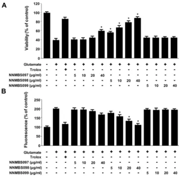
각 시료를 농도별로 12 시간 처리한 결과 건강 추출물의 비수용성 분획물 (NNMBS098)이 세포 독성이 나타나지 않 은 농도 (5-40 µg/ml)에서 글루타메이트로 인한 독성으로부 터 유의한 뇌 세포보호 활성을 보였으며, 동시에 동일한 농 도에서 강력한 ROS 소거 활성을 나타냈다. 뇌 세포 보호 효과물질로 알려진 trolox 50 µM을 양성 대조약물로 사용 하였다 (Fig. 1). 한편, 뇌 세포 보호 기전에 있어 중요한 단 백질인 Heme oxygenase (HO)-1의 발현 여부에 관하여 실 험한 결과 건강의 추출물 (NNMBS097)이 40 µg/ml 농도에서
Fig. 1. Effects of NNMBS097, NNMBS098 and NNMBS099 on glutamate-induced oxidative neurotoxicity and inhibition of reactive oxygen species generation. (A) Cells were treated with samples and then incubated for 12 h with glutamate (5 mM).
(B) Exposure of HT22 cells to 5 mM glutamate for 12 h increased reactive oxygen species production. NNMBS098 effectively inhibited glutamate-mediated reactive oxygen species production. Each bar represents the mean±S.D. of three independent experiments, *P<0.05. Trolox (50µM) was used as the positive control.
Fig. 2. Effects of NNMBS097, NNMBS098 and NNMBS099 on HO-1 expression and HO activity in HT22 cells. Cells were incubated for 12 h with NNMBS097 (A), NNMBS098 (B) and NNMBS099 (C). Expression of HO-1 was determined by western blot analysis, and representative blots of three independent experiments are shown. HO activity was determined via bilirubin formation at 12 h after treatment with various concentrations of NNMBS097 (D), NNMBS098 (E) and NNMBS099 (F). Each bar represents the mean±S.D. of three independent experiments. *P<0.05 vs. control. CoPP (20µM) was used as the positive control.
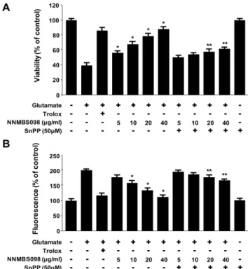
약간의 HO-1 발현이 관찰되고 수용성 분획물 (NNMBS099) 은 아무런 작용이 없는 반면, 비수용성 분획물인 NNMBS098 은 HO-1단백질을 농도 의존적으로 발현시키는 것을 확인 하였다. 또한 HO activity 역시 NNMBS098에 의해 증가하 는 것을 확인하였다 (Fig. 2). 40 µg/ml 농도에서 NNMBS098 에 의한 HO-1 단백질의 발현을 시간별로 측정한 결과 6시 간 에서부터 발현이 시작되어 18시간에서 제일 많이 발현 되었으며 HO activity 역시 18 시간에서 가장 높은 활성을 나타냈다 (Fig. 3). NNMBS098에 의해 증가 했던 세포 보 호활성과 ROS 소거 활성이 HO-1의 억제제인 SnPP로 인해 감소 되는 것을 통하여 NNMBS098은 HO-1을 발현 함으 로서 뇌세포 보호활성과 ROS 소거 활성을 나타냈다 (Fig.
4).
현재 많은 연구에 의하면 HO-1 단백질의 발현에 대한 메 커니즘 중 nuclear factor-E2-related factor 2 (Nrf2) 의 핵내 전사가 가장 직접적인 관련이 있는것으로 알려져 있다. Nrf2 는 전사인자로서 HO-1과 같은 항산화 단백질의 유전자에 존재하는 ARE(Antioxidant Response Element)에 결합하여 이들 유전자의 발현을 조절하고 단백질발현을 항진시킴으 로서 산화스트레스에 대한 생체방어기구에 중심적 역할을
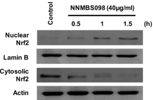
담당하고 있다.15,16) NNMBS098을 40 µg/ml 처리하여 시간 별로 측정한 결과 Nrf2가 핵내로 전사 되면서 nuclear의 Nrf2 가 증가하는 반면cytosol의 Nrf2가 현저히 줄어드는 양상을 보였다 (Fig. 5). 기존의 연구결과와 마찬가지로 NNMBS098 는 Nrf2의 핵내전사를 통하여 HO-1 단백질의 발현을 조절 하는 것을 실험을 통하여 확인하였다.
MAPK전달계는 세포 외부로부터의 자극을 전달하여 세 포내 반응을 일으키는데 있어서 아주 중요한 역할을 하고 있으며 extracellular signal-regulated kinase (ERK), p38 MAPK와 c-Jun N-terminal kinase (JNK) 등 3가지 계열의 MAPK family가 있다.17,18) 여러 연구들에 의하면mitogen- activated protein kinase (MAPK) 경로 역시 HO-1의 발현에 직접적으로 관여하는것으로 밝혀졌다.19,20) NNMBS098을 40µg/ml 농도로 처리한 결과 ERK와 p38 경로는 변화가 없 는 반면, JNK는 30분에서부터 인산화과정이 이루어 지는 것을 확인하였으며 NNMBS098 40 µg/ml과 JNK 억제제인 SP600125를 함께 처리했을 때 HO-1의 발현이 억제되고 Fig. 3. Effects of NNMBS098 on HO-1 expression and HO
activity in HT22 cells. (A) Cells were incubated for indicated periods with 40µg/ml of NNMBS098. Expression of HO-1 was determined by western blot analysis, and representative blots of three independent experiments are shown. (B) HT22 cells were treated with 40µg/ml of NNMBS098, and HO activity was measured at the indicated time points. Each bar represents the mean±S.D. of three independent experiments.
*P<0.05 vs. control.
Fig. 4. Effects of NNMBS098-induced HO-1 on glutamate- induced oxidative neurotoxicity and reactive oxygen species generation. (A) Cells were treated with 40µg/ml of NNMBS098 in the presence or absence of 50µM SnPP and then exposed to glutamate (5 mM) for 12 h. (B) Exposure of HT22 cells to 5 mM glutamate for 12 h increased reactive oxygen species production. NNMBS098 induced HO-1 effectively inhibited reactive oxygen species production. Each bar represents the mean±S.D. of three independent experiments,
*P<0.05 vs. control. **p<0.05 vs. group treated with NNMBS098.
NNMBS098의 세포보호 효과 역시 감소하는 것을 확인하였 다. 따라서 본연구를 통하여 건강 추출물의 비수용성 분획 물인 NNMBS098은 HT22 세포에서 Nrf2 핵내전사와 JNK 인산화를 통하여 HO-1 단백질을 발현함으로써 글루타메이 트에 의한 독성으로부터 뇌 세포보호활성과 ROS소거 활성 이 있음을 확인하였으며, 향후 NNMBS098의 뇌세포 보호 기전과 지표물질의 분리 및 성분분석에 관한 더 많은 연구 가 진행되어야 할 것으로 판단된다.
결 론
본 연구는 건강 추출물의 뇌 세포 보호활성과 그 메커니 즘을 탐색하였으며 그 결과 비수용성 분획물인 NNMBS098 이 글루타메이트로 유발한 HT22 세포주에 대해 훌륭한 보 호 활성을 나타냈으며, Nrf2 핵내전사와 JNK 인산화를 통 하여 항산화 및 세포보호 효소인 HO-1 단백질을 발현함으
Fig. 6. Effects of NNMBS098-induced MAPK activation on HO-1 expression and glutamate-induced neurotoxicity in HT22 cells.
(A) Cells were treated with 40µg/ml of NNMBS098 for the indicated times. Cell extracts were analyzed by western blot with antibodies specific for phosphorylated ERK1/2 (p-ERK), phosphorylated JNK (p-JNK), or phosphorylated p38 (p-p38). Membranes were stripped and re-probed for total form of each MAPK antibody as a control, and the representative blots of three independent experiments are shown. (B) Cells were pretreated for 1 h with the specific inhibitor PD98059 (40µM), SP600125 (25 µM) and SB203580 (20µM), and then treated with NNMBS098 (40 µg/ml) for 12 h. Western blot analyses for HO-1 expression were performed as described in materials and methods and representative blots of three independent experiments are shown. (C) Cells untreated or treated with NNMBS098 (40µg/ml) in the presence or absence of each specific inhibitor for 12 h were exposed to 5 mM glutamate for 8 h. Each bar represents the mean±S.D. of three independent experiments. *P<0.05 compared to the group treated with glutamate. Trolox (50µM) was used as positive control.
Fig. 5. Effects of NNMBS098 on Nrf-2 nuclear translocation in HT22 cells. Cells were treated with 40µg/ml of NNMBS098 for 0.5, 1, and 1.5 h. The nuclei were fractionated from the cytosol using PER-Mammalian Protein Extraction buffer as described in materials and methods. Nrf-2 protein was detected by western blot analysis, and representative blots of three independent experiments are shown.
로써 뇌 세포보호활성과 ROS소거 활성을 나타내는 메커니 즘을 규명하였다.
사 사
본 연구는 한국연구재단 지원 연구소재지원사업 연구비 (20100000675)에 의해 이루어 졌으며 이에 감사를 드립니다.
인용문헌
1.생약학교재편찬위원회(2007) 생약학, 218-220. 동명사, 서울.
2. Suekawa, M., Ishige, A., Sudo, K., Aburada, M. and Hosoya, E. (1984) Pharmacological studies on ginger. I. Pharmaco- logical actions of pungent constitutents, (6)-gingerol and (6)- shogaol. J. Pharmacobiodyn. 7: 836-848.
3.전국한의과대학 본초학 공동교재편찬위원회(2004) 본초학, 378-380. 영림사, 서울.
4. Coyle, J. T. and Puttfarcken, P. (1993) Oxidative stress, glutamate and neurodegenerative disorders. Science. 262:
689-695.
5. Satoh, T., Enokido, Y., Kubo, K., Yamada, M. and Hatanaka, H. (1999) Oxygen toxicity induces apoptosis in neuronal cells. Cell Mol. Neurobiol. 18: 649-666.
6. Satoh, T. and Lipton, S. A. (2007) Redox regulation of neu- ronal survival mediated by electrophilic compounds. Trends.
Neurosci. 30: 37-45.
7. Choi, D. W. (1988) Glutamate neurotoxicity and diseases of the nervous system. Neuron. 1: 623-634.
8. Siesjö, B. K. (1981) Cell damage in the brain: a speculative synthesis. J. Cereb. Blood Flow Metab. 1: 155-185.
9. Lee, M. S., Lee, J., Kwon, D. Y. and Kim, M. S. (2006) Ondamtanggamibang protects neurons from oxidative stress with induction of heme oxygenase-1. J. Ethnopharmacol.
108: 294-8.
10. Choi, B. M., Kim, H. J., Oh, G. S., Pae, H. O., Oh, H. C., Jeong, S. J., Kwon, T. O., Kim, Y. M. and Chung, H. T.
(2002) 1,2,3,4,6-Penta-O-galloyl-beta-D-glucose protects rat neuronal cells (Neuro 2A) from hydrogen peroxide-mediated cell death via the induction of heme oxygenase-1. Neurosci.
Lett. 328: 185-189.
11. Jeong, G. S., Li, B., Lee, D. S., Byun, E., Kang, D. K., Lee, H. S. and Kim, Y.C. (2007) Cytoprotective constituents of
Alipinia katsumadai seeds against glutamate-induced oxi- dative injury in HT22 cells. Nat. Prod. Sci. 13: 268-272.
12. Tenhunen, R., Marver, H. S. and Schmid, R. (1970) The enzymatic catabolism of hemoglobin: stimulation of microso- mal heme oxygenase by hemin. J. Lab. Clin. Med. 75: 410- 421.
13. Tan, S., Schubert, D. and Maher, P. (2001) Oxytosis: a novel form of programmed cell death. Curr. Top. Med. Chem. 1:
497-506.
14. Rössler, O. G., Bauer, I., Chung, H. Y. and Thiel, G. (2004) Glutamate-induced cell death of immortalized murine hip- pocampal neurons: neuroprotective activity of heme oxy- genase-1, heat shock protein 70, and sodium selenite.
Neurosci Lett.. 362: 253-257.
15. Balogun, E., Hoque, M., Gong, P., Killeen, E., Green, C. J., Foresti, R., Alam, J. and Motterlini, R. (2003) Curcumin acti- vates the heme oxygenase-1 gene via regulation of Nrf2 and the antioxidant-responsive element. Biochem. J. 371: 887- 895.
16. Itoh, K., Chiba, T., Takahashi, S., Ishii, T., Igarashi, K., Katoh, Y., Oyake, T., Hayashi, N., Satoh, K., Hatayama, I., Yama- moto, M. and Nabeshima, Y. (1997) An Nrf2/small Maf het- erodimer mediates the induction of phase II detoxifying enzyme genes through antioxidant response elements. Bio- chem. Biophys. Res. Commun. 236: 313-322.
17. Choi, B. H., Hur, E. M., Lee, J. H., Jun, D. J. and Kim, K. T.
(2005) Protein kinase C delta-mediated proteasomal degra- dation of MAP kinase phosphatase-1 contributes to glutamate- induced neuronal cell death. J. Cell Sci. 119: 1329-1340.
18. Oh, H. L., Seok, J. Y., Kwon, C. H., Kang, S. K. and Kim, Y.
K. (2006) Role of MAPK in ceramide-induced cell death in primary cultured astrocytes from mouse embryonic brain.
Neurotoxicology, 27: 31-38.
19 Elbirt, K. K., Whitmarsh, A. J., Davis, R. J. and Bonkovsky, H. L. (1998) Mechanism of sodium arsenite-mediated induc- tion of heme oxygenase-1 in hepatoma cells. Role of mito- gen-activated protein kinases. J. Biol. Chem. 273: 8922-8931.
20. Kietzmann, T., Samoylenko, A. and Immenschuh, S. (2003) Transcriptional regulation of heme oxygenase-1 gene expres- sion by MAP kinases of the JNK and p38 pathways in pri- mary cultures of rat hepatocytes. J. Biol. Chem. 278: 17927- 17936.
(2010. 7. 23 접수; 2010. 8. 16 심사; 2010. 8. 18 게재확정)