OriginalArticle
DOI: http://dx.doi.org/10.3831/KPI.2012.15.015
Received: Oct 19, 2012 Accepted:Nov 09, 2012
This is an Open-Access article distributed under the terms of the Creative Commons Attribution Non-Commercial License (http://creativecommons.org/licenses/by-nc/3.0/) which permits unrestricted noncommercial use, distribution, and reproduction in any medium, provided the original work is properly cited.
This paper meets the requirements of KS X ISO 9706, ISO 9706-1994 and ANSI/NISO Z39.48-1992 (Permanence of Paper).
*Corresponding Author
Cheol-Hong Kim. Department of Acupuncture & Moxibustion, Dong-Eui University College of Oriental Medicine and Research Institute of Oriental Medicine, San 45-1, Yangjung 2-dong, Busanjin-gu, Busan, 614-710, Korea.
Tel: +82-51-850-8613 Fax: +82-51-867-5162 E-mail: kmdkch@deu.ac.kr
ⓒ 2012 Korean Pharmacopuncture Institute http://www.journal.ac
Rehmannia Glutinosa Pharmacopuncture Solution Regulates Functional Activation, Fc ε RI Expression, and Signaling Events in Mast Cells
Kyung-Hwa Kang
1, Kyung-Hee Lee
2, Hyun-Min Yoon
2, Kyung-Jeon Jang
2, Chun- Ho Song
2, Cheol-Hong Kim
2*
1Department of Oriental Physiology, Dong-Eui University College of Oriental Medicine and Research Institute of Oriental Medicine, Busan, Korea
2Department of Acupuncture & Moxibustion, Dong-Eui University College of Oriental Medicine and Research Institute of Oriental Medicine, Busan, Korea
atory cytokines (IL-1β, IL-6, and GM-CSF) in stimu- lated RBL-2H3 cells significantly (p < 0.05). RGPS also suppressed mRNA expression of inflammatory enzymes (HDC2, COX-1, COX-2, 5LO). In addition, mRNA expression levels of FcεRIα, FcεRIβand FcεRIγ were lowered by treatment with RGPS. Finally, RGPS prevented phosphrylation of Lyn, Syk, LAT, Gab2, PLC γ1/2, PI3K, Akt, cPLA2 and IκBα.
Conclusions: RGPS effectively suppresses mast cell activations such as degranulation and inflammatory response via down-regulation of the FcεRI-mediated signaling pathways in IgE/Ag-stimulated mast cells.
1. Introduction
Mast cells are key effector cells in IgE-associated im- mune responses, including allergic disorders such as anaphylaxis, atopic dermatitis, allergic rhinitis, and allergic asthma. Mast cell activation through FcεRI and preformed and newly synthesized mediator release results in allergic inflammatory conditions: erythema, edema, and itching in the skin; sneezing, rhinorrhea, cough, bronchospasm, and mucous secretion in the respiratory tract; and nausea, vomiting, diarrhea, and cramping in the gastrointestinal tract [1].
Abstract
Objectives: Rehmannia glutinosa pharmacopuncture solution (RGPS) was investigated to determine both its anti-allergic inflammatory effects on mast cells and its detailed mechanism of actions.
Methods: We investigated whether RGPS suppress cytokines, enzymes, FcεRI expression and FcεRI- mediated signaling in RBL-2H3 cells stimulated with anti-DNP IgE/DNP-HSA. The suppressive effects of RGPS on the levels of cytokines such as IL-1β, IL-6 and GM-CSF were measured using emzyme-linked immunospecific assay (ELISA). The mRNA expression levels of cytokines, enzymes (HDC2, COX-1, COX-2 and 5LO) and FcεRI αβγsubunits were measured using reverse transcription polymerase chain reaction (RT- PCR) method. The activation of FcεRI-mediated signaling was examined using Western blot analyses.
Results: RGPS suppressed production of proinflamm- Key Words
Rehmannia glutinosa pharmacopuncture solution (RGPS); cytokine, enzyme; FcεRI expression; FcεRI- mediated signaling
Interleukin (IL)-1 is a major proinflammatory cytokine that induces acute and chronic inflammation through activation of the innate and the acquired immune systems.
IL-1 can enhance cytokine secretion [2-5] and histamine [6] in the development of IgE/Ag-mediated allergic diseases such as allergic asthma. IL-6, a multifunctional cytokine, is produced by a variety of hematopoietic and non-hematopoietic cell types in response to diverse stimuli [7]. Recent studies have shown that IL-6 plays a role in the development and the exacerbation of Th2-mediated diseases such as allergic airway inflammation and asthma [8]. Granulocyte-macrophage colony-stimulating factor (GM-CSF) has been implicated as an important mediator in the pathogenesis of asthma [9]. In particular GM-CSF is pivotal in eosinophil maturation and survival [10], a key effector cell in asthma.
Inflammatory mediators such as histamine, leukotrienes and prostaglandins are synthesized by enzymes such as L- histidine decarboxylase (HDC), cyclooxygenase (COX)-1, COX-2 and 5-lipoxygenase (LO) in mast cells activated with IgE/antigen (Ag). As HDC is the sole enzyme that produces histamine by decarboxylation of L-histidine in mammals, HDC is an important regulator of histamine signaling [11]. COX contributes to the synthesis of prostaglandins, such as prostaglandin D2 (PGD2) and prostaglandin E2 (PGE2), which are inflammatory lipid mediators from arachidonic acid. COX-1 is associated with the immediate PGE2 and PGD2 generation and COX-2 is associated with the delayed PGD2 generation [12-14] 5-LO synthesizes leukotriene, another class of inflammatory lipid mediators from arachidonic acid [15].
Mast cells express a high-affinity receptor for IgE (FcεRI) that binds to Ag-specific IgE on the cell surface. FcεRI consists of an IgE binding αchain, a signal-amplifying, receptor-stabilizing β‚ chain, and two disulfide-bonded γ chains that are the main signal transducers [16]. IgE/Ag- induced aggregation of FcεRI initiates a cascade event, leading to allergic inflammation via intracellular signal transduction. The activation of signaling cascades depe- nds initially on the interaction of FcεRI with the Src kinase Lyn and Fyn and subsequently on the activation of spleen tyrosine kinase (Syk) and other tyrosine kinases [17].
Phosphorylated Syk leads to downstream signaling with phosphorylation of linker for activation of T cells (LAT).
LAT functions as a signal platform providing binding sites for phospholipase Cγ(PLCγ) and other adaptor proteins [18]. PLCγin this complex hydrolyzes phosphatidylinositol 4,5-bisphosphate (PIP2) to produce diacylglycerol (DAG), activating protein kinase C (PKC) and inositol 1,4,5- triphosphate (IP3), increasing intracellular Ca2+ concen- trations [19]. Fyn and Syk phosphorylate adapters like Grb- associated binder 2 (Gab2), which are essential for
phosphatidylinositol-3 kinase (PI3K) /Akt activation [20].
All these events involved in Ca2+mobilization, PKC activation and PI3K/Akt activation are crucial for mast cell degranulation, phosphorylation of mitogen-activated protein kinase (MAPK), and activations of nuclear factor for T-cell activation (NFAT) and nuclear factor κB (NFκB), both of which lead to cytokine synthesis [17].
The root of Rehmannia glutinosa is mainly used for Yin deficiency syndrome in traditional medicine in the East Asian region. It has been frequently used to reduce inflammation and is included in various formulas [21-25].
It inhibits inflammation in the development of atopic dermatitis [25] and in the diabetic foot ulcer rat model [23].
The objective of the present study was to examine the proinflammatory cytokines and enzymes responsible for the anti-allergic inflammatory activity of RGPS. In addition, the underlying anti-allergic inflammatory mechanism of RGPS was investigated.
2. Materials and methods
2.1. Preparation of RGPS
RGPS was purified from the roots of Rehmannia glut- inosa. The preparation of ethanol extracts of Rehmannia glutinosa took place at Dong-Eui University Oriental Medicine Hospital. The roots of Rehmannia glutinosa (800 g) were extracted with 25% ethanol (4 L) for 10 h at room temperature. The extracted solution (RGPS) was filtrated and concentrated to a 400-ml ethanol extract. RGPS was diluted in Dulbeco's Modified Eagle's Medium (DMEM) media containing ethanol, and the final concentration of ethanol was adjusted to 1% (v/v) in the cell culture system.
The control cells were treated with media containing 1%
ethanol.
2.2. Cells
Rat basophilic leukemia RBL-2H3 cells were obtained from the Korea Cell Line Bank (Seoul, Korea). The cell line was cultured in DMEM supplemented with 10% heat- inactivated fetal bovine serum (FBS) and 100 U/ml of penicillin and streptomycin in an atmosphere of 5% CO2at 37°C. Cells were detached with trypsin-EDTA solution.
After the cells had been washed, they were resuspended in fresh medium and used for subsequent experiments.
2.3. Materials
Chemicals and cell culture materials were obtained from the following sources: anti-dinitrophenyl (DNP) IgE, DNP- human serum albumin (HSA), p-nitrophenyl-N-acetyl-β- D-glucosaminide, Igepal CA-630, dimethylsulfoxide (DMSO), sodium deoxycholate, NaCl, Tris-HCl, sodium
pyrophosphate, Na3VO4, NaF, leupeptin and phenylme- thylsulfonyl fluoride from Sigma-Aldrich Co. (St. Louis, MO, USA); fetal bovine serum (FBS), penicillin, streptom- ycin, DMEM, and trypsin-EDTA solution from Gibco BRL (Grand Island, NY, USA); ELISA kits were obtained from BD Biosciences Pharmingen (BD OptEIATM, San Diego, CA, USA); reverse transcription polymerase chain reaction (RT-PCR) kits from iNtRON Biotechnology (one-step RT- PCR PreMix, Kyunggido, Korea); primary antibodies against phospho-Lck/yes-related novel tyrosine kinase (p- Lyn), Lyn, phospho-LAT (p-LAT), LAT, phospho-Gab2 (Gab2), phospho-PLCγ(p-PLCγ)1/2, phospho-PI3K (p- PI3K), PI3K, phospho-AKT (p-AKT), AKT, phospho- cytosolic phospholipase2 (p-cPLA2), cPLA2, phospho- inhibitory-kappa B alpha (p-IκBα), IκBαand β-actin from Cell Signaling Technology (Beuerly, MA, USA); phospho- Syk (p-Syk), Syk and Gab2 from Santa Cruz Biotechnology (Santa Cruz, CA, USA); horseradish peroxidase (HRP)- conjugated secondary antibody from Kirkegaard & Perry Laboratories, Inc. (KPL, Gaithersburg, MD, USA); the ECL chemiluminescence system from Amersham; and the nitrocellulose transfer membrane from Whatman GmbH.
TRIzol was from Invitrogen (Carlsbad, CA, USA);
polymerase chain reaction (PCR) oligonucleotide primers were custom-synthesized by Bioneer Co. (Korea). All other reagents were of the highest grade commercially available.
2.4. Cytokine assays
RBL-2H3 cells (5×10⁵cells/ml) were sensitized with anti- DNP IgE (0.5 μg/ml) overnight. The cells were washed twice with Siraganian buffer I; the cells were replaced in Siraganian buffer II. Cells were pretreated with or without RGPS (10-4and 10-3dilution) dissolved in Siraganian buffer II for 1 h and were then stimulated with 10 μg/ml DNP- HSA for 4 h at 37°C in 5% CO2. To stop the reaction, we put the plate on ice for 10 min. We then transferred the super- natants to e-tubes. The supernatants were centrifuged at 5,000 rpm for 10 min and transferred to new e-tubes. IL-1β, IL-6 and GM-CSF concentrations were determined by enzyme-linked immunosorbent assays (ELISAs) (BD Biosciences, Franklin Lakes, NJ, USA) according to the manufacturer’s instructions.
2.5. RNA preparation and RT-PCR
RBL-2H3 cells (1×10⁶cells/ml) were sensitized with anti DNP-IgE overnight. The cells were pretreated with or without RGPS for 1 h and were then stimulated with DNP- HSA for 4 h at 37°C in 5% CO2. The cells were then chilled with ice to terminate the stimulation. Thereafter, the cells were washed twice with ice-cold PBS; then, the total RNA was extracted from the cells by using TRIzol reagent acco- rding to the manufacturer’s instructions. The PCR
products were electrophoresed in 2% (w/v) agarose gels and were stained with ethidium bromide (EtBr) (Amresco). The detection and the densitometric analyses of the bands were performed with a Scion Image System (Scion Corporation). The sizes of bands were confirmed with reference to molecular size markers (100 bp DNA Ladder Marker, Invitrogen). The amount of mRNA for each cytokine was normalized to the amount of GAPDH mRNA, which was utilized as a housekeeping gene for each experimental condition
2.6. Protein preparation and western blotting
RBL-2H3 cells (1×10⁶cells/ml) were sensitized with anti DNP-IgE overnight. The cells were pretreated with or without RGPS for 1 h and were then stimulated with DNP- HSA for 15 min at 37°C in 5% CO2. The cells were then chilled with ice to terminate the stimulation. Thereafter, the cells were washed twice with ice-cold PBS and lysed in 0.5 ml with an ice-cold lysis buffer (20 mmol/L HEPES [pH 7.9), 0.4 mmol/L NaCl, 1% Igepal CA-630, 10% glycerol, 5 mmol/L NaF, 1 mmol/L Na3VO4, 1 mmol/L DTT, 1 mmol/L EDTA, 1 mmol/L EGTA, and 0.5 mmol/L PMSF).
The lysates were kept on ice for 30 min, followed by centrifugation at 15,000 g for 15 min at 4°C. The proteins were separated by using sodium dodecyl sulfate- polyacrylamide gel electrophoresis with 8% polyacry- lamide gels and were transferred to nitrocellulose transfer membranes (Whatman, GmbH). Subsequent to blocking in a TBS-T buffer (10 mmol/L Tris-HCl [pH 7.5], 150 mmol/L NaCl, and 0.05% Tween 20) containing 5%
skimmed milk powder, the membrane was incubated with individual antibodies. The primary antibodies were diluted 1:1000-fold unless otherwise noted and were incubated at 4°C overnight. The membranes were washed 3 times for 5 min each with the TBS-T buffer. The immunoreactive proteins were incubated using horseradish peroxidase- coupled secondary antibodies diluted 1:2000-fold for 1 h at room temperature, were subsequently washed 3 times (10 min each wash) with the TBS-T buffer, and were develop- ed with enhanced chemoluminescence, according to the manufacturer’s protocols (Amersham Biosciences, Piscataway, NJ).
2.7. Statistical analysis
Data are presented as means ± standard deviations (SDs). The data were evaluated by using the one way analysis of variance (ANOVA) followed by the least significant difference. Differences among groups were analyzed using Dunnett’s test, and those at p < 0.05 were accepted as significant.
3. Results
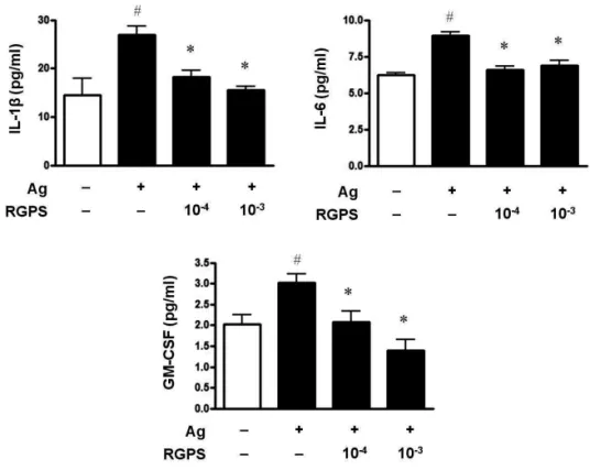
3.1. RGPS suppresses proinflammatory cytokines, IL-1β, IL-6, and GM-CSF
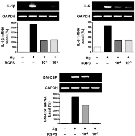
To determine whether RGPS suppresses the secretion of proinflammatory cytokines, IL-1β, IL-6 and GM-CSF, in mast cells activated with IgE/Ag, we conducted cytokine ELISA on cell supernatants. Compared to the basal levels of IL-1β, IL-6 and GM-CSF, the levels of those cytokines in IgE-sensitized RBL-2H3 cells was markedly increased after Ag stimulation. RGPS (10-4and 10-3dilution) decreased the levels of IL-1β, IL-6 and GM-CSF in a dose-dependent manner (Fig. 1). We also investigated the expressions of the IL-1β, IL-6 and GM-CSF genes by using RT-PCR. RGPS (10-4 and 10-3dilution) significantly suppressed the induction of those cytokine genes by Ag stimulation (Fig. 2).
3.2. RGPS suppresses induction of inflammatory enzymes
Inflammatory enzymes, such as HDC2, COX-1, COX-2 and 5LO mRNA, are involved in the synthesis of typical allergic mediators, such as histamine, prostaglandins and leukotriene. Thus, we measured whether RGPS suppresses the induction of these genes by using RT-PCR. Compared to the basal levels of HDC2, COX-1, COX-2 and 5LO
mRNA, the induction levels of those enzymes in IgE- sensitized RBL-2H3 cells was markedly increased after Ag stimulation. RGPS reduced induction of those enzyme genes by Ag stimulation (Fig. 3).
3.3. RGPS suppresses the gene expressions of FcεRI α, FcεRIβ‚ and FcεRIγ
Recent studies have shown that receptors with bound IgE appears to be permanently expressed on the surface of the cells while empty receptors are internalized and degraded [26]. Thus, the density of FcεRI expression correlates with the IgE level, where binding of IgE stabilizes the receptor at the cell surface. Compared to the basal levels of FcεRIα, Fc εRIβ‚ and FcεRIγ, mRNA, the induction levels of the enzymes in IgE-sensitized RBL-2H3 cells were clearly elevated after Ag stimulation. RGPS reduced induction of receptor genes by Ag stimulation (Fig. 4).
3.4. RGPS suppresses FcεRI-mediated signaling events in IgE/Ag-stimulated mast cells
To understand the mechanism for the inhibitory effect of mast cell activation by RGPS, we examined its effects on Fc εRI-mediated signaling events. IgE/Ag-mediated FcεRI aggregation induces activation of a Src family kinase, Lyn, which phosphorylates Syk and LAT, or a second Src family
Figure 1 Effects of RGPS on the secretion of IL-1β, IL-6 and GM-CSF in IgE/Ag-stimulated RBL-2H3 cells. The cells (5 × 10⁵cells/ml) were sensitized with anti-DNP IgE (0.5 μg/ml) for 24 h and stimulated with DNP-HSA (10 μg/ml). RGPS (10-4and 10-3dilution) was pretreated for 1 h prior to DNP-HSA stimulation for 4 h. IL-1β, IL-6 and GM-CSF concentrations were measured in the cell supernatant using ELISA method. The absorbance was measured at 450 nm using an ELISA reader. The results represent as the mean ± SD. # p < 0.05 vs vehicle group, *p < 0.05 vs stimulated group.
Figure 2 Effects of RGPS on the mRNA expression levels of IL-1β, IL-6 and GM-CSF in IgE/Ag-stimulated RBL-2H3 cells. IgE-sensitized cells were treated with RGPS (10-4and 10-3dilution) for 1 h and stimulated with DNP-HSA for 4 h. Detection of mRNA was examined by using RT-PCR analyses. GAPDH was used as an internal control gene.
Figure 3 Effects of RGPS on the mRNA expression levels of inflammatory enzymes, HDC2, COX-1, COX-2 and 5LO in IgE/Ag-stimulated RBL-2H3 cells. IgE-sensitized cells were treated with RGPS (10-4and 10-3dilution) for 1 h and stimulated with DNP-HSA for 4 h.
Detection of mRNA was examined by using RT-PCR analyses. GAPDH was used as an internal control gene.
Figure 4 Effects of RGPS on the mRNA expression levels of FcεRIα, FcεRIβ‚ and FcεRIγ, in IgE/Ag-stimulated RBL-2H3 cells. IgE-sensitized cells were treated with RGPS (10-4and 10-3dilution) for 1 h and stimulated with DNP-HSA for 4 h. Detection of mRNA was examined by using RT-PCR analyses. GAPDH was used as an internal control gene.
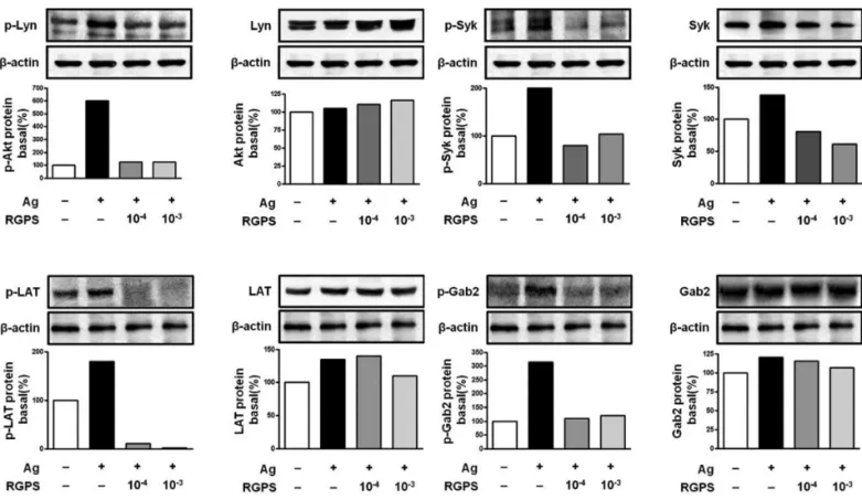
Figure 5 Effects of RGPS on the phosphorylation of Lyn, Syk, LAT and Gab2 in FcεRI-mediated signal transduction of IgE/Ag-stimulated RBL-2H3 cells. IgE-sensitized cells were treated with RGPS (10-4and 10-3dilution) for 1 h and stimulated with DNP-HSA for 15 min.
The expressions of Lyn, Syk LAT, Gab2 and β-actin and phosphorylations of Lyn, Syk, LAT and Gab2 were assayed by using western blot analyses as described in Materials and methods.
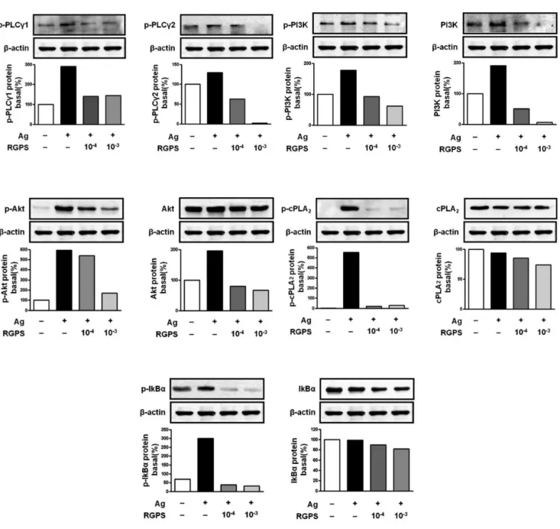
kinase, Fyn, which phosphorylates Gab2. We found that RGPS suppressed Ag-induced phospho-activation of Lyn, Syk, LAT and Gab2 in proximal signaling events of IgE/Ag- stimulated mast cells (Fig. 5). The adaptor molecule LAT regulates activation of the PLCγ, PI3K and other pathways.
In addition, the adaptor molecule Gab2 activates the PI3K/
Akt pathway. cPLA2, a downstream regulator of Syk, regul- ates arachidonic acid metabolism. Phosphoylation of IκBα, a downstream regulator of PI3K/Akt, with NF-βB is related to cytokine gene transcription. We found that RGPS dose- dependently suppressed Ag-induced phos-pho-activation of PLCγ1, PLCγ2, PI3K, Akt, cPLA2, and IκBα(Fig. 5).
4. Discussion
We already demonstrated in previous studies that RGPS could suppress cytokine production via MAPKs and NF-κB pathway in the late-phase response to mast cell activation [27]. In the present study, we focused on the early-phase response to mast cell activation initiated by IgE/Ag- stimulation and on the late-phase response. We identified if RGPS regulated FcεRI αβγsubunit expression, Lyn and Fyn downstream signaling pathways related to initiation of FcεRI-mediated mast cell activation, and signaling pathways related to the productions of cytokines and arachidonic acid metabolites.
RGPS suppressed the production and gene expression of proinflammatory cytokines and the gene expression of enzymes associated with the production of histamine, prostaglandins and leukotrienes (Figs. 1, 2 and 3). The res- ults indicate that RGPS can effectively regulate functional activations, such as degranulation and inflammatory response triggered by cytokines and lipid mediators, in IgE/Ag-stimulated mast cells.
RGPS suppressed the gene expression of FcεRI αβγ subunits on IgE/Ag-stimulated mast cells (Fig. 4). FcεRI is expressed exclusively on mast cells and basophils and initiates IgE/Ag-mediated mast cell activation for triggering the degranulation [28]. The FcεRIαsubunit binds to the constant region of IgE and is essential for the functioning of the mast cell surface receptor for IgE [29].
The FcεRIβsubunit amplifies signaling through the receptor (1). The FcεRIγsubunit enables the stable expression of the FcεRIαsubunit on the cell surface and transfers the signal to induce degranulation via immu- noreceptor tyrosine-based activation motif (ITAM) [30].
Down-regulation of FcεRI expression by RGPS may inhibit degranulation of mast cells.
The FcεRI signaling event is thought to be mediated by at least two major signaling pathways [18]. First, FcεRI aggregation activates Lyn, which mediates ITAM
phosphorylation of the β- and the γ-subunits. This promotes Syk recruitment to the phosphorylated γ-chains.
Activated Syk phosphorylates the adaptor proteins LAT.
LAT phosphorylation leads to the activation of PLCγa calcium regulator and activator of PKC, which is involved in mast cell degranulation. LAT also recruits the exchange factors responsible for the subsequent activation of the MAPK pathway that leads to the transcription of target genes including cytokines important in the late-phase mast cell response. Second, FcεRI aggregation also can activate Fyn. Fyn recruits and phosphorylates the adaptor protein Gab2. Gab2 recruits and phosphorylates PI3K, which PIP2to make phosphatidylinositol 3, 4, 5-trisphos- phate (PIP3). PIP3induces PLCγactivation, leading to calci- um flux and degranulation through IP3. These pathways also elicit transcription of inflammatory cytokine genes by activating transcription factors including NFAT, activator protein 1 (AP-1), NF-κB, and signal transducer and activ- ator of transcription (STAT) family members [17,31,32].
Therefore, degranulation of mast cells stimulated with IgE is induced by activity of Lyn/Syk/LAT/ PLCγor Fyn/Gab2/
PI3K/PLCγpathway. Ag-induced phospho-activation of the two signaling pathways was concurrently suppressed by RGPS in IgE-sensitized cells (Figs. 5 and 6). These results indicated that RGPS could act as a suppressor of degranulation. Lyn/Syk/LAT/MAPK or Fyn/Gab2/PI3K/
Akt/NF-κB pathway is associated with cytokine gene transcription. We found that RGPS concurrently suppr- essed Ag-induced phospho-activation of the two signaling pathways in IgE-sensitized cells (Figs. 5 and 6). These results indicated that RGPS could act as a suppressor in cytokine production. Furthermore, RGPS suppressed phospho-activation of cPLA2, which regulates arachidonic acid metabolism (Fig. 6). cPLA2is activated by the increase of Ca2+mobilization and extracellular signal-regulated kinase (ERK) activation and releases arachidonic acid from membrane phospholipids. The released arachidonic acid is catalyzed by COX and LO to produce prostaglandins and leukotriens. ERK and cPLA2act as downstream regulators of Syk. These results indicated that RGPS could act as a suppressor through the regulation of ERK/cPLA2pathway in arachidonic acid cascades. Taken together, RGPS suppressed FcεRI αβγsubunit expression and two major signaling pathways, Lyn and Fyn downstream pathways, related to degranulation and production of cytokines and arachidonic acid metabolites in IgE/Ag-stimulated mast cells.
5. Conclusion
In summary, RGPS down-regulates both the gene
expression of FcεRI αβγsubunits on cell surfaces and two major downstream signaling pathways initiated by phospho-activation of Lyn and Fyn via FcεRI aggregation in IgE/Ag-stimulated mast cells. Thus, RGPS effectively suppresses functional activations such as degranulation triggered by histamine and inflammatory response triggered by cytokines and lipid mediators via down- regulation of the FcεRI-mediated signaling pathways in IgE/Ag-stimulated mast cells.
Acknowledgments
This work was supported by Dong-Eui University (2011AA126).
References
1. Stone KD, Prussin C, Metcalfe DD. IgE, mast cells, basophils, and eosinophils. J Allergy Clin Immunol.
2010;125(2 Suppl 2):S73-S80.
Figure 6 Effects of RGPS on the phosphorylation of LAT, PLCγ1, PLCγ2 PI3K, Akt, cPLA2and IκBαin FcεRI-mediated signal transduction of IgE/Ag-stimulated RBL-2H3 cells. IgE-sensitized cells were treated with RGPS (10-4 and 10-3 dilution) for 1 h and stimulated with DNP-HSA for 15 min. The expressions of LAT, PI3K, Akt, cPLA2, IκBαand β-actin and phosphorylations of LAT, PLCγ1, PLCγ2 PI3K, Akt, cPLA2and IκBαwere assayed by using western blot analyses as described in Materials and methods.
2. Stassen M, Arnold M, Hu..
ltner L, Mu..
ller C, Neudo..
rfl C, Reineke T, et al. Murine bone marrow-derived mast cells as potent producers of IL-9: costimulatory function of IL-10 and kit ligand in the presence of IL-1. J Immunol. 2000;164(11):5549-55.
3. Hu..
ltner L, Ko..
lsch S, Stassen M, Kaspers U, Kremer J-P, Mailhammer R, et al. In activated mast cells, IL-1 up- regulates the production of several Th2-related cytokines including IL-9. J Immunol.
2000;164(11):5556-63.
4. Kandere-Grzybowska K, Letourneau R, Kempuraj D, Donelan J, Poplawski S, Boucher W, et al. IL-1 induces vesicular secretion of IL-6 without degranulation from human mast cells. J Immunol. 2003;171(9):4830-6.
5. Lee SA, Fitzgerald SM, Huang SK, Li C, Chi DS, Milhorn DM, et al. Molecular regulation of interleukin-13 and monocyte chemoattractant protein-1 expression in human mast cells by interleukin-1β. Am J Respir Cell Mol Biol. 2004;31(3):283-91.
6. Subramanian N, Bray MA. Interleukin 1 releases histamine from human basophils and mast cells in vitro. J Immunol. 1987;138(1):271-5.
7. Kishimoto T. 2005. INTERLEUKIN-6: from basic science to medicine-40 years in immunology. Annu Rev Immunol. 2005;23:1-21.
8. Walker C, Bauer W, Braun RK, Menz G, Braun P, Schwarz F, et al. Activated T cells and cytokines in bron- choalveolar lavages from patients with various lung diseases associated with eosinophilia. Am J Respir Crit Care Med. 1994;150(4):1038-48.
9. Saha S, Doe C, Mistry V, Siddiqui S, Parker D, Sleeman M, et al. Granulocyte-macrophage colony-stimulating factor expression in induced sputum and bronchial mucosa in asthma and COPD. Thorax. 2009;64(8):671-6.
10. Lamas AM, Leon OG, Schleimer RP. Glucocorticoids inhibit eosinophil responses to granulocyte- macrophage colony-stimulating factor. J Immunol.
1991;147(1):254-9.
11. Hirata, Takeuchi, Ukai, Sakakura. Expression of histidine decarboxylase messenger RNA and histamine N-methyltransferase messenger RNA in nasal allergy.
Clin Exp Allergy. 1999;29(1):76-83.
12. Reddy ST, Tiano HF, Langenbach R, Morham SG, Herschman HR. Genetic evidence for distinct roles of COX-1 and COX-2 in the immediate and delayed phases of prostaglandin synthesis in mast cells.
Biochem Biophys Res Commun. 1999;265(1):205-10.
13. Tanioka T, Nakatani Y, Semmyo N, Murakami M, Kudo I. Molecular identification of cytosolic prostaglandin E2 synthase that is functionally coupled with cyclo- oxygenase-1 in immediate prostaglandin E2 biosy- nthesis. J Biol Chem. 2000;275(42):32775-82.
14. M, Nakashima K, Kamei D, Masuda S, Ishikawa Y, Ishii T, et al. Cellular prostaglandin E2 production by membrane-bound prostaglandin E synthase-2 via both cyclooxygenases-1 and -2. J Biol Chem.
2003;278(39):37937-47.
15. Lewis RA, Austen KF, Soberman RJ. Leukotrienes and other products of the 5-lipoxygenase pathway.
biochemistry and relation to pathobiology in human diseases. N Engl J Med. 1990;323(10):645-55.
16. Kinet J-P. THE HIGH-AFFINITY IgE RECEPTOR (Fcε RI): from physiology to pathology. Annu Rev Immunol.
1999;17(1):931-72.
17. Gilfillan AM, Tkaczyk C. Integrated signalling pathways for mast-cell activation. Nat Rev Immunol.
2006;6(3):218-30.
18. Rivera J, Gilfillan AM. Molecular regulation of mast cell activation. J Allergy Clin Immunol.
2006;117(6):1214-25.
19. Vig M, Kinet JP. Calcium signaling in immune cells. Nat Immunol. 2009;10(1):21-7.
20. Alvarez-Errico D, Lessmann E, Rivera J. Adapters in the organization of mast cell signaling. Immunol Rev.
2009;232(1):195-217.
21. Han Y, Jung HW, Lee JY, Kim JS, Kang SS, Kim YS, et al.
2,5-Dihydroxyacetophenone isolated from Rehma- nniae Radix Preparata inhibits inflammatory responses in lipopolysaccharide-stimulated RAW264.7 macro- phages. J Med Food. 2012;15(6):505-10.
22. Liu CL, Cheng L, Ko CH, Wong CW, Cheng WH, Cheung DW, et al. Bioassay-guided isolation of anti- inflammatory components from the root of Rehman- nia glutinosa and its underlying mechanism via inhibition of iNOS pathway. J Ethnopharmacol.
2012;143(3):867-75.
23. Lau TW, Lam FF, Lau KM, Chan YW, Lee KM, Sahota DS, et al. Pharmacological investigation on the wound healing effects of Radix Rehmanniae in an animal model of diabetic foot ulcer. J Ethnopharmacol.
2009;123(1):155-62.
24. Baek GH, Jang YS, Jeong SI, Cha J, Joo M, Shin SW, et al.
Rehmannia glutinosa suppresses inflammatory responses elicited by advanced glycation end products.
Inflammation. 2012;35(4):1232-41.
25. Sung YY, Yoon T, Jang JY, Park SJ, Kim HK. Topical application of Rehmannia glutinosa extract inhibits mite allergen-induced atopic dermatitis in NC/Nga mice. J Ethnopharmacol. 2011;134(1):37-44.
26. MacGlashan DW. Endocytosis, recycling, and degra- dation of unoccupied FcεRI in human basophils. J Leukoc Biol. 2007;82(4):1003-10.
27. Kang KH, Kim CH. Inhibitory effect of rehmannia glutinosa pharmacopuncture solution on β-hexo-
saminidase release and cytokine production via FcεRI signaling in RBL-2H3 Cells. Journal of Pharma- copuncture. 2011;14(2):15-24.
28. Wu LC. Immunoglobulin E receptor signaling and asthma. J Biol Chem. 2011;286(38):32891-7.
29. Nishiyama C. Molecular mechanism of allergy-related gene regulation and hematopoietic cell development by transcription factors. Biosci Biotechnol Biochem.
2006;70(1):1-9.
30. Garman SC, Kinet JP, Jardetzky TS. The crystal structure of the human high-affinity IgE receptor (Fcε RIα). Annu Rev Immunol. 1999;17(1):973-6.
31. Gilfillan AM, Rivera J. The tyrosine kinase network regulating mast cell activation. Immunol Rev.
2009;228(1):149-69.
32. Kalesnikoff J, Galli SJ. New developments in mast cell biology. Nat Immunol. 2008;9(11):1215-23.