Vol. 44, No. 3, September 2018, 295-302 http://dx.doi.org/10.15230/SCSK.2018.44.3.295
1)
1. Introduction
Natural resources have been applied in cosmetic for- mula for many years. Since many natural resources have been used as traditional medicines, using them as in-
† 주 저자 (e-mail: [email protected]) call: 02)6360-0662
gredients for cosmetic products is considered as an effec- tive marketing strategy[1-3]. In fact, many scientific re- search papers have reported that natural resources have positive effects on the human skin such as anti-oxidant, anti-inflammatory, whitening, and anti-wrinkle effect[4-9].
While having these benefits, natural ingredients are fac- ing to extinction and pollution. The global climate change caused by destruction of forests and consuming of fossil
접시꽃 캘러스 추출물의 천연화장품 원료로서의 효능 평가
이 기 복†⋅염 아 름⋅김 동 원⋅박 창 민⋅정 민 석⋅이 기 용⋅정 철 승* (주)한국화장품제조 기술개발연구소
*충북 대학교
(2018년 7월 9일 접수, 2018년 9월 7일 수정, 2018년 9월 14일 채택)
Evaluation of Alcea rosea L. Callus Extract as a Natural Cosmetic Ingredient
Gibok Lee†, Areum Yeom, Kim Dong Won, Chang-Min Park, Min-Seok Joung, Gi Yong Lee, and Cheol-seung Jeong* R&D Center, Hankook Cosmetics Manufacturing Co., Ltd., 35 Cheonggyecheon-ro, Jongno-gu, Seoul 03188, Korea
*Chungbuk National University, Cheong-ju, Chungchenonbuk-Do 28644, Korea (Received July 9, 2018; Revised September 7, 2018; Accepted September 14; 2018)
요 약: 본 연구에서는 천연 화장품 소재 개발을 위하여 접시꽃 캘러스 생리활성 효능에 대해서 평가하였다. 접시 꽃캘러스 추출물의 항산화 효능은 DPPH, ABTS, FRAP assay를 통해서 확인하였다. 그 결과 접시꽃캘러스 추출물은 농도 의존적으로 강력한 항산화 능력을 확인되었다. 더불어, 접시꽃 캘러스 추출물은 10 mg/mL 의 농도에서 세포 내 ROS를 효과적으로 감소시키는 것으로 확인되었다. 접시꽃 캘러스 추출물의 미백효능을 평가 하기 위해 tyrosinase 저해 효과를 확인한 결과, 접시꽃 캘러스 추출물은 10 mg/mL 의 농도에서 tyrosinase 활성을 51% 감소시키는 것으로 확인되었다. 이상의 결과를 바탕으로, 접시꽃 캘러스 추출물은 항산화 및 미백 효능을 지닌 화장품 기능성 소재로서 적용 가치가 높은 천연소재로 사료된다.
Abstract: In this study, we investigated the biological effects of Alcea rosea L. callus extract for the development of natural cosmetics ingredients. The antioxidant activities of A. rosea L. callus extract was measured through DPPH, ABTS and FRAP assay. As a result, A. rosea L. callus extract were found to have a strong antioxidant ability in a dose dependent manner. In addition, A. rosea L. callus effectively reduced the intracellular oxidative stress induced by AAPH at a concentration of 10 mg/mL. In a tyrosinase activity assay, we found that A. rosea L. callus extract reduced tyrosinase activity by 51% at 10 mg/mL. Based on these results, A. rosea L. callus extract is considered as a promising natural ingredients for cosmetics with antioxidant and whitening functions.
Keywords: callus, Alcea rosea L., antioxidant, cosmetic
fuels directly affects the habitat of natural resources.
Pollutions such as toxic chemicals, air pollution, and wa- ter contamination have spread to natural resources and may alter their physiology[10,11]. In fact, air pollutions, specially sulfur dioxide, ozone, and oxides of nitrogen af- fect stomatal conductance and suppress plant growth[12].
Therefore, it is now becoming a more important issue to retain and safe natural cosmetic ingredient[13].
Plant tissue culture is based on the theory of plant cell totipotency suggested by Gottlieb Haberlandt in 1902[14].
The term “callus” originates from the Latin word callum, which means hard, and in medicine it refers to the thick- ening of dermal tissue. In plant science, callus was firstly introduced as plant tissues which develop over a cut or damaged plant surface[15]. The plant callus can be differ- entiated into whole plant through addition of hormones to the culture medium. Plant callus culture system has bene- fit of the stable and quick plant cell generation on the constant culture condition[16]. Furthermore, valuable compounds such as plant-derived secondary metabolites and recombinant protein can be produced during the scale up culture condition which is conducted in liquid medium in a bioreactor[17,18]. Thus, plant callus culture method has been considered as the sustainable and effective tech- nology in order to get natural resources.
Alcea rosea L., commonly called hollyhock, is a per- ennial ornamental plant that is included in the member of the Malvaceae family. The root and flowers of A. rosea L. have been used as folk medicine in asia countries for many years. Indeed, several reports discovered the phar- macological and biological effects of A. rosea L. such as anti-oxidant, anti-inflammatory, immunomodulatory, an- ti-urolithiatic and anti-cancer activities[19-22]. However, it has rarely been investigated the cosmetic effectiveness of A. rosea L. callus extract.
We induced callus of A. rosea L. in a bioreactor and extracted to investigate the skin benefit effect. In this re- search, we found that A. rosea L. callus extract has strong possibility as an effective and sustainable natural cosmetic ingredient.
2. Materials and Methods
2.1. Reagents and Antibodies
1,1-Diphenyl-2-picrylhydrazyl (DPPH), 2,2’-azino -bis-3-ethyl- benzthiazoline-6-sulphonic acid (ABTS), 2’,7’-dichlorofluorescin diacetate (DCF-DA), 2,2-azobis di- hydrochloride (AAPH), potassium ferricyanide, α-MSH, L-DOPA and mushroom tyrosinase were purchased from sigma (USA). Antibodies against MITF (#12590) were pur- chased from Cell Signaling Technology (USA). TRP1 (sc-514900), Tyrosinase (sc-7833), β-actin (sc-47778), was purchased from Santa Cruz Biotechnology (USA).
2.2. Preparation A. rosea L. Callus Extract
A. rosea L. callus powder (10 g) was extracted with 60% ethanol (500 mL) for 3 days at 37 ℃. A. rosea L.
callus extract was filtered through Whatman No.1 filter paper (GE Healthcare UK Limited, UK) and then evapo- rated under reduced pressure in a rotary evaporator. The final yield was 5 g of 60% ethanol extract. The extracts were dissolved in phosphate-buffered saline for long-term storage at -20 ℃.
2.3. Cell Culture and Cell Viability Assay
HaCaT (human keratinocyte), HDF (human dermal fi-
broblast), CCD-986sk (human skin fibroblast), B16F10
(mouse melanoma cells), cells were cultured in Dulbecco’s
modified Eagle’s medium (DMEM, WelGENE, Korea)
supplemented with 10% heat-inactivated calf serum (CS)
(Gibco BRL), 100 U/mL penicillin (Gibco BRL) and 100
µg/mL streptomycin (Gibco BRL) at 37 ℃ in a humidi-
fied atmosphere containing 5% CO
2. Cell viability was
evaluated using cell proliferation kit I (Applied Science,
Germany) and crystal violet staining method. Cells into
96-well plates at densities of 1 × 10
5cells well
-1, washed
twice with phosphate buffer saline (PBS). The sample
pre-treatment was performed with different concentrations
for 48 h. Following day, MTT solution was then added
to each well and plates were re-incubated at 37 ℃. After
4 h, cell viability was measured at an excitation wave-
length of 540 nm using a microplate reader (SpectraMax
i3, Molecular devices, USA).
Crystal violet staining method was followed by pre- vious report. The crystal violet was dissolved by 1% SDS solution and measured at an excitation wavelength of 595 nm using a microplate reader (SpectraMax i3, Molecular devices, USA).
2.4. DPPH Radical Scavenging Assay
DPPH radical scavenging assay was performed as pre- viously described[23]. 100 µL of DPPH solution (0.1 mM) was mixed with 100 µL of the sample and incubated 10 min in dark room. The DPPH radical scavenging activity was measured at a wavelength of 517 nm using a micro- plate reader (SpectraMax i3, Molecular devices, USA).
2.5. ABTS Radical Scavenging Assay
ABTS radical scavenging assay was performed as pre- viously described[24]. ABTS (7 mM) aqueous solution with 2.45 mM potassium persulfate was prepared and stor- ed in the dark at room temperature for 12-16 h before use.
The solution was diluted in PBS and equilibrated to obtain an absorbance of 0.700 at 734 nm. 0.2 mL of the sample and blank were mixed with 0.8 mL of ABTS solution.
Finally, the absorbance was measured at 734 nm against a blank after a 5 min of reaction time at room temperature.
2.6. Ferric Reducing Antioxidant Power (FRAP) Assay Ferric reducing antioxidant power of extract was de- termined using the method previously described[25]. A serial dilution of the extract was performed with 0.2 M sodium phosphate buffer (pH 6.6) and mixed with 2.5 mL of 1% potassium ferricyanide. The mixture was incubated at 50 ℃ for 20 min. 10% trichloroacetic acid (2.5 mL) was then added and centrifuged at 3,000 g for 10 min.
The supernatant was collected and mixed with distilled water (2.5 mL) containing 1% ferric chloride (0.5 mL).
The FRAP was measured at an excitation wavelength of 700 nm using a microplate reader (SpectraMax i3, Molecular devices, USA).
2.7. Intracellular ROS Detection
Intracellular ROS was detected using DCF-DA fluorescence. Cells were incubated with (20 mM) AAPH for 1 h in the presence or absence of sample, then washed with PBS and stained with 10 µM DCF-DA solution for 15 min. The cells were washed with PBS and DCF-DA flu- orescence was detected by fluorescence microplate reader (SpectraMax i3, Molecular devices, USA) with excitation and emission of 495 nm and 529 nm, respectively.
2.8. Tyrosinase Inhibition Assay
Tyrosinase inhibition assay was performed as pre- viously described[27]. L-DOPA was used as substrate.
The reaction mixture consisted of 0.1 M PBS (pH 6.5), purified mushroom tyrosinase and the sample. After add- ing 2 mM of L-DOPA, tyrosinase inhibition activity was measured at 492 nm using a microplate reader (SpectraMax i3, Molecular devices, USA).
2.9. Western Blotting
Cell lysate was prepared using RIPA lysis buffer and the protein extracts were separated via SDS-poly- acrylamide gels, and then transferred to a PVDF mem- brane (EMD Millipore, USA). Blocking process was con- ducted with 5% non-fat dry milk in TTBS and incubated overnight at 4 ℃ with primary antibodies. The flowing day, membranes were incubated with HRP-conjugated secondary antibodies for 1 h at room temperature. Protein detection was performed using an ECL prime kit (GE healthcare, USA).
2.10. Statistical Analysis
For two experimental comparisons, data were analyzed using the unpaired Student’s t-test. For multiple compar- isons, data were analyzed using one-way ANOVA with Tukey posttest. Values represent means ± SD of three in- dependent experiments performed in triplicate;
*p < 0.05,
#
p < 0.01
3. Result
3.1. Preparation of A. rosea L. Callus
The tissue cultured callus of A. rosea L. were provided by PhD. Ceol-seung Jeong, Research Center for the Development of Advanced Horticultural Technology in Chungbuk National University. Briefly, using young leaves of A. rosea L., we initiated callus formation in cul- ture tubes containing an SH basic medium supplemented with 3% sucrose and 0.5 mg/L-(2,4-D). Cultures were maintained at 25 ± 2 ℃ under a photoperiod and with a light intensity. These cell suspensions were sub-cultured at 2 week intervals. The cultures were transferred to con- ical flasks containing 100 mL of the SH basic medium and were grown for 15 days in the dark on a gyratory shaker (95 rpm) at 20 ± 2 ℃ and were subcultured in SH basic medium with plant growth regulators. These proc- esses are shown at Figure 1.
3.2. Cytotoxicity of A. rosea L. Callus Extract Because cosmetic products are designed for applying
on the skin surface, it is crucial point to evaluate whether the A. rosea L. callus extract affects cell viabilities on hu- man normal skin cell lines. Crystal violet staining assay and MTT assay were performed to investigate whether A.
rosea L. callus extract is toxic on HaCaT, HDF, CCD-986sk (Figure 2A, B). We found that the A. rosea L. callus extract does not reduce cell viabilities, even at high dose (10 mg/mL). It was shown that cell viability were slightly reduced from above 80 mg/ml of A. rosea L. callus extract.
3.3. Antioxidant Activities of A. rosea L. Callus Extract
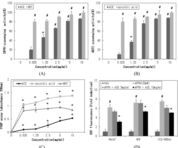
Oxidative stress generated by ultraviolet radiation, pol- lutants, smoking etc. is considered as major reason of skin aging. Therefore, most of natural product is normally ap- plied to cosmetic formula as the purpose of antioxidant agent[28,29]. The antioxidant effect of A. rosea L. callus
(A)(B)
(C)
Figure 1. Scheme of the whole procedure of A. rosea L. callus cultured by the bioreactor culture system (A) A. rosea L.
callus induction, (B) Liquid medium culture, (C) A. rosea L.
callus harvest.
(A)
(B)
Figure 2. A. rosea L. callus extract dose not reduce cell viabilities (A) Cell viabilities was measured by MTT assay, (B) Cell viabilities was measured by MTT crystal violet staining assay, ACE: A. rosea L. callus extract, Values represent means ± SD of three independent experiments performed in triplicate; *p < 0.05.
extract was evaluated by measuring DPPH, ABTS radial scavenging activities. The results indicate that the A. ro- sea L. callus extract can reduce DPPH, ABTS radical in a dose dependent manner (Figure 3A, B). The A. rosea L. callus extract showed a maximum of 81.51% DPPH radical scavenging activity at 5 mg/mL. ABTS radical scavenging activity of the A. rosea L. callus extract was shown a maximum of 86.51% at 5 mg/mL. These results were compared to DPPH (1.25 mg/mL) activities of buty- lated hydroxytoluene (BHT) and ABTS (2.5 mg/mL) ac- tivities of ascorbic acid.
FRAP assay was performed to measure the ability of Ferric-ferricyanide (Fe
3+) to be reduced to Ferrous (Fe
2+).
The higher the measured absorbance value is, the higher
the Fe
3+reducing activity is. We found that A. rosea L.
callus extract showed Fe
3+reducing activity in a dose de- pendent manner. However, A. rosea L. callus extract showed low activity compare to BHT and ascorbic acid (Figure 3C). Furthermore, we investigated whether A. ro- sea L. callus extract can reduce intracellular reactive oxy- gen species (ROS) level on human normal skin cell lines.
The ROS generated by an oxidative stress model, 2,2-azo- bis(2-amidinopropane) dihydrochloride (AAPH) was sig- nificantly reduced in the presence of A. rosea L. callus extract in Figure 3D. Together, A. rosea L. callus extract has strong antioxidant effect and reduce intracellular oxi- dative stress.
(A) (B)
(C) (D)
Figure 3. Antioxidant effect of A. rosea L. callus extract (A) DPPH radical scavenging activity of A. rosea L. callus extract, (B) ABTS radical scavenging activity of A. rosea L. callus extract, (C) Ferric Reducing Antioxidant Power of A. rosea L. callus extract, (D) Intracellular ROS scavenging activity of A. rosea L. callus extract, ACE: A. rosea L. callus extract, Values represent means ± SD of three independent experiments performed in triplicate; *p < 0.05, #p < 0.01.
3.4. Tyrosinase Inhibition Activities of A. rosea L.
Callus Extract
Tyrosinase is the key enzyme on melanogenesis which mediates the conversion of amino acid L-tyrosine to DOPA quinone. Therefore, tyrosinase inhibitors from nat- ural sources have been used as a whitening agent in cos- metic formula[6,8]. We found that the A. rosea L. callus extract has strong inhibition activity of mushroom tyrosinase. A. rosea L. callus extract reduced tyrosinase activity by 51% at 10 mg/mL (Figure 4A).
Furthermore, we investigated whether the A. rosea L.
callus extract also affects melanogenesis related protein expressions in α-MSH stimulated B16F10 cells.
Micropthalmia-associated transcription factor (MITF), Tyrosinase (TYR), tyrosinase-related protein 1 (TRP-1) expression were evaluated. MITF, Tyrosinase, TRP-1 ex- pressions are strongly upregulated after α-MSH treatment. The A. rosea L. callus extract slightly inhibit
TYR, and TYRP1 protein levels. However, the statistical significance could not be ascertained (Figure 4B).
4. Discussion and Conclusion
It has become an important problem that environmental changes resulting from rapid industrial development threatens the ecosystem of natural resources and causes them to die out[10]. Therefore, plant callus culture meth- od is now considered effective method to solve those matters. Furthermore, several reported discovered that plant secondary metabolites such as phenol, flavonoids, derivatives of cinnamic acid are increased during the plant callus culture[30]. We also induced the callus of A. rosea L. and investigated the biological effects. In this present study, we focused on antioxidant and whitening effects.
The antioxidant effect of A. rosea L. callus extract was measured by DPPH radical scavenging, ABTS radical
(A)(B)
Figure 4. A. rosea L. callus extract inhibits tyrosinase activities, but not regulates melanogenesis related protein expressions (A) Tyrosinase inhibition activity of A. rosea L. callus extract, (B) A. rosea L. callus extract dose not regulate melanogenesis related protein expression on B16F10, Graphs were subjeted to densitometric scanning using the Scion image NIH image software. ACE: A.
rosea L. callus extract, Values represent means ± SD of three independent experiments performed in triplicate; *p < 0.05, #p < 0.01.