Journal of Minimally Invasive Surgery Vol.18, No.4, 2015 http://dx.doi.org/10.7602/jmis.2015.18.4.137
INTRODUCTION
Port site hernias are a rare complication after laparoscopic and robotic surgery. Hernias at a port site less than 10 mm are a very rare complication.
1Tounichi et al.
2classified port site hernias into three groups according to the timing of presentation and the fascia defect involved. In the early-onset type, dehiscence of the anterior and posterior fascial layers as well as the peritoneum presents immediately post surgery and usually also presents with small bowel obstruction. These hernias require urgent surgical intervention and hernia site repair. Late-onset type refers to dehiscence of the anterior and posterior fascial layers alone, and hernias usually develop several months to years after surgery. These are not associated with small bowel obstruction and often present as a palpable abdominal swelling by the wound site. These hernias can be
repaired in an elective surgery. Lastly, special type hernias refer to complete dehiscence of the abdominal wall and external evisceration of the peritoneal contents (intestine and/or omentum) without a hernia sac, usually occurring in the immediate postoperative period. These hernias require emergent surgery and immediate repair. The present case is an 8-mm port site hernia that occurred after robot-assisted colorectal surgery. According to the Tounichi classification of port site hernias, it is the late-onset type.
CASE REPORT
A 70-year-old woman with a body mass index of 25.7 was undergoing follow-up at an outpatient clinic after lower anterior resection for rectal cancer. She had no medical history and no operative history. She underwent a robot-assisted low
Trocar Site Hernia after Use of an 8-mm Bladeless Trocar in Robotic Colorectal Surgery
Dae Ro Lim, M.D., Hyuk Hur, M.D., Byung Soh Min, M.D., Seung Hyuk Baik, M.D., Nam Kyu Kim, M.D.
Section of Colon and Rectal Surgery, Department of Surgery, Yonsei University College of Medicine, Seoul, Korea
Port site hernias are a rare complication after laparoscopic and robotic surgery. The current case is an 8-mm port site hernia which occurred after robot-assisted colorectal surgery. A 70-year-old female with a BMI (body mass index) of 25.7 was diagnosed as rectosigmoid colon cancer. She underwent a robot-assisted low anterior resection with double-stapled anastomosis for AJCC (American Joint Committee on Cancer) stage IIIB. After the main procedure, fascial defects in the supra-pubic and the supra-umbilical site were closed with a routine procedure (12 mm). Thirty two months after surgery, she developed an incisional hernia in the left mid abdomen (8-mm port). Recognition of the potential for a port site hernia using an 8-mm bladeless trocar port as well as a 12-mm port is essential in robotic surgery. Patients with risk factors for a port site hernia may require complete port site closure.
Keywords:
Low anterior resection, Incisional hernia, Robotic surgeryReceived September 8, 2015 Revised 1st October 12, 2015
2nd October 12, 2015 Accepted October 15, 2015
Corresponding author Nam Kyu Kim
Section of Colon and Rectal Surgery, Department of Surgery, Yonsei University College of Medicine, Yeonseiro 50-1, Seodaemun-gu, Seoul 03722, Korea
Tel: +82-2-2228-2105 Fax: +82-2-313-8289 E-mail: [email protected]
This is an Open Access article distributed under the terms of the Creative Commons Attribution Non-Commercial License (http://
creativecommons.org/licenses/by-nc/4.0/) which permits unrestricted non-commercial use, distribution, and reproduction in any medium, provided the original work is properly cited.
Copyright © 2015 The Journal of Minimally Invasive Surgery. All rights reserved.
Journal of Minimally Invasive Surgery
pISSN 2234-778X •eISSN 2234-5248 J Minim Invasive Surg 2015;18(4):137-140
CASE REPORT
Dae Ro Lim et al.
Journal of Minimally Invasive Surgery Vol. 18. No. 4, 2015
138
anterior resection with total mesorectal resection and double- stapled anastomosis for AJCC stage IIIB (pT3N1bM0) on April 2010. The procedure was completed with the 4-arm da Vinci Si Surgical System (Intuitive Surgical, Sunnydale, CA). After the main procedure, fascia defects of the 12-mm port in the supra-pubic area and the 12-mm port at the supra-umbilical site (camera port) were closed in a routine procedure. Other fascia defects of the 8-mm port and the 5-mm port were not closed. On the tenth postoperative day, she was discharged in a satisfactory condition. She then received 12
thoxaliplatin-based adjuvant chemotherapy. Thirty-two months after surgery, she developed an incisional hernia in the left mid abdomen at the site of the 8-mm port, which was also used for the drainage tube. She presented with abdominal pain, a palpable
mass, and tenderness. CT scan showed a focal mesenteric fat hernia extruding through the incision site in the left abdomen (Fig. 1). She received elective surgery, in which the hernia sac was removed and the hernia was repaired (Fig. 2). She was discharged on the second postoperative day.
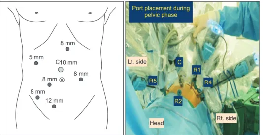
Patient setup and port placement
We used the following patient setup and port placement for robotic colorectal surgery. A 10-mm trocar was placed through an incision approximately 3 cm above the umbilicus after achieving pneumoperitoneum. Next, a 30
ostandard robotic laparoscope was inserted through the 10-mm trocar.
The first 8-mm da Vinci
®trocar was placed at a point two- thirds of the distance between the supraumbilical 12-mm trocar (camera port) and the right anterior superior iliac supine. The second 8-mm da Vinci
®trocar was inserted 2 cm below and 2 cm to the left of the xiphoid process. The third 8-mm da Vinci
®trocar was inserted at a point one-third of the distance between the camera port and the right anterior superior iliac supine. The fourth 8-mm trocar was placed in the left mid abdomen approximately 8 cm lateral to the umbilicus. The 12-mm trocar was placed 2 cm above and 2 cm to the right of the suprapubic area. The 5-mm trocar was placed in the right upper abdomen (Fig. 3).
DISCUSSION
The incidence of port site hernias is 1.47%, occurring within an average follow-up period of 71.5 months after laparoscopic colorectal surgery.
3Lumley et al.
4reported port site hernia after laparoscopic colorectal surgery for cancer with an incidence of 0.55%. Fuller et al.
5reported port site hernia after robot-assisted radical prostatectomy with an incidence of 4.8%
Fig. 1. Focal mesenteric fat hernia through the incision site in the left abdomen.
Fig. 2. Incisional hernia at the left mid abdomen; 8-mm trocar site and
drainage tube used.
Trocar Site Hernia after Use of an 8-mm Bladeless Trocar in Robotic Colorectal Surgery
www.e-jmis.org
139
(12/250 cases) during a follow-up period of 35 months. In the present study, the incidence of port site hernias after robot- assisted rectal cancer surgery was 0.54% (2/370 cases) between January 2006 and December 2010, with a mean follow-up of 36.5 months (Table 1). There are several factors that affect port site hernias. These factors can be divided into two categories:
patient factors and technical factors. Patient factors include nutritional status, chronic obstructive pulmonary disease,
5and diabetes.
6Technical factors include placement of the port, size of the port, the presence of a port site defect closure, and manipu- lation of the port site.
The placement of ports is a risk factor for port site hernias.
Midline ports, particularly umbilical ports, are by far the most common sites for herniation after a laparoscopic procedure.
3Matin et al.
7reported six of seven patients with hernias in the right lower quadrant after laparoscopic or robotic urologic surgery. The patients in the present cases developed a port site hernia in the left lateral region.
The size of the port used is associated with a tendency to develop a port site hernia. A 1994 survey by the American Association of Gynecological Laparoscopists reported that 725 (86.3%) of 840 hernias in which the size of the original defect were occurred at the sites of ports 10 mm in diameter or greater. Only 23 (2.7%) patients had hernias that occurred in port sites smaller than 8 mm.
1Tounichi et al.
2reported 78.3%
8 mm
8 mm
8 mm 8 mm 5 mm
10 mm
12 mm C
Port placement during pelvic phase
Lt. side
Head Rt. side
R5
R2
R4 R1 C
Fig. 3. Patient setup and port placement.
Table 1. Literature review of robotic trocar site hernias
Author Surgery Incidence Number of
cases Location of hernia Size of
trocar Onset type Contents Seamon et al.
(2008)
8Gynecologic cancer surgery
- 1 Left abdominal wall 8 mm Early Small bowel
evisceration Matin et al.
(2010)
7Prostate cancer surgery
0.66%
(7/1055)
4 case of robotic
Right lower quadrant of the abdominal wall
5, 8, & 12 mm Late (median, 13 months)
Small bowel incarceration &
mesentery Fuller et al.
(2011)
5Prostate cancer surgery
4.8%
(12/250)
12 Midline and left abdominal wall
12 & 8 mm Late (mean, 14.8 months)
Small bowel incarceration &
mesentery Park MG et al.
(2012)
9Colorectal cancer surgery
- 1 Right lower quadrant of the abdominal wall
12 mm Early Small bowel
incarceration Kim IY et al.
(2012)
10Prostate cancer surgery
0.4%
(2/498)
2 Supraumbilical abdominal wall
12 mm Late
(12/18 months)
Omentum
Present case Colorectal cancer surgery
0.54%
(2/370)
1 Left abdominal wall 8 mm Late (32 months)
Omentum &
mesentery fat
Dae Ro Lim et al.
Journal of Minimally Invasive Surgery Vol. 18. No. 4, 2015
140
of port site hernias occurred at the 10~12 mm trocar site, while 21.7% occurred at the site of a trocar whose diameter was ≤ 5 mm. Unlike laparoscopic surgery, in robotic surgery, a trocar is mostly used with the 8-mm port. Seamon et al.
8reported a case of small bowel obstruction after herniation through an 8-mm robotic port defect at the left mid abdomen after robotic gynecologic cancer surgery. Fuller et al.
5reported that 10 of 12 patients had trocar site hernias at the 12-mm umbilical port site, and one patient had a hernia at the left lateral 8-mm port site after robot-assisted radical prostatectomy. The 8-mm port site was used occasionally for the wound drainage tube insertion site, and herniation at this site occurred in association with removal of the wound drainage tube. The present case was a left lateral 8-mm port site hernia, which was the site also used for the wound drainage tube.
Whether to close the port site defect or leave it open is still controversial. Most surgeons have advised fascial closure for trocar sites greater than 10 mm due to literature reporting incidence. Sanz-Lopez et al.
6recommended routine fascial closure for laparoscopic port sites greater than 5 mm and for all defects in children regardless of size.
Manipulation of the trocar port site may widen a port site incision. Multiple reinsertions of the trocar and advanced surgical difficulty requiring increased force and torque on the fascia can also widen the initial port site. The robotic arm moves around a fixed pivoting point through the 8-mm port site. If the pivot point of the robotic port is inadequately distanced from the fascial layer, the robotic arm can damage the fascia around the port site because of its constant large motion and force. The robotic arms are prone to creating larger fascial defects due to the inadequate location of the robotic port. Because of that, the pivoting point is painted on the tip of the robotic port, surgeon should check whether the pivoting point is at the adequate position. In addition force of torque of robotic arms is strong and range of motion is wide. In these reasons, we think that potentialities of trocar site herniation development are higher by using robotic arm at same size small trocar site comparing by using other instrument such like laparoscopic device. The reason is that robotic arms have more large torque and range of motion than laparoscopic instrument. Based on our incidence data, we think that routine closure of fascial defects of the 8-mm port site is not necessary; however, if operations require reinsertion of the trocar, a longer operation time, or excessive motion and traction of the robotic arm during surgery, surgeons should
check the fascial defect and closure of the port site. Due to force of torque of robotic arms is strong and range of motion is wider, defect size of 8 mm trocar site could become more than 12 mm defect size at finishing operation time. And then it will be occurred hernia through this defect site widening more, if missed the shift which surgeon did not check. In conclusion, recognition of the potential for port site herniation using an 8-mm bladeless trocar port as well as a 12-mm trocar port is essential in robotic surgery. If a patient has risk factors, port site closure may be necessary.
REFERENCES
1) Montz FJ, Holschneider CH, Munro MG. Incisional hernia following laparoscopy: a survey of the American Association of Gynecologic Laparoscopists. Obstet Gynecol 1994;84:881-884.
2) Tonouchi H, Ohmori Y, Kobayaschi, Kusunoki M. Trocar site hernia. Arch Surg 2004;139:1248-1256.
3) Owens M, Barry M, Januua AZ, Winter DC. A systemic review of laparoscopic port site hernias in gastrointestinal surgery. The Surgeon 2011;9:218-924.
4) Lumley J, Stitz R, Stevenson A, Fielding G, Luck A. Laparoscopic colorectal surgery for cancer: intermediate to long-term outcomes.
Dis Colon Rectum 2002;45:867-872.
5) Fuller A, Fernandez A, Pautler SD. Incisional hernia after robot- assisted radical prostatectomy predisposing factors in a prospective cohort of 250 cases. Journal of Endourology 2011;24:1021-1024.
6) Sanz-Lopez R, Martinez-Ramos C, Nunez-Pena JR, Ruiz de Gopegui M, Pastor-Sirera L, Tamames-Escobar S. Incisional hernias after laparoscopic vs open cholecystectomy. Surg Endosc 1999;13:922-924.
7) Chiong E, Hegarty PK, Davis JW, Kamat AM, Pisters LL, Martin SF. Port-site hernias occurring after the use of bladeless radially expanding trocars. Urology 2010;75:574-580.
8) Seamon LG, Backes DF, Resnick K, Cohn DE. Robotic trocar site small bowel evisceration after gynecologic cancer surgery. Obstet Gynecol 2008;112:462-4.
9) Park MG, Kang JK, Kim JY, Hur H, Min BS, Lee KY, Kim NK.
Trocar site hernia after the use of 12-mm bladeless trocar in robotic colorectal surgery. Surg Laparosc Endo Percutan Tech 2012;22:34-36.
10) Kang DI, Woo SH, Lee DH, Kim IY. Incidence of port site hernias after robot assisted radical prostatectomy with the fascial closure of only the midline 12-mm port site. Journal of endourology 2012;
26:848-851.