․Correspondence to: Joung-jo An 621 Dujeong-dong, Seobuk-gu, Cheonan-si, Chungcheongnam-do
Cheonan Oriental Hospital of Daejeon Univ.
TEL: 042-521-7000 E-mail: [email protected]
Lipid-lowering and Antioxidant Effects of Curcuma Radix in Poloxamer 407-induced Hyperlipidemia Model Rat Models
So-ae Park, Hyun-kyung Jo, Ho-ryong Yoo, Yoon-sik Kim, In-chon Seol, Joung-jo An
Division of Circulatory System, Dept. of Internal Medicine, College of Oriental Medicine, Dae-Jeon University
ABSTRACT
Objectives :
This study investigated the hypolipidemic and antioxidant effects of Curcuma radix using a rat model induced by poloxamer 407 injection.
Methods :
Serum lipid parameters and oxidative stress-associated biomarkers were determined. Additionally, hepatic cholesterol and triglyceride as well as lipid metabolism-associated gene expressions were observed in hepatic tissue.
Results :
1. Curcuma radix ameliorated elevation of serum cholesterol, triglyceride, LDL-cholesterol, MDA, hepatic cholesterol level, and reduction of serum TAC, SOD, GSH, GSH-reductase level.
2. Curcuma radix augmented up-regulated ACAT gene expression.
3. Curcuma radix almost completely ameliorated down-regulated CYP-7A1 but up-regulated HMG-CoA gene expression.
Conclusions :
The hypolipidemic and antioxidant properties of Curcuma radix were evidenced. This study provides a scientific basis for the clinical application of Curcuma radix and development of hypolipidemics using this herb in the future.
Key words :
Curcuma Radix, hyperlipidemia, poloxamer 407, cholesterol
Ⅰ. Introduction
Hyperlipidemia is a pathologically pre-disease status characterized as an excess of fatty substances including cholesterol, triglycerides or lipoproteins in the blood
1,2. Hyperlipidemia is well known as one
of the critical risk factors in developing heart disease and stroke which are leading causes of death in the most developed countries
3,4.
The hypolipidemic agents provide benefits lowering lipid level. However, the current drugs had a limitation because the final goal reducing the risk of heart disease and stroke is doubt, but all drugs have notable adverse effects such as hepatotoxicity, myopathy or noncardiovascular death
5,6.
On the other hand, it is well known that oxidative
stress is an early event in the evolution of hyperlipidemia, and antioxidant supply help preventing the course of the hyperlipidemia-derived diseases
7,8. Accordingly, there are many investigations to develop novel drugs having both pharmaceutical properties of hypolipidemic activity and antioxidant effect
9. In particular, many researchers have paid attentions on natural products as candidates of hypolipidemics which have not side effects but antioxidant effect
10,11.
Curcuma radix is a herbal medicine which as been prescribed for patients with insufficient circulation of Qi and blood, menstrual pain, irregular menstruation, or flank pain. Many reported the pharmaceutical properties of Curcuma Radix, such as antioxidant and anti-inflammatory activity, angiogenic action, and protection of vascular endothelial cells
12,13. The traditional clinical applications and above experimental reports indicate the possibility of Curcuma radix as a herbal-derived hypolipidemics which guarantee both improving blood lipidemic levels and reducing its complications.
However there is no research to determine the hypolipidemic effect and antioxidant activity in hyperlipidemia animal models.
The present study aimed to evaluate the effects of Curcuma radix on serum lipid levels, and oxidative stress-related biomarkers, and lipid metabolism -associated gene expressions.
Ⅱ. Materials and Methods
1. Preparation of Curcuma radix water extract Curcuma radix (root of Curcuma aromatica SALISB.) was purchased from Jeongseong Herbal Company (Daejeon, Korea). After multiple cleaning process and drying, 500 g of sliced Curcuma radix
was mixed with 5 L of distilled water and then was boiled for 2 h. The decoction was filtered and then centrifuged at 2,000 rpm for 20 mins to remove the unsolved and coarse materials. After evaporation (Rotary evaporator, Buchi B-480, Switzerland), the decoction was freeze-dried (Freeze dryer, Eyela FDU-540, Japan). Finally 9.525 g of Curcuma radix dried extract was obtained, and then the yield was 1.905 % (w/w). The extract was stored in deep freezer (- 70 ℃) for experimental use.
2. High performance-thin layer chromatography (HP-TLC)-based fingerprinting
In order to produce fingerprint of Curcuma radix, HP-TLC procedure was adapted using the CAMAG application system (Muttenz, Switzerland). Aqueous extracts of Curcuma radix and its main compositional ingredient, curcumine (Sigma Chemical Co., St.
Louis, MO, USA) as standard, were dissolved in HPLC-grade methanol and applied to pre-washed 60 F
254HP-TLC plates (silica gel thickness 2 ㎜, from Merck, Darmstadt, Germany) with an automated applicator (Linomat IV; CAMAG). Curcuma radix and curcumine were separated (migration distance 65 ㎜) using HPLC-grade chloroform (CHCl3) : methanol (MeOH) : formic acid (CH
2O
2) /96 : 4:0.7 and then were visualized under UV of wave length 366 ㎚. Thereafter photos were taken using Reprostar 3 with a digital camera (CAMAG).
3. Total antioxidant capacity (TAC) for Curcuma radix
Total antioxidant capacity (TAC) of Curcuma
radix itself in vitro was determined according to
the modified method of Kambsyashi
14. Gallic acid
was used as control and antioxidant activity was
expressed as gallic equivalent antioxidant capacity
(GEAC).
4. Animal and experimental design
Seven-week old male Sprague-Dawley rats were purchased from commercial animal breeder (Daehan BioLink, Korea). After one week of acclimation, rats were used in this experiment. The rats were housed in an environmentally controlled room at 22±2 ℃, relative humidity at 55±10% and 12 h light/dark and fed with commercial pellets (Samyang Feed Co, Korea) and tap water ad libitum.
Forty rats were divided into 5 groups of 8 animals each. (normal, pre-treatment with Curcuma radix 50 ㎎/㎏ and Curcuma radix 100 ㎎/㎏, Lipitor
®or distilled water) Hyperlipidemia was induced by 30% poloxamer 407 (Sigma Chemical Co., St. Louis, MO, USA) injection. Rats were pre-treated with Curcuma radix (50 or 100 ㎎/㎏ body weight), Lipitor
®(Pfrizer, USA, 10 ㎎/㎏ body weight) or distilled water by orally administration for seven days. The rats were intraperitoneally injected with 2 ㎖ of poloxamer 407, and then orally fed with Curcuma radix, Lipitor
®or distilled water respectively once again at 12 h after poloxamer 407 injection.
On 24 h-time point of poloxamer-407 injection as status of 12 h fasting, animals were sacrificed by whole blood collection from abdominal aorta under ether anesthesia.
Liver was removed and weighted, and then was stored separately for RNA expression analysis in RNAlater® solution (Invitrogen, USA), and biochemical evaluation in deep freezer (-70 ℃) respectively. After clotting by standing blood at room temperature for 1 h, serum was separated by centrifugation at 3000 rpm for 15 min.
5. Measurement of serum cholesterol and triglyceride
The levels of serum total cholesterol, high density lipoprotein (HDL)-cholesterol, low density lipoprotein (LDL)-cholesterol, and triglyceride were determined using Olympus optical reply (Olympus, Japan).
6. Measurement of serum oxidative stress parameters 1) Determination of malondialdehyde (MDA) Lipid peroxidation levels in the serum were determined using the method of thiobarbituric acid reactive substances (TBARS)
15.
2) Determination of total antioxidant capacity (TAC)
TAC level were determined according to the method of Kambsyashi
16.
3) Determination of superoxide dismutase (SOD) Briefly, SOD activity in the serum was determined using a SOD assay kit (Dojindo Laboratories, Kumamoto, Japan). Bovine erythrocyte SOD (Sigma) was diluted serially from 100 to 0.001 U/㎖ and used as a standard.
4) Determination of total glutathione (GSH) content
Total GSH content was determined according to the method of Ellman
17.
5) Determination of glutathione reductase (GSH-Rd)
GSH-reductase activity was determined according to the slightly modified method of Worthington
18. mM
-1㎝
-1.
7. Measurement of Cholesterol and triglyceride liver tissue
The determination of hepatic cholesterol and triglyceride was performed. A liver specimen (200
㎎) was homogenized with a 3:1 ethanol:ether
mixture. The samples were centrifuged and
supernatant were then evaporated at 90 ℃ to
dryness. Residues were solublized with 1.5 ㎖ of 2-propanol. The level of cholesterol and triglyceride were determined with an Auto Chemistry Analyzer (Chiron, Emeryville, CA, USA).
8. RT-PCR for analysis gene expression
Total RNA was extracted from homogenized liver sample of SD female rats. Total cellular RNA was isolated by the TRIzol
○Rreagent (Gibco, maryland, USA) according to the manufacturer's instructions.
The mRNA levels were quantified at 260 ㎚ by
spectrophotometer (Cary50, Varian, USA).
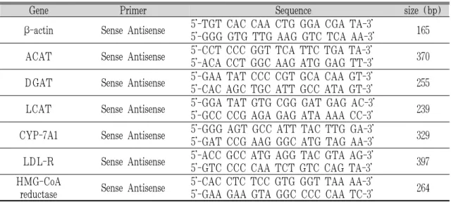
To determine the expression pattern of acyl-CoA :cholesterol acyltransferase (ACAT), diacylglycerol acyltransferase (DGAT), lecithin cholesterol acyltransferase (LCAT), Cholesterol 7 alpha hydroxylase (CYP-7A1), low density lipoprotein receptor (LDLR) and 3-hydroxy-3-methylglutaryl -Coenzyme A (HMG-CoA) reductase, 1 ㎕ of cDNA was amplified by a thermal cycler using the primers (Table 1).
Gene Primer Sequence size (bp)
β-actin Sense Antisense 5'-TGT CAC CAA CTG GGA CGA TA-3'
5'-GGG GTG TTG AAG GTC TCA AA-3' 165
ACAT Sense Antisense 5'-CCT CCC GGT TCA TTC TGA TA-3'
5'-ACA CCT GGC AAG ATG GAG TT-3' 370
DGAT Sense Antisense 5'-GAA TAT CCC CGT GCA CAA GT-3'
5'-CAC AGC TGC ATT GCC ATA GT-3' 255
LCAT Sense Antisense 5'-GGA TAT GTG CGG GAT GAG AC-3'
5'-GCC CCG AGA GAG ATA AAA CC-3' 239
CYP-7A1 Sense Antisense 5'-GGG AGT GCC ATT TAC TTG GA-3'
5'-GAT CCG AAG GGC ATG TAG AA-3' 329
LDL-R Sense Antisense 5'-ACC GCC ATG AGG TAC GTA AG-3'
5'-GTC CCC CAA TCT GTC CAG TA-3' 397
HMG-CoA
reductase Sense Antisense 5'-CAC CTC TCC GTG GGT TAA AA-3'
5'-GAA GAA GTA GGC CCC CAA TC-3' 264
Table 1. Oligonucleotide Sequences of Primers
9. Statistical analysis
Results were expressed as the mean ± standard deviation. Statistical analysis of the data was carried out by Student's t-test. A difference from the respective control data at the levels of p<0.05 and p<0.01 was regarded as statistically significant.
Ⅲ. Results
1. Fingerprint of Curcuma radix
The fingerprint of Curcuma radix with curcumine
as a reference component was produced with using
HP-TLC system (Fig. 1). The semi-quantity of
curcumine was approximately 5.25 % of Curcuma
radix extract which used in present study.
Fig. 1. HP-TLC-based fingerprint for Curcuma radix
HP-TLC analysis was performed to produce the fingerprint of Curcuma radix. 2 μL of the Curcuma radix extract and curcumine as a reference component were subjected to HP-TLC.2. Total antioxidant capacity (TAC) of Curcuma radix
In order to examine the antioxidant capacity of Curcuma radix itself, total antioxidant acpacity (TAC) was determined using in vitro assay. When the value was expressed as gallic equivalent antioxidant capacity (GEAC), the capacity of Curcuma radix showed a very high GEAC value from the lowest volume 5 ㎍ (61.5 uM GEAC) to the hight volume 1,000 ㎍ (279.2 uM GEAC) of Curcuma radix respectively (Fig. 2).
Fig. 2. Antioxidant capacity of Curcuma radix
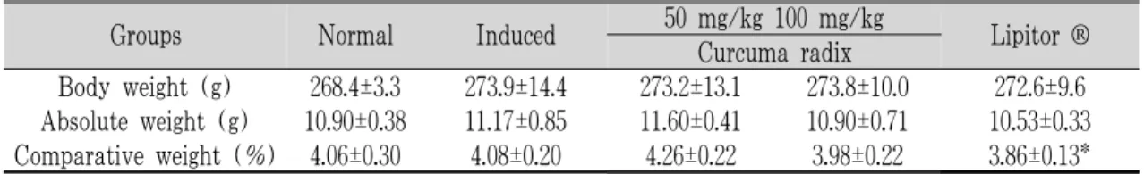
Antioxidant capacity of Curcuma radix itself was determined via (through) vitro assay, and expressed as GEAC.3. Comparison of liver weight
On the last day of experiment, the removed liver weight was compared among groups. Poloxamer 407-induced group showed a slight increase of both body weight and absolute liver weight without statistical significance. Curcuma radix 100㎎-treated group and Lipitor
®group showed a decreased pattern of absolute liver weight, however no statistical significance was observed. Only Lipitor
®group showed the significant decrease of comparative liver weight as p<0.05 (Table 2).
Groups Normal Induced 50 mg/kg 100 mg/kg
Lipitor ® Curcuma radix
Body weight (g) 268.4±3.3 273.9±14.4 273.2±13.1 273.8±10.0 272.6±9.6 Absolute weight (g) 10.90±0.38 11.17±0.85 11.60±0.41 10.90±0.71 10.53±0.33 Comparative weight (%) 4.06±0.30 4.08±0.20 4.26±0.22 3.98±0.22 3.86±0.13*
Data are expressed as mean ± standard deviation.
* : p<0.05. significant differences compared with the induced group.
Table 2. Comparison of liver weight
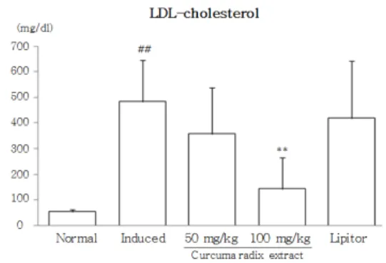
4. Serum cholesterol and triglyceride
On the last day of experiment, the serum levels of lipid parameters were compared among groups.
Poloxamer 407-injection induced severe elevation of
total cholesterol by 3 folds and especially triglyceride
by the 20 fold. Accordingly, LDL-cholesterol was
elevated by about 10 folds. Curcuma radix treatment
(100 ㎎/㎏) significantly prevented the elevation
of total cholesterol, triglyceride, and LDL-cholesterol, especially in high-dose group as p<0.01 (Fig. 3-5).
Low-dose Curcuma radix (50 ㎎/㎏) and Lipitor
®group showed a positive trend, but was not significant.
Fig. 3. Serum total cholesterol level
Rats were pre-treated with water, Curcuma radix (50, 100 ㎎/㎏) or Lipitor® (10 ㎎/㎏) before poloxamer 407 injection. On the last day of experiment, serum total cholesterol level was determined. Data are expressed as mean ± standard deviation.
##: p<0.01, significant differences compared with the normal group.
**: p<0.01, significant differences compared with the induced group.
Fig. 4. Serum triglyceride level
Rats were pre-treated with water, Curcuma radix (50, 100 ㎎/㎏) or Lipitor® (10 ㎎/㎏) before poloxamer 407 injection. On the last day of experiment, serum triglyceride level was determined.
Data are expressed as mean ± standard deviation.
##: p<0.01, significant differences compared with the normal group.
**: p<0.01, significant differences compared with the induced group.
Fig. 5. Serum LDL-cholesterol level
Rats were pre-treated with water, Curcuma radix (50, 100 ㎎/㎏) or Lipitor® (10 ㎎/㎏) before poloxamer 407 injection. On the last day of experiment, serum LDL-cholesterol level was determined. Data are expressed as mean ± standard deviation.
##: p<0.01, significant differences compared with the normal group.
**: p<0.01, significant differences compared with the induced group.
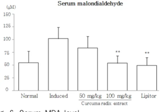
5. Serum malondialdehyde (MDA)
On the last day of experiment, the serum MDA levels were compared among groups. Poloxamer 407 injection elevated serum MDA level by 2 folds, and then pre-treatment with Curcuma radix 50 ㎎ /㎏ and 100 ㎎/㎏ ameliorated this elevation by 83
% and 53 % respectively compared to induced group while Lipitor also inhibited the elevation of MDA level by 39 % (Fig. 6).
6. Serum total antioxidant capacity (TAC)
On the last day of experiment, the serum TAC levels as gallic equivalent antioxidant capacity (GEAC) were compared among groups. Poloxamer 407 injection depleted serum TAC level by 60 % of normal level. Curcuma radix 50 ㎎/㎏ and 100
㎎/㎏ ameliorated this depletion slightly, but didn't
reach the statistical significance (Fig. 7).
Fig. 6. Serum MDA level
Rats were pre-treated with water, Curcuma radix (50, 100 ㎎/㎏) or Lipitor® (10 ㎎/㎏) before poloxamer 407 injection. On the last day of experiment, serum MDA level was determined.
Data are expressed as mean ± standard deviation.
**: p<0.01, significant differences compared with the induced group.
Fig. 7. Serum SOD level
Rats were pre-treated with water, Curcuma radix (50, 100 ㎎/㎏) or Lipitor® (10 ㎎/㎏) before poloxamer 407 injection. On the last day of experiment, serum SOD level was determined.
Data are expressed as mean ± standard deviation.
#: p<0.05, significant differences compared with the normal group.
*: p<0.05, significant differences compared with the induced group.
7. Serum superoxide dismutase (SOD)
On the last day of experiment, the serum SOD levels were compared among groups. Poloxamer 407 injection depleted serum TAC level by 18 % of normal level. Curcuma radix ameliorated significantly
this depletion especially 100 ㎎/㎏group, but Lipitor
®didn't show any changes.
8. Serum total glutathione (GSH) content
On the last day of experiment, the serum GSH content levels were compared among groups. Poloxamer 407 injection depleted serum GSH content by 6 % of normal level. Curcuma radix ameliorated slightly this depletion without statistical significance, but Lipitor
®showed significant availability as 20 % of normal level (Fig. 8).
9. Serum total glutathione reductase (GSH-Rd) On the last day of experiment, the serum GSH-Rd levels were compared among groups. Poloxamer 407 injection slightly reduced serum GSH-Rd level.
Curcuma radix completely ameliorated significantly this decrease in especially 100 ㎎/㎏ group (Fig.
9).
Fig. 8. Serum total GSH content level
Rats were pre-treated with water, Curcuma radix (50, 100 ㎎/㎏) or Lipitor® (10 ㎎/㎏) before poloxamer 407 injection. On the last day of experiment, serum GSH content was determined.
Data are expressed as mean ± standard deviation.
##: p<0.01, significant differences compared with the normal group.
*: p<0.05, significant differences compared with the induced group.
Fig. 9. Serum total GSH-Rd level
Rats were pre-treated with water, Curcuma radix (50, 100 ㎎/㎏) or Lipitor® (10 ㎎/㎏) before poloxamer 407 injection. On the last day of experiment, serum GSH-Rd was determined.
Data are expressed as mean ± standard deviation.
*: p<0.05, significant differences compared with the induced group.
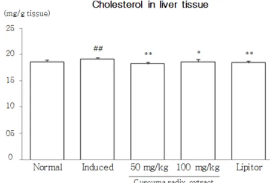
10. Cholesterol and triglyceride in liver tissue On the last day of experiment, the hepatic total cholesterol and triglyceride levels were compared among groups. poloxamer 407 injection induced slight elevation of total cholesterol and triglyceride with statistical significance (p < 0.01). Curcuma radix pre-treatment (50 ㎎/㎏ or 100 ㎎/㎏) significantly prevented the elevation of total cholesterol while didn't change the triglyceride (Fig. 10).
Fig. 10. Total cholesterol in liver tissue
Rats were pre-treated with water, Curcuma radix (50, 100 ㎎/㎏) or Lipitor® (10 ㎎/㎏)
before poloxamer 407 injection. On the last day of experiment, heaptic total cholesterol was determined. Data are expressed as mean
± standard deviation.
##: p<0.01, significant differences compared with the normal group.
*: p<0.05, **: p<0.01, significant differences compared with the induced group.
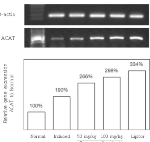
11. Gene expression of ACAT, DGAT, LCAT, LDL -receptor, and CYP-7A1 and HMG-CoA reductase To examine the effects of Curcuma radix on lipid metabolism, six gene expressions such as ACAT, DGAT, LCAT, CYP-7A1, LDL-R, and HMG-CoA reductase in liver tissues were measured. These gene expressions were calculated by comparing to expression of corresponding house keeping gene, β-actin. Overall pattern of above gene expressions didn't show any specific changes among groups treated with Curcuma radix (50 or 100 ㎎/㎏) or Lipitor
®, except ACAT gene expression.
ACAT gene expression was drastically up-regulated by poloxamer 407 injection, and then pre-treatment with Curcuma radix or Lipitor
®augmented the high expression of ACAT gene (Fig. 11). The expressions of DGAT, LCAT, and LDL-R gene were not notably changed by poloxamer 407 injection or administration of Curcuma radix. CYP-7A1 was drastically down-regulated by 407 injection, and then pre-treatment with Curcuma radix or Lipitor
®moderately ameliorated this down-regulation (Fig.
12). HMG-CoA reductase gene expression were
slightly up-regulated by 407 injection while Curcuma
radix pre-treatment normalized this gene expression
(Fig. 13).
Fig. 11. ACAT gene expression in liver tissue
Rats were pre-treated with water, Curcuma radix (50, 100 ㎎/㎏) or Lipitor® (10 ㎎/㎏) before poloxamer 407 injection. On the last day of experiment, RT-PCR was performed to compare the gene expression of ACAT in hepatic tissue. The values were expressed as ratio of ACAT to β-actin, and then normal group.Fig. 12. CYP-7A1 gene expression in liver tissue
Rats were pre-treated with water, Curcuma radix (50, 100 ㎎/㎏) or Lipitor® (10 ㎎/㎏) before poloxamer 407 injection. On the last day of experiment, RT-PCR was performed to compare the gene expression of CYP-7A1 in hepatic tissue.The values were expressed as ratio of CYP-7A1 to β-actin, and then normal group.
Fig. 13. HMG-CoA reductase gene expression in liver tissue
Rats were pre-treated with water, Curcuma radix (50, 100 ㎎/㎏) or Lipitor® (10 ㎎/㎏) before poloxamer 407 injection. On the last day of experiment, RT-PCR was performed to compare the gene expression of HMG-CoA reductase in hepatic tissue. The values were expressed as ratio of HMG-CoA reductase to β-actin, and then normal group.
Ⅳ. Discussion
In bloodstream, the lipids should be combined with apoprotein resulting in forming one of lipoproteins: chylomicrons, very low-density lipoproteins (VLDL), intermediate-density lipoproteins (IDL), low-density lipoproteins (LDL), or high- density lipoproteins (HDL)
19. The pathologic changes of blood vessels result from both the quantitative overload of lipid or qualitative unbalance of lipoproteins, for example, HDL- to LDL-cholesterol
20. The current major lipid lowering drugs are belonged in one of four classes; 3-hydroxy-3-methylglutaryl coenzyme A (HMG-CoA) reductase inhibitors commonly referred to statins, bile acid sequestrants, nicotinic acid, and fibric acids
21,22.
However, these drugs have limitations due to
low clinical efficacy as well as often adverse effects associated with impairing quality of life. The statins are the most potent lipid-lowering agents currently available, which can lower LDL and triglyceride levels, but may induce myopathy and an elevation of liver enzyme level
23,24. The bile acid sequestrants, nicotinic acid, and fibric acids lower lipid levels but they show adverse effects such as gastrointestinal distress, constipation, flushing, hyperglycemia, hyperuricemia, or hepatotoxicity
25,26.
In traditional Korean medicine, hyperlipidemia is regarded as retention of phlegm and fluid disease, and its etiology and pathogenesis is explained as status of spleen-deficiency and phlegm-stagnation, accumulation and stasis of damp-heat, and Qi and blood stagnation
27,28. Recently, many herbal formula or medicinal plants have been studied as strong candidates for lipid-lowering drugs like GamiSamgieum, Ojung-hwan, and Typhae Pollen
29-31. These drugs are supposed to improve quality of life as well as lowering lipid levels in blood.
In present study, investigated the lipid-lowering action of Curcuma radix using poloxamer 407 injection rat model. Poloxamer 407 is a general lipase inhibitor, alters the normal elimination of lipid into bile
32. As expectation, poloxamer 407 injection induced drastic elevation of lipid levels, total cholesterol, LDL- cholesterol, and triglyceride by about 3 folds, 10 folds, and about 20 folds respectively in this experiment. However, pre-treatment with Curcuma radix significantly attenuated the abnormality of all those lipid parameters, especially in high-dose group (100 mg /kg). Low-dose of Curcuma radix (50 mg /kg) didn't reach the statistical significance.
Lipitor
®was herein adapted as a positive control drug. Lipitor
®is the trade name of Atorvastatin (INN). This drug is a member of statins, which is
the top-selling branded pharmaceutical in the world
33. Interestingly, this drug however couldn't show any significant efficacy, so Curcuma radix was better than Lipitor
®at least in this result.
In the observation of hepatic lipids, cholesterol and triglyceride was significantly increased by poloxamer 407 injection. And, pre-treatment with Curcuma radix significantly prevented the abnormal accumulation of cholesterol but not triglyceride.
These above results support that Curcuma radix has anti-lipidemic effects. One group investigated the lipid-lowering effect of a mixture of Curcuma radix and Angelica keiskei Koidzumi using poloxamer 407-induced hyperlipidemia model
34. This group also reported the clinical efficacy of the same supplement focussed hypolipidemic action and antioxidant effect
35.
In order to further mechanism study of Curcuma radix, six lipid metabolism-associated gene expression in hepatic tissue were examined. The lipid supply come from the diet in the small intestine being esterified to form triglyceride and cholesterol, or from endogenous fatty acid synthesis, primarily in the liver
36. Statins was known to inhibit HMG-CoA reductase which is a rate-limiting enzyme of cholesterol synthesis in the liver. Statins also stimulate LDL receptor in the liver resulting in clearance of LDL from the bloodstream
37. In this study, Lipitor
®as well as Curcuma radix drastically low-regulated the gene expression of HMG-CoA reductase in liver tissue. This proposes that Curcuma radix may play a role as statins.
CYP-7A1 is the rate-limiting enzyme in the
synthesis of bile acid from cholesterol, then it
plays a key role eliminating cholesterol normally
into bile in liver
38,39. This result is correspondence
to the result from of lowered hepatic total cholesterol
level. Strangely, ACAT is the primary enzyme in the intestinal cholesterol absorption and synthesis of cholesterol via ACAT-mediated esterification of cholesterol in liver
40,41Unlikely a expectation, Curcuma radix pre-treatment enhanced ACAT gene expression, and its cause is not explained yet. The expressions of DGAT, LCAT, and LDL-R gene were not notably changed by poloxamer 407 injection or administration of Curcuma radix.
On the other hand, poloxamer 407-induced hyperlipidemia caused the status of intensive oxidative stress, evidenced by high level of serum MDA and depletion of TAC, SOD activity, and GSH system.
Oxidative stress represents an imbalance between the production of reactive oxygen species (ROS) and an ability to readily detoxify the reactive intermediates
42. Oxidative stressors are normally eliminated by various antioxidant systems including catalase, SOD, and the glutathione oxidation/reduction system
43,44. MDA is major maker of lipid peroxidation by ROS. In this study, Curcuma radix pre-treatment significantly normalized or attenuated the alteration of antioxidant bioparameters. Oxidative stress is an early event in the evolution of hyperlipidemia, and affects the development of arteriosclerosis and myocardial infarction
45. This result is in accordance with others reports which used curcumin, an active compound of Curcuma radix
46,47.
This study is the first to explore the hypolipidemic effect and antioxidant activity of Curcuma radix in one animal model. And, this study produced a scientific basis for more appropriate clinical application of Curcuma radix and may provide a notable information for drug development of using this herb in the future.
Ⅴ. Conclusion
This study investigated the hypolipidemic and antioxidant effects of Curcuma radix using rat model induced by poloxamer 407 injection. Serum lipid parameters and oxidative stress-associated biomarkers were determined. Additionally, hepatic cholesterol and triglyceride as well as lipid metabolism -associated gene expressions were observed in hepatic tissue.
1. Poloxamer 407 elevated serum cholesterol level by 3 folds, and then pre-treatment with Curcuma radix 50 ㎎/㎏ and 100 ㎎/㎏ ameliorated this elevation as 85 % and 50 % of induced group respectively.
2. Poloxamer 407 elevated serum triglyceride level by 20 folds, and then pre-treatment with Curcuma radix 50 ㎎/㎏ and 100 ㎎/㎏ ameliorated this elevation by 67 % and 23 % of induced group respectively.
3. Poloxamer 407 elevated serum LDL-cholesterol level by 9 folds, and then pre-treatment with Curcuma radix 50 ㎎/㎏ and 100 ㎎/㎏ ameliorated this elevation as 74 % and 30 % of induced group respectively.
4. Poloxamer 407 elevated serum MDA level by 2 folds, and then pre-treatment with Curcuma radix 50 ㎎/㎏ and 100 ㎎/㎏ ameliorated this elevation as 83 % and 53 % of induced group respectively.
5. Poloxamer 407 reduced serum TAC level by 60%
of normal, and then pre-treatment with Curcuma radix 50 ㎎/㎏ and 100 ㎎/㎏ moderately ameliorated this reduction.
6. Poloxamer 407 reduced serum SOD level by 18
% of normal level, and then pre-treatment with
Curcuma radix significantly ameliorated this
reduction especially in 100 ㎎/㎏ group.
7. Poloxamer 407 reduced serum GSH level by 6
% of normal level, and then pre-treatment with Curcuma radix 50 ㎎/㎏ and 100 ㎎/㎏ slightly ameliorated this reduction without statistical significance.
8. Poloxamer 407 moderately reduced serum GSH -redauctase level, and then Curcuma radix completely ameliorated this reduction.
9. Poloxamer 407 elevated hepatic cholesterol level, and then pre-treatment with Curcuma radix 50
㎎/㎏ and 100 ㎎/㎏ significantly ameliorated this elevation.
10. Poloxamer 407 elevated hepatic triglyceride level, but pre-treatment with Curcuma radix didn't affect the elevation of triglyceride in hepatic
tissue.
11. Poloxamer 407 drastically up-regulated ACAT gene expression, and then Curcuma radix augmented this change.
12. Poloxamer injection drastically down-regulated CYP-7A1 but up-regulated HMG-CoA gene expression, and then Curcuma radix almost completely ameliorated these changes.
From above results, the hypolipidemic and
antioxidant properties of Curcuma radix were
evidenced. This study would produce the scientific
basis for the clinical application of Curcuma radix
and development of hypolipidemics using this herbal
plant in the future.
鬱金 水抽出物의 Poloxamer 407로 유발시킨 모델에서의 抗高脂血症 및 抗酸化 效果
박소애, 조현경, 유호룡, 김윤식, 설인찬, 안정조 대전대학교 대학원 한의학과 한방내과학 교실
초록
목적 :
본 실험에서는 울금의 항고지혈증 효과와 항산화 작용을 증명하고 향후 항고지혈증 약물로서의 개발 가능성을 연 구하고자 하였다.
방법 :
울금의 수추출물을 선처치한 후, poloxamer 407의 주사로 유발시킨 고지혈증 모델에서 혈중의 지질 및 산화적 스트 레스 관련 생물학적 지표들, 지질대사에 관련된 유전자들의 변화를 관찰하였다.
결과 :
1. Poloxamer 407은 혈중 콜레스테롤과 중성지방, LDL-콜레스테롤, MDA의 농도를 상승시켰으며, 울금 수추출물은 대조군 에 비하여 농도를 억제시켰다.
2. Poloxamer 407은 혈중 TAC의 활성도, SOD의 활성도, GSH, GSH-reductase를 감소시켰으며, 울금 수추출물은 활성도를 억제시켰다.
3. Poloxamer 407은 간 조직에서 총 콜레스테롤을 유의하게 축적시켰으며, 울금 수추출물은 총 콜레스테롤의 축적을 유의하 게 억제시켰다.
4. Poloxamer 407은 간 조직에서 ACAT의 유전자 발현을 유의하게 증가시켰는데, 울금 수추출물은 이를 더욱 촉진하였다.
5. Poloxamer 407은 간 조직에서 CYP-7A1의 발현은 감소시키고 HMG-CoA는 증가시켰는데, 울금 수추출물은 이러한 변화 를 매우 유의하게 억제시켰다.
결론 :
위 연구 결과들을 통하여 울금의 항고지혈증과 항산화 효능을 동물모델에서 증명하였으며, 향후 울금을 이용한 고 지혈증 및 관련 질환에 대한 임상적용 및 예방과 치료를 위한 약물의 개발에 응용할 수 있을 것으로 사료된다.
중심단어 :