紫河車 藥鍼이 실험적으로 유발된 흰쥐의 자궁내막증에 미치는 영향
1 세명대학교 한의학과 한방부인과교실, 2 세명대학교 한의학과 사상체질의학교실 유영기 1 , 김형준 1 , 신미란 2 , 이동녕 1
ABSTRACT
Effects of Hominis Placenta Pharmacopuncture Therapy on the Experimentally-induced Endometriosis in the Rats Yung-Ki Yoo 1 , Hyung-Jun Kim 1 , Mi-Ran Sin 2 , Dong-Nyung Lee 1
1 Dept. of Oriental Gynecology, Se-Myung University
2 Dept. of Sasang Constitutional, Se-Myung University
Objectives: This study was performed to investigate the effects of Hominis Placenta pharmacopuncture (HPP) therapy on the experimentally-induced endometriosis in the rats.
Materials and Methods: Endometriosis was induced in rats by autotransplanting uterine tissue to the peritoneum and divided them into three groups: (1) sham-operated group (n=8), (2) surgically induced endometriosis and untreated control group (n=8), (3) surgically induced endometriosis and HPP treated group. Sham-operated group and control group were inject with normal saline once a every other day for 30days, while treated group was injected with HPP extract once a every other day for same duration. Injected point of HPP and normal saline were subcutaneous tissue at Gwanwon (CV4) acupoint. Then we measured the body weight, the volume of endometriotic implants, the weigh of uterus and ovaries, and investigated the concentration of cytokines (MCP-1, TNF-α) in peritoneal fluids. Histopathology, immunohistochemisty for COX-2 and VEGF, and histochemistry for mast cell in transplanted uterine tissue were performed.
Results: The volume (mm 2 ) of endometriotic implants in HPP treated group (55.4±41.6) was significantly decreased (p<0.01) compared with control group (140±66.1).
And the concentration (pg/ml) of MCP-1 in peritoneal fluids in HPP treated group (1117.6±60.5) was significantly decreased (p<0.01) compared with control group (1446.2±280.3). The concentration (pg/ml) of TNF-α in peritoneal fluids in HPP treated group (80.6±31.4) was decreased (p<0.01) compared with control group (145.3±86.9).
Histopathologically, proliferation of endometriotic epithelia, infiltration of inflammatory cell and angiogenesis in transplanted uterine tissue of HPP treated group were weakly observed than those of control group. The COX-2 expression in endometrial, epithelial and stromal cells in transplanted uterine tissue of HPP treated group was decreased compared with control group. The VEGF expression of endometriotic epithelia, neovascular endothelia and stromal cell in transplanted uterine tissue of HPP treated group were weakly observed than those of control goup.
Conclusions: HPP is effect on Endometriosis of rats by Experimentally-induced.
Key Words: Hominis Placenta Pharmacopuncture, Endometriosis, MCP-1 TNF-α, COX-2, VEGF
7)
Corresponding author(Dong-Nyung Lee) : Se-Myung University Chung-Ju Oriental Medical Hospital, Bong-Bang-dong, Chung-Ju-si, Chungcheongbuk-do, Korea
Tel : 043-841-1733 E-mail : [email protected]
Ⅰ. 서 론
자궁내막증은 자궁내막조직(선과 기질) 이 자궁강 이외의 부위에 위치하여 1) , 월 경곤란증, 성교통, 배변통 등과 함께 만성 골반통을 초래하는 질환으로, 종종 황체 기 출혈과 불임을 유발하고 2) , 만성화되거 나 침윤성으로 발전할 수 있어 조기진단과 함께 적절한 치료가 필요한 질환이다 3,4) .
자궁내막증의 원인은 월경역류설과 함 께, 자연 발생적 또는 유도된 체강상피 의 화생에 의해 발생한다는 증거도 있으 며, 유전적 소인, 환경적 요인, 면역학적 및 내분비적 기능의 변화 등도 중요한 역할을 하는 것으로 알려져 있다 5) .
서양의학적인 치료는 주로 진통제와 함께 혈중 에스트로겐을 억제하는 약물 치료를 하고 있지만 경우에 따라 골반강 내 유착된 내막조직을 제거하는 외과적 치료를 하고, 이 외에 기대요법 및 병합 요법 등을 하고 있다 1) . 그러나 수술은 재 발이 잦고 약물치료는 체중증가, 유방축 소, 다모증, 위축성질염, 질출혈, 질건조 증, 안면홍조, 여드름, 골다공증, 우울증, 피로 등의 부작용 등을 나타내며 6,7) , 또한 약물치료로 무월경 상태가 되면 환자는 불임에 대한 두려움도 가질 수 있다 8) .
자궁내막증 치료와 관련된 한의학 연 구로는 活血化瘀작용하는 약물을 이용한
연구 10-13) , 항염증 및 항종양 작용이 있는
약물을 이용한 연구 14-16) 그리고, 면역감 시기능과 밀접한 관계가 있다 9) 는 점에서 八珍湯 17) , 黃芪 18) 등이 연구되고 있다.
또한 치료방법으로는 주로 약물복용을 전제로 한 복합처방 또는 단일 약물에 대한 연구가 많지만, 보류관장법 14) , 약침
19,20)
처럼 다양한 치료법이 시도되고 있 다. 그런데 보류관장법은 실제 임상에 적용하기 힘든 치료법이지만 약침은 임 상적 활용면에서 좋은 점이 있을 것으로 사료된다.
이에 저자들은 태반에서 추출한 紫河 車 21) 가 補益藥으로 補氣・養血・益精・治虛 損 등 인체의 저항력과 면역력을 향상시 키는 효능이 있고, 임상에서 다양한 증 상에 藥鍼으로 많이 활용되고 있기에
22-25) , 자궁내막증에 미치는 영향을 확인
하기 위하여 본 연구를 실시하였다.
자궁내막증에 미치는 영향을 조사하기 위해, 자궁의 자가이식법으로 자궁내막 증을 유발된 흰쥐를 대조군과 紫河車 藥 鍼을 투여한 처치군으로 분류하고, 각 군의 체중, 이식자궁의 체적, 자궁과 난소 의 중량, 복강 세척액내 cytokine(MCP-1, TNF-α)의 함량을 측정하고, 병리조직학 적검사, 면역조직학적검사(COX-2, VEGF), 비만세포 조직학적 검사를 실시한 결과, 紫河車 藥鍼이 자궁내막증 발생 억제에 미치는 기전을 확인하고, 유의한 결과를 얻었기에 보고하는 바이다.
Ⅱ. 재료 및 방법
1. 실험동물 및 자궁내막증 유발
8주령의 SPF 암컷 Sprague-Dawley계
흰쥐(나라바이오텍, 한국)를 구입하여 세
명대학교 청정동물사육실(온도 23±1℃, 습
도 55±5%, 명암주기 12시간)에서 일주일
간 적응시킨 후 실험에 사용하였다. 전 실
험기간동안 사료(제일제당, 한국)와 음수
는 충분한 양을 제공하였다. 본 동물실
험은 세명대학교 동물실험윤리위원회의
승인(smeac 11-06-02)하에 실시되었다.
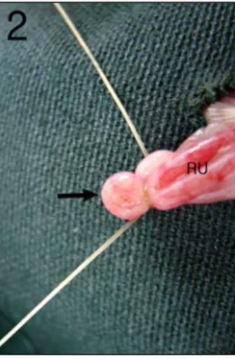
실험적 자궁내막증은 Vernon and Wilson 26) 의 방법에 준하여 자궁자가이식수술을 실시하여 유발시켰다. 실험개시일 오전 8시에 흰쥐의 질 도말표본(vaginal smear) 을 만들어 Giemsa 염색하고, 유핵상피세 포가 주로 관찰되는 발정전기인 개체를 선별하였다(Fig. 1). Zoletil 50(Virbac Lab., France) 0.2 ml을 복강주사하여 마취시 킨 후 개복수술하고 자궁각(uterine horn) 을 기준으로 우측 자궁을 고리모양으로 묶고(Fig. 2) 자궁의 일부를 적출하였다.
적출된 자궁조직을 37℃ Creb's sol.에 담 근 상태에서 종축으로 절단하고 4×4 mm 크기의 절편을 만들었다. 절편의 자궁외 막이 복벽으로 향하도록 좌측복벽에 위 치시키고 catgut(B. Braun Surgical Sdn.
Bhd., Malaysia)으로 자궁조직의 중앙부 위를 1회 결찰 하여(Fig. 3) 자궁내막증 을 유발시켰다.
Fig. 1. Cytology of vaginal smear of rat.
Nucleated epithelia (arrows) are major cell type at proestrus stage. Giemsa's stain, bar=20 μm.
Fig. 2. Surgical procedure of partial right uterectomy.
A portion of right uterine horn (arrow) was ligated and taked out. RU : right uterine horn.
Fig. 3. Surgical procedure of auto- transplantation of uterus.
Fragment (4×4 mm) of ectomized right uterus (arrow) was transplanted to left abdominal wall by single tie of catgut suture.
2. 약침액 및 약침액의 처치
약침액은 100g의 紫河車를 둥근 flask
에 2ℓ의 증류수와 함께 넣어 수증기 증
류법으로 1600ml의 증류액을 만들고 냉
각, 여과하고, 이 여과액을 100ml로 감압,
농축한 후 pH METER를 이용해 pH 7
로 조정해 멸균과정을 거쳐 냉장 보관하
였다.
紫河車 藥鍼은 사람의 關元穴(CV4)에 해당하는 좌우 상전장골극(anterior superior iliac spine) 사이의 정중 복부 피하에 주 사하였다.
3. 실험군의 분류
아래와 같이 실험군을 분류하고 각 군 당 8마리의 흰쥐를 배정하였다. 음성군 및 대조군에는 생리식염수 0.2 ml를, 처 치군에는 紫河車 藥鍼 0.2 ml를 2일 간 격으로 30일간 15회 피하 주입하였다.
음성군(sham group) : 우측 자궁의 일부를 적출하고, 이식술은 실시하지 않 고 생리식염수를 처치한 군.
대조군(control group) : 자궁의 자가 이식술을 실시하여 자궁내막증을 유발하 고 생리식염수를 처치한 군.
처치군(treated group) : 자궁의 자가 이식술을 실시하여 자궁내막증을 유발하 고 紫河車 藥鍼액을 처치한 군.
4. 체중의 측정
실험 개시일(0), 10, 20, 종료일(30일) 에 모든 개체의 체중을 측정하였다.
5. 이식자궁조직의 체적(volume) 측정 이식자궁조직의 크기 차이는 그 체적 으로 평가하였다. 실험종료일에 부검하 여 이식자궁조직 및 주변의 복벽을 넓게 적출하여 이식자궁조직의 크기를 digital calipper(Mitutoyo, CD-15CP, Japan)로 장경 및 단경을 측정하고 아래와 같은 공식으로 그 체적을 구하였다.
체적(volume)= 장경×단경×단경 2
6. 자궁 및 난소 무게의 측정
실험종료일에 부검하여 좌측 자궁 및 양측 난소를 적출하고 주변의 지방조직 및 결합조직을 세밀하게 분리한 후 무게 를 측정하였다.
7. 복막강 세척액내 cytokine 함량 측정 실험종료일에 흰쥐를 가볍게 ether로 마취시키고 심장을 통해서 전혈을 채취 하고 원심하여 혈청을 분리하였다. 전혈 을 채취한 후 복강내로 동일 개체에서 채취한 분리 복혈청 0.2 ml를 주입하고 복강을 노출시켰다. 멸균 복막대봉으로 복막강이 충분히 세척되도록 복강장기를 저은 다음 흡입관을 이용하여 복막강 세 척액을 회수하였다. 회수된 복막강 세척 액은 검사 전까지 -70℃에 보관하였다.
1) Monocyte Chemoattractant Protein-1 (MCP-1) 함량 측정
복막강 세척액내 MCP-1 함량은 Enzyme -Linked ImmunoSorbent Assay(ELISA) kit(Pierce, ERMCP1, USA)로 측정하였 다. 표준액 및 검액은 1시간, biotinylated antibody reagent는 1시간, streptavidin-HRP solution은 30분간 반응시켰다. 각 반응 사이에는 0.05M PBS(pH 7.5) 용액으로 충분하게 세척하였다. 최종적으로 TMB substrate solution을 30분간 반응시켜 발 색시킨 후 stop solution으로 반응을 정지 시켰으며, ELISA 판독기(Molecular Devices, E10514, USA)로 450 nm에서 흡광도를 측정하였다.
6단계의 MCP-1 표준액(1500, 600, 240,
96, 38, 0 pg/ml)에서 측정된 흡광도로부
터 다음과 같은 직선회귀방정식이 구해
졌으며, 검액의 흡광도를 이 방정식에 적
용하여 MCP-1의 함량을 구하였다.
MCP-1 함량=(검액의 흡광도×1431)+94 2) Tumor Necrosis Factor-α(TNF-α)
함량측정
복막강 세척액내 TNF-α 함량은 ELISA kit로 측정하였다. 표준액 및 검액은 1시 간, biotinylated antibody reagent는 1시 간, streptavidin-HRP solution은 30분간 반응시켰다. 각 반응사이에는 0.05M PBS (pH 7.5) 용액으로 충분하게 세척하였다.
최종적으로 TMB substrate solution을 30분간 반응시켜 발색시킨 후 stop solution 으로 반응을 정지시켰으며, ELISA 판독 기(Molecular Devices, E10514, USA)로 450 nm에서 흡광도를 측정하였다.
6단계의 TNF-α 표준액(2500, 833, 278, 93, 31, 0 pg/ml)에서 측정된 흡광도로부 터 다음과 같은 직선회귀방정식이 구해 졌으며, 검액의 흡광도를 이 방정식에 적용하여 TNF-α의 함량을 구하였다.
TNF-α 함량=(검액의 흡광도×755)-32
8. 병리조직학적 검사
실험 종료일에 모든 개체를 부검하여 얻은 이식자궁조직, 좌측 자궁 및 좌측 난소를 10% 중성 포르말린에 2일간 고정 하였다. 고정된 조직을 알콜탈수과정을 거 쳐서 파라핀 포매하고 5 μm의 박절편을 제작하였다. 제작된 박절편을 Hematoxylin -Eosin 염색하여 광학현미경으로 병리조 직학적 소견을 관찰하였다.
9. COX-2 면역조직화학적 검사
이식자궁조직의 박절편을 poly-L-lysine (Sigma, USA)으로 처리된 슬라이드에 부착하여 ABC 법으로 COX-2 면역조직 화학 염색을 다음과 같이 실시하였다.
탈파리핀과정과 함수과정을 거친 이식
자궁조직 절편을 protease K(Roche, Germany) 용액으로 20분간 처리하여 조 직내 항원을 부활시켰다. 내인성 과산화 반응을 억제하기 위해서 3% H 2 O 2 용액 에 60분간 처리한후 0.5M PBS(pH 7.5) 로 3회 세척하였다. 비특이 반응을 억제 하기 위해서 0.3% Triton-X100, 1% BSA 및 3% normal rabbit serum이 함유된 PBS로 30분간 처리한 후 1:200으로 희 석된 goat polyclonal anti-COX-2(Santa Cruz Biotechnology, USA)를 냉장상태 에서 24시간 반응시켰다. 조직을 PBS로 3회 세척한 후 biotinylated anti-goat antibody 및 avidin-biotin peroxidase complex(Vector Laboratories, USA)를 각각 차례로 1시 간씩 반응시키고 diaminobenzidine(Sigma, USA)으로 발색시켜 광학현미경으로 이 식자궁조직의 점막상피세포 및 간질에 침윤된 세포에서의 양성반응 정도를 관 찰하였다.
10. Vascular Endothelial Growth Factor (VEGF)의 발현검사
이식자궁조직의 박절편을 poly-L-lysine 으로 처리된 슬라이드에 부착하여 ABC 법으로 VEGF 면역조직화학 염색을 다 음과 같이 실시하였다.
탈파리핀과 함수과정을 거친 이식자궁 조직 절편을 protease K 용액으로 20분 간을 처리하여 조직내 항원을 부활시켰 다. 내인성 과산화 반응을 억제하기 위 해서 3% H 2 O 2 용액에 60분간 처리한 후 0.5M PBS(pH 7.5)로 3회 세척하였다.
비특이 반응을 억제하기 위해서 0.3%
Triton-X100, 1% BSA 및 3% normal
horse serum이 함유된 PBS로 30분간 처리
한 후 1:200으로 희석된 mouse monoclonal
anti-VEGF(Santa Cruz Biotechnology, USA)를 냉장상태에서 24시간 반응시켰다.
조직을 PBS로 3회 세척한 후 biotinylated anti-mouse antibody 및 avidin-biotin peroxidase complex를 각각 차례로 1시간 씩 반응시키고 diaminobenzidine으로 발 색시켜 광학현미경으로 이식자궁조직에서 증식된 신생혈관 및 간질에 침윤된 세포 에서의 양성반응 정도를 관찰하였다.
11. 비만세포 수의 측정
탈파리핀과 함수과정을 거친 이식자궁 조직 절편을 Toludine Blue 염색법으로 비만세포를 표지하고 이식자궁조직에 침 윤된 비만세포수를 모두 계수하였다.
12. 통계처리
측정된 항목의 결과에 대한 통계는 SPSS(SPSS 10.1 for Windows, USA)를 이용하여 student's t-test를 실시하여 검 증하였다.
Ⅲ. 결 과
1. 체중의 차이
음성군 및 대조군 모두에서 실험개시 일 부터 실험종료일 까지 완만한 체중의 증가가 있었으며, 군간에 유의성 있는 차이는 없었다. 처치군은 대조군에 비해 서 실험 10일에 유의성 있게 체중이 높 았으나 그 이후부터 실험종료일까지 유 의성 있는 차이는 없었다(Table 1).
Group Body weight (g) 0 10 20 30 days Sham
(n=8) 208±6* 243±9 256±8 268±9 Control
(n=8) 211±3 239±7 254±3 269±5 Treated
(n=8) 209±5 248±8 # 259±9 272±10
* : data expressed as Mean±S.D.
Sham : right partial uterectomy and saline pharmacopuncture therapy
Control : right partial uterectomy and uterine autotransplantation and saline pharmacopuncture therapy
Treated : right partial uterectomy and uterine autotransplantation and Hominis Placenta Pharmacopuncture Therapy
# : Statistically significant compared with control group (# : p<0.05)
Table 1. Effects of Hominis Placenta Pharmacopuncture Therapy on the Body Weight of Rats with Experimentally- induced Endometriosis
2. 이식 자궁조직의 육안 소견
대조군 및 처치군의 모든 예에서 이식 자궁조직은 좌측 복벽에 비교적 둥근 형 태로 증식되어 있었다.
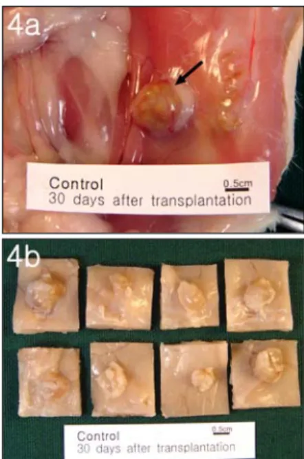
대조군의 8예 중 1예는 황회색의 내용 물이 차 있는 덩어리 형태로 관찰되었으 며(Fig. 4a), 7예는 장액성 물질이 차 있 는 낭포상 구조를 하고 있었다(Fig. 4b).
처치군의 이식자궁조직은 대조군에 비해
서 전반적으로 작은 크기로 관찰되었으며
(Fig. 5a), 8예 중 7예는 장액이 차 있는
낭포상의 형태를, 그리고 1예는 치밀한
덩어리 형태로 증식되어 있었다(Fig. 5b).
Fig. 4. Gross Structure of Transplanted Uterine Tissue of Control Group.
(4a) : Presentative case, (4b) : All cases (formalin fixed) of control group. Note cystic development of the implanted tissues.
Fig. 5. Gross Structure of Transplanted Uterine Tissue of Treated Group (Hominis Placenta Pharmacopuncture Therapy).
(5a) : Presentative case, (5b) : All cases (formalin fixed) of pharmacopuncture with Jahageo group.
Compare the sizes with three of Fig. 4b.
3. 이식자궁조직 체적(volume)의 차이 실험종료일에 측정한 이식자궁조직의 체적은 처치군이 대조군에 비해서 유의 성 있게 작았다(Table 2).
Group Volume of Transplanted Uterine Tissue (mm 3 ) Control (n=8) 140.2±66.1*
Treated (n=8) 55.4±41.6 ##
* : data expressed as Mean±S.D.
Control : right partial uterectomy and uterine autotransplantation and saline pharmacopuncture therapy
Treated : right partial uterectomy and uterine autotransplantation and Hominis Placenta Pharmacopuncture therapy
## : Statistically significant compared with control group (## : p<0.01)
Table 2. Effects of Hominis Placenta Pharmacopuncture Therapy on the Volume of Transplantated Uterine Tissue in Rats with Experimentally-induced Endometriotiosis
4. 자궁 및 난소 중량의 차이
실험종료일에 측정한 좌측 자궁의 중량 은 대조군이 음성군에 비해서 낮았으나, 군간에 유의성 있는 차이는 없었다. 또한 처치군은 대조군에 비해서 높았으나, 군 간에 유의성 있는 차이는 없었다(Table 3).
난소의 중량은 대조군이 음성군에 비 해서 낮았으나, 군간에 유의성 있는 차 이는 없었다. 또한 처치군은 대조군에 비해서 높았으나 군간에 유의성 있는 차 이는 없었다(Table 3).
5. Tumor Necrosis Factor-α(TNF-α) 함량의 차이
실험종료일에 측정한 복막강세척액내
TNF-α의 함량은 대조군이 음성군에 비해
서 유의성 있게 높았다. 처치군의 TNF-α
의 함량은 대조군에 비해서 현저히 낮았
으나 군간에 유의성 있는 차이는 관찰되 지 않았다(Table 4).
Group Left Uterus (mg)
Ovaries (mg) Sham (n=8) 171±28* 88±14 Control (n=8) 161±27 79±7 Treated (n=8) 177±28 86±7
* : data expressed as Mean±S.D.
Sham : right partial uterectomy and saline pharmacopuncture therapy
Control : right partial uterectomy and uterine autotransplantation and saline pharmacopuncture therapy
Treated : right partial uterectomy and uterine autotransplantation and Hominis Placenta Pharmacopuncture therapy
Table 3. Effects of Hominis Placenta Pharmacopuncture Therapy on the Weight of Left Uterus and Ovaries in Rats with Experimentally-induced Endometriosis
Group Tumor Necrosis Factor-α (pg/ml) Sham (n=8) 39.3±25.3*
Control (n=8) 145.3±86.9 ##
Treated (n=8) 80.6±31.4
* : data expressed as Mean±S.D.
Sham : right partial uterectomy and saline pharmacopuncture therapy
Control : right partial uterectomy and uterine autotransplantation and saline pharmacopuncture therapy
Treated : right partial uterectomy and uterine autotransplantation and Hominis Placenta Pharmacopuncture therapy
## : Statistically significant compared with sham group (## : p<0.01)
Table 4. Effects of Hominis Placenta Pharmacopuncture Therapy on the TNF-α Values in Peritoneal Fluids of Rats with Experimentally-induced Endometriosis
6. Monocyte Chemoattractant Protein-1 (MCP-1) 함량의 차이
실험 종료일에 측정한 복막강세척액내
MCP-1 함량은 대조군이 음성군에 비해 서 유의성 있게 높았다. 처치군의 MCP-1 함량은 대조군에 비해서 유의성 있게 낮 았다(Table 5).
Group Monocyte Chemoattractant Protein-1 (pg/ml) Sham (n=8) 714.7±216.8*
Control (n=8) 1446.2±280.3 ###
Treated (n=8) 1117.6±60.5 ##
* : data expressed as Mean±S.D.
Sham : right partial uterectomy and saline pharmacopuncture therapy
Control : right partial uterectomy and uterine autotransplantation and saline pharmacopuncture therapy
Treated : right partial uterectomy and uterine autotransplantation and Hominis Placenta Pharmacopuncture therapy
### : Statistically significant compared with sham group (### : p<0.001)
## : Statistically significant compared with control group (## : p<0.01)
Table 5. Effects of Hominis Placenta Pharmacopuncture Therapy on the MCP-1 Values in Peritoneal Fluids of Rats with Experimentally-induced Endometriosis
7. 병리조직학적 소견 1) 이식자궁조직
대조군의 모든 예에서 이식자궁조직은
낭(cyst)의 형태로 증식되어 있었다. 단
층 또는 중층으로 이루어진 자궁내막,
자궁중막 및 자궁외막의 모든 구조가 관
찰되었다. 대조군 8례 중 2례는 낭내에
호중구가 가득 차 있었으며 나머지 6예
는 소수의 호중구 및 탈락상피세포가 관
찰되었다. 대조군 8례중 5례는 내막의
기질내에 자궁선이 발달되어 있었으며,
3례에서는 기질의 증식이 두드러지게 관
찰되었다. 대조군의 모든 예에서 이식자
궁조직과 복벽사이의 유착부위에는 결합
조직세포 및 신생모세혈관의 증식이 관
찰되었으며, 이부위에는 호중구, 대식세 포, 림프구 및 형질세포의 침윤이 현저 하였다. 대조군 8례 중 6례에서는 이식 자궁조직이 복벽의 근층까지 침윤 증식 되어 있었다(Fig. 6).
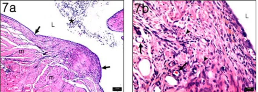
처치군에서도 이식자궁조직은 대조군 에서와 같이 자궁내막, 자궁중막 및 자 궁외막의 구조를 모두 유지하고 있었으 나 대조군에 비해서 자궁 점막상피세포, 기질세포 및 자궁선의 발달은 미약하였 다. 이식자궁조직과 복벽이 유착된 부위 에서의 신생모세혈관 증식과 호중구, 대 식세포, 림프구 및 형질세포의 침윤 등
은 대조군에 비해서 미약하게 관찰되었 다(Fig. 7).
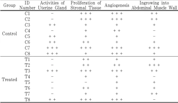
주요 병리조직학적 소견은 처치군은 대조군에 비해서 자궁선의 발달, 기질의 증식, 신생혈관의 증식 및 복벽근층내로 의 성장 등의 소견이 완화되어 관찰되었 다(Table 6).
2) 자궁 및 난소
음성군, 대조군 및 처치군의 모든 예 에서 좌측 자궁은 정상범위의 소견을 나 타내었으며, 난소도 모든 군에서 다양한 단계의 난포 및 황체가 발달되고 있는 정상범위의 소견만이 관찰되었다.
Fig. 6. Histological Structure of Transplanted Uterine Tissue of Control Group.
(6a) : Stromal hyperplasia and proliferation of connective tissue (arrow heads) into abdominal muscle layer (m) are prominent. L : lumen of transplanted uterine tissue. * : desquamated epithelia and neutrophils. bar=100 μm
(6b) : Higher magnification of Fig. 6a. Proliferation of connective tissue, infiltration of inflammatory cells including macrophages (arrow), and prominent angiogenesis (*) are shown. L : lumen of transplanted uterine tissue. bar=20 μm
Fig. 7. Histological Structure of Transplanted Uterine Tissue of Treated Group (Hominis Placenta Pharmacopuncture therapy).
(7a) : The stroma of transplanted uterine tissue is poorly developed. And proliferation of connective tissue at the adherent region of abdominal wall (m) is observed weakly. L : lumen of transplanted uterine tissue. * : desquamated epithelia and neutrophils. bar=100 μm
(7b) : Higher magnification of Fig. 7a. Proliferation of stromal connective tissue, infiltration of inflammatory cells (arrow heads) and angiogenesis (arrows) are not prominent. L : lumen of transplanted uterine tissue. bar=20 μm
Group ID Number
Activities of Uterine Gland
Proliferation of
Stromal Tissue Angiogenesis Ingrowing into Abdominal Muscle Wall
Control
C1 - +++ +++ ++
C2 - +++ +++ ++
C3 ++ + + +
C4 - + ++ -
C5 ++ + + -
C6 ++ ++ + +
C7 +++ +++ +++ +++
C8 +++ + +++ +
Treated
T1 - ++ + +
T2 - ++ ++ +++
T3 +++ +++ +++ ++
T4 - + + -
T5 - - + +
T6 - ++ + +
T7 - + + ++
T8 ++ +++ +++ -
+: Mild, ++: Moderate, +++: Severe
Control : right partial uterectomy and uterine autotransplantation and saline pharmacopuncture therapy Treated : right partial uterectomy and uterine autotransplantation and Hominis Placenta Pharmacopuncture therapy
Table 6. Effects of Hominis Placenta Pharmacopuncture Therapy on the Histopathological Lesions of Transplanted Uterine Tissues of Rats with Experimentally-induced Endometriosis
8. COX-2 발현 양상 및 차이
대조군의 이식자궁조직에서 COX-2 양 성반응은 점막상피세포, 낭내에 산재된 탈락상피세포 및 호중구의 세포질에서 강하게 나타났으며, 기질내 침윤된 호중 구, 대식세포 및 형질세포의 세포질에서 도 강하게 관찰되었다(Fig. 8).
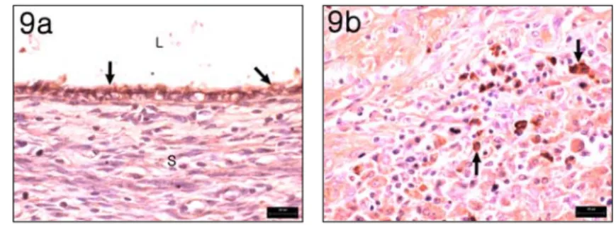
처치군에서의 COX-2 양성반응은 대 조군에서 관찰된 곳과 동일한 부위에서 확인되었다(Fig. 9).
대조군 및 처치군의 점막상피세포 및 간질에 침윤된 세포에서의 COX-2 양성 반응 정도는 처치군이 대조군에 비해서 미약하게 관찰되었다(Table 7).
Fig. 8. COX-2 Immunohistochemistry in Control Group.
(8a) Strong positive reactions of COX-2 are expressed in cytoplasms of transplanted endometrial epithelia (arrows). L : lumen of transplanted uterine tissue. ABC immunohistochemistry, bar=20 μm.
(8b) Strong positive reactions of COX-2 are expressed in cytoplasms of infiltrated inflammatory cells (arrows). ABC immunohistochemistry, bar=20 μm.
Fig. 9. COX-2 Immunohistochemistry in Hominis Placenta Pharmacopuncture Therapy Group.
(9a) Positive reactions of COX-2 are expressed in cytoplasms of transplanted endometrial epithelia (arrows). Compare the intensity of reaction with Fig. 8a. L : lumen of transplanted uterine tissue. ABC immunohistochemistry, bar=20 μm.
(9b) Positive reactions of COX-2 are expressed in cytoplasms of infiltrated inflammatory cells (arrows). Compare the intensity of reaction with Fig. 8b. ABC immunohistochemistry, bar=20 μm.
Group ID Number
Mucosal Epithelia
Cells in Stroma
Control
C1 ++ +++
C2 ++ ++
C3 +++ ++
C4 + +
C5 + ++
C6 ++ ++
C7 +++ ++
C8 +++ +++
Treated
T1 ++ +
T2 + ++
T3 + +
T4 ++ +
T5 + ++
T6 +++ +
T7 ++ +
T8 ++ +
-: No, +: Mild, ++: Moderate, +++:
Strong
Control : right partial uterectomy and uterine autotransplantation and saline Pharmacopuncture therapy
Treated : right partial uterectomy and uterine autotransplantation and Hominis Placenta Pharmacopuncture therapy
Table 7. Effects of Hominis Placenta Pharmacopuncture therapy on the Activities of COX-2 Expression in Transplanted Uterine Tissues of Rats with Experimentally -induced Endometriosis
9. VEGF 발현 양상 및 차이
대조군의 이식자궁조직에서 VEGF 양 성반응은 간질에 침윤된 대식세포, 형질 세포 등에서 강하게 나타났으며, 신생혈 관에서도 강하게 관찰되었다(Fig. 10).
처치군에서의 VEGF 양성반응은 대조 군에서 관찰된 곳과 동일한 부위에서 확 인되었다(Fig. 11)
대조군 및 처치군의 신생혈관 및 간질 에 침윤된 세포에서의 VEGF 양성반응 정도는 처치군이 대조군에 비해서 미약 하게 관찰되었다(Table 8).
Fig. 10. VEGF Immunohistochemistry in Control Group.
Strong positive reactions of VEGF are expressed in cytoplasms of infiltrated inflammatory cells (arrows) and endothelia of newly formed capillaries (arrow heads). ABC immunohistochemistry, bar=20 μm.
Fig. 11. VEGF immunohistochemistry in Hominis Placenta Pharmacopuncture therapy group.
Positive reactions of VEGF are expressed in cytoplasms of infiltrated inflammatory cells (arrows) and endothelia of newly formed capillaries (arrow heads). Compare the intensity of reaction with Fig. 10. ABC immunohistochemistry, bar=20 μm.
Group ID Number
Vascular Endothelia
Cells in Stroma
Control
C1 +++ ++
C2 ++ +++
C3 +++ ++
C4 ++ ++
C5 +++ ++
C6 +++ ++
C7 +++ +++
C8 ++ +++
Treated
T1 + +
T2 + ++
T3 + ++
T4 ++ +
T5 ++ ++
T6 + ++
T7 +++ +
T8 +++ +++
-: No, +: Mild, ++: Moderate, +++:
Strong
Control : right partial uterectomy and uterine autotransplantation and saline pharmacopuncture therapy
Treated : right partial uterectomy and uterine autotransplantation and Hominis Placenta Pharmacopuncture therapy
Table 8. Effects of Hominis Placenta Pharmacopuncture Therapy on the Activities of VEGF Expression in Transplanted Uterine Tissues of Rats with Experimentally -induced Endometriotiosis
10. 비만세포 수의 차이
대조군 및 처치군의 이식자궁조직에서 비만세포는 자궁내막의 기질 및 자궁외 막의 결합조직에서 주로 관찰되었다(Fig.
12, Fig. 13).
이식자궁조직에 침윤된 비만세포 수는 처치군이 대조군에 비해서 적었으나 군간 에 유의성 있는 차이는 없었다(Table 9).
Fig. 12. Mast cells in transplanted uterine tissue of control group.
Mast cells (arrows) are observed abundantly in adjacent tissue of transplanted uterine tissue. L; lumen of uterine tissue. arrows : endometrial epithelia. Toludine blue stain.
bar=30 μm.
Fig. 13. Mast cells in transplanted uterine tissue of Hominis Placenta Pharmacopuncture therapy group.
A number of mast cells (arrow heads) are observed in adjacent tissue of transplanted uterine tissue. Compare with Fig. 12. arrow;
endometrial epithelia, Toludine blue stain.
bar=30 μm.
Group No. of Mast Cells Control (n=8) 89.40±28.5*
Treated (n=8) 63.3±33.2
* : data expressed as Mean±S.D.
Control : right partial uterectomy and uterine autotransplantation and saline pharmacopuncture therapy
Treated : right partial uterectomy and uterine autotransplantation and Hominis Placenta Pharmacopuncture therapy