서 론
영상진단 기술의 발전으로 종양과 정상조직을 명확히 구 별 할 수 있게 되었다. 이러한 기술을 바탕으로 방사선치료 에서는 삼차원적인 영상을 이용한 정밀한 치료법이 시행되 어지고 있다. 종양조직에 치유방사선량의 정확한 전달이 치 료성적에 중요한인자로 작용하는 세기변조방사선 치료 및 정위적 방사선치료에서는 환자의 내부정보를 이용한 영상 유도하의 환자 위치 확인이 반드시 선행되어야 한다.1)부정 확한 위치확인은 정상조직의 허용 가능한 선량을 넘겨 중대 한 부작용을 초래 할 수 있다. 영상유도방법으로는 단편적 인 영상정보를 비교하는 방법과 삼차원적인 Cone-Beam Computed Tomography(CBCT)을 이용하여 정확한 내부위
치를 확인 및 교정 하는 방법이 사용되고 있으며, 단편적인 두 방향 이미지와 삼차원적인 볼륨을 조합하여 더욱 정확한 영상유도방사선치료를 수행하는 Novalis TX system(Novalis TX system, Varian, CA, USA and BrainLAB, Herimstetten, Germany)있다.2)위 장비는 영상유도 방식은 BrainLab사에 서 나온 ExacTrac과 Varian사의 On-Board Imager (OBI)로 구성되어있다. ExacTrac장비의 기능은 환자 표면이나 reference에 부착된 marker를 적외선 카메라로 인식하여 치료계획용 CT의 digitally reconstructed radiographs (DRR)이미지와 경사각 엑스선 이미지를 비교 분석하여 로 봇식 6개 방향으로 이동할 수 있는 치료 테이블에서 조정함 으로써 기존의 4방향 비해 더 정확한 영상유도방사선치료 가 가능하다.3)OBI 장비를 이용한 CBCT 재구성방식으로써 는 치료실에서 pencil beam을 이용하여 관심영역의 볼륨이 미지의 횡단면을 획득하여 치료계획용 CT의 횡단면과 내부 위치를 정확히 비교 분석해줄 수 있는 장비이다.4) 본 논문 에서는 ExacTrac 6D couch의 영상유도의 정확도를 알아보
본 논문은 2016년 10월 27일 접수하여 2016년 12월 10일 채택되었음.
책임저자 : 박호춘, 화순전남대학교병원 방사선종양학과 전라남도 화순군 화순읍 서양로 332, 58115 Tel : 061) 379-7209
E-mail : [email protected]
CBCT을 이용한 ExacTrac 6D 영상유도방사선치료법의 정확도 평가
화순전남대학교병원 방사선종양학과, 전남대학교병원 방사선종양학 교실
ExacTrac 6D couch를 이용한 영상유도 방사선 치료의 정확도를 검증하기 위하여 여섯 개의 방향의 오차 값을 임의로 부여하여 교정 한 후 CBCT 이미지와 비교 분석하여 ExacTrac의 정확도를 평가 하고자 하였다.
Rando head Phantom의 치료좌표 값을 X, Y, Z 방향으로 이동시킨 Translationgroup과 pitch, Roll, Yaw방향으로 이동시킨 Rotationgroup으로 나 누어 교정하였다. 교정 수치는 couch의 여섯 개의 방향으로 복합적으로 상호 작용하여 이동 하였다. 교정 값은 최소 1mm, 최대 23mm까지 다양하 게 나타났다. 치료좌표로 수정된 Phantom를 CBCT 촬영한 이미지와 3D/3D matching 오차 값의 분석에서는 Rotation group에서 높은 오차 값이 나타났다. CBCT로 교정된 치료좌표 오차 값에 대한 선량분포의 비교에서는 정상조직에 선량 제한치 값은 처방선량에 충족되었으며 종양조직의 선 량 균질성 지표인 PHI, PCI 값은 Rotation group에서 저 선량 분포영역이 다소 높음으로 나타났다.
본 연구에서는 ExacTrac 6D couch 의 정확도에 대한 평가를 CBCT를 이용하여 검증해 보았다. 단편적인 이동에 대한 오차 값은 비교적 정확한 교 정 능력을 갖추고 있었지만, couch에 각이 들어가는 이동에서는 부정확한 교정 수치를 보였다. 환자의 체위가 Rotation방향으로 많은 변화가 예상 되어지거나 ExacTrac 교정하였을 때 pitch, Roll, Yaw값의 오차가 크게 나타나면 CBCT 영상유도를 시행하여 정확히 치료 좌표를 교정하여 예상치 못한 부작용을 최소화해야 된다고 여겨진다.
핵심용어 :ExacTrac 6D couch, 영상유도방사선치료, CBCT, pitch, Roll, Yaw
박호춘・김효중・김종덕・지동화・송주영
기 위해서 가상의 표적을 Phantom모사한 후 couch의 위치 및 각도를 6개 방향으로 움직여서 실험을 하였다. 위치 및 각도에 따른 couch의 변화를 ExacTrac 영상유도로 교정하 였으며, 영상유도의 정확도를 파악하기 위하여 CBCT촬영 을 시행하여 치료계획용 단층촬영의 볼륨이미지와 비교분 석하였다. Plan CT 와 CBCT 횡단면이미지의 교정 오차에 따른 값을 couch좌표 값에 반영하여 치료계획을 재 수정하 였으며 선량분포에 미치는 영향을 알아보고자 하였다.
대상 및 방법
Navalis TX에는 영상유도방사선기법으로 주로 사용되는 OBI system을 이용한 CBCT와 BrainLAB사의 ExacTrac이 있다.
1. OBI system use to CBCT
Varian사의 OBI system은 kv X-ray source와 40×30 cm2
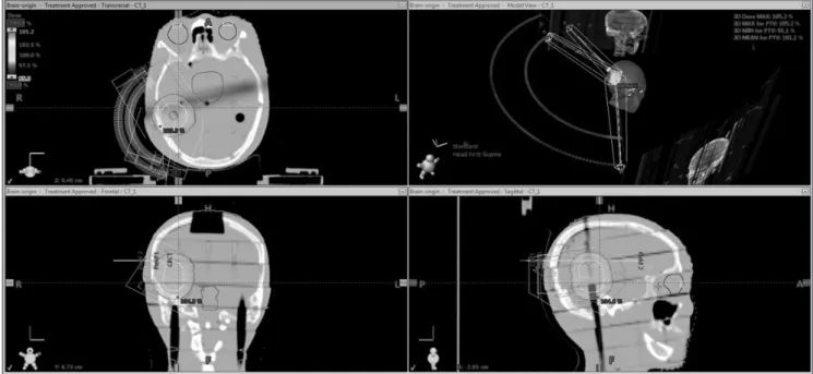
Fig 1.
Image of the Phantom Setup.
immobilization masks were used to fix the head in the translational (lateral, longitudinal, and vertical) and rotational (pitch, roll, and yaw) dimensions.
Fig 2.
Image Registration Using kV Tubes 1 and 2 with BrainLAB ExacTrac for Phantom. (a) ExacTrac image, and (b) image registration.
감광영역을 가진 kv amorphop silicon detector로 구성된 다. 2D 이미지와 3D CBCT 이미지를 구현할 수 있으며, CBCT 이미지 획득과정은 선형가속기에 부착된 roboric arm을 360도을 일정한 속도로 회전하여 2mm슬라이스 간 격으로 3차원 체적 데이터를 획득 할 수 있다. CBCT 이미지 획득 모드 설정 중 본 실험에서는 Half-fan의 Standard- Dose Head 모드를 사용하였으며, 슬라이스 두께 2mm, 해 상도 512*512 방식을 사용하여 3차원이미지를 획득 하였다 [그림 1].
2. Brainlab of 6D ExacTrac couch using calibration error
치료실 바닥에 부착된 2개의 X-ray tube에서 발생한 source로부터 천장에 부착된 2개의 kv X-ray detector에서 이미지를 획득하며[그림 2(a)], Plan CT DRR 이미지에서 재 구성된 bony anatomy 로 auto matching 6D Fusion algorithms로 교정하였다[그림 2(b)].
3. 치료계획용 삼차원체적과 치료계획수립
인체조직과 유사한 물질로 만들어진 Rando head & neck phantom에 가상의 종양을 우측 측두엽에 삽입 후 Philips Medical System(Brilliance CT Big Bore, OH, USA) CT 에서
2mm 슬라이스 두께로 치료계획용 삼차원 체적과 치료 좌표 값을 획득하였다. Eclipse version13 에서 RTOG(Radioation Terapy Oncology Group) 가이드에 따라서 처방선량은 PTV: 1.8Gy*28fx = 50.4Gy, OAR(Organ At Risk) 제한 값은 eyeball : V4<30%, Dmax< 25Gy, lens : Dmax < 5Gy.
pituitary : V20<40%, Dmax<30Gy, brainstem : Dmax<42Gy, optic nerve : Dmax<42Gy 로 설정하여 Novalis 6MV의 Half Two Arc IMRT plan을 하였다[그림 3].
4. BrainLAB 6D couch 6개 방향의 오차 값 설정 및 CBCT를 이용한 교정 정확도 재검증
본 실험 시행 전에 couch isocenter calibration 을 이용하 여 Exactrac 6D couch 중심축을 교정하였으며, In room beam lager calibration을 시행하여 치료좌표의 정확도를 높였다. Rando head phantom을 치료계획에서 설정된 치 료좌표로 정확히 위치시킨다. 치료좌표 중심점을 기준으로 AP, SI, LR 3개 방향으로 0.5mm, 1mm, 2mm , pitch, Roll, Yaw 0.1°, 0.5°, 1°2°(Yaw) 각각 이동시킨 후 ExacTrac 6.1 system을 이용하여 치료좌표를 재 교정한다. 6개 방향 의 조건에서 교정된 phantom을 CBCT을 이용하여 삼차원 영상을 획득하였으며 치료계획용 삼차원 영상과 비교하는 3D/3D auto matching 방식으로 오차 값을 측정하였다. 이 와 같은 방법으로 일주일 간격으로 총 3회 실험하여 오차
Fig 3. Novalis 6MV of Two Arc Brian Rando Phantom IMRT plan
값을 측정 하였다..
5. 3D matching 오차에 따른 치료계획 재구성
6개의 방향을 단편적인 움직임(LR, AP, SI) Translation group과 couch에 경사가 임의로 들어가는(pitch, Roll, Yaw) Rotation group으로 나누어 3번 측정된 3D/3D matching 오차 값을 바탕으로 오차 치료 좌표 값의 평균과 표 준 편 차 를 구 하 여 치 료 계 획 을 분 석 하 였 다. Eclipse version13을 이용한 치료계획 프로그램에 couch X, Y, Z, Rtn의 최대 오차 값을 기존의 치료계획 좌표에 변경하여 동
일한 처방선량과 OAR 조건으로 치료계획 계산을 다시 하였 다. 단 오차 값에 의한 분포를 확인하기 위해서 치료계획 최 적화 과정을 생략하고 마지막 계산 과정만 수행하였다.
PTV 선량분포의 균일성을 분석에는 객관적인 도구 사용되 고 있는 PTV_HI(radical dose homogeniety index = dMin/dMax in PTV)와 일치성지수(CI, conformity index) 는 Nedzi 등5)에 의해 다음과 같이 정의 되었으며 표적 범위 에서의 품질을 평가하기 위해 사용하였다.
PTV V RI
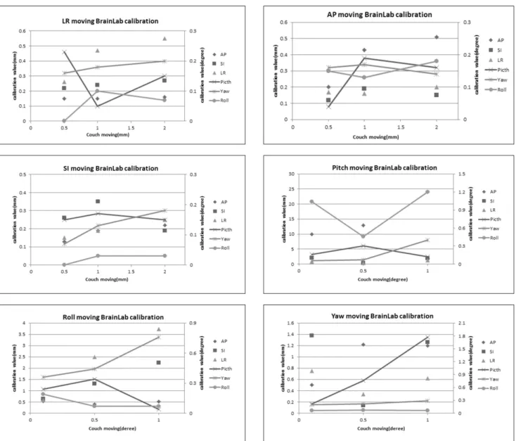
Fig 4. couch moving in six directions compared ExacTrac 6D couch calibration
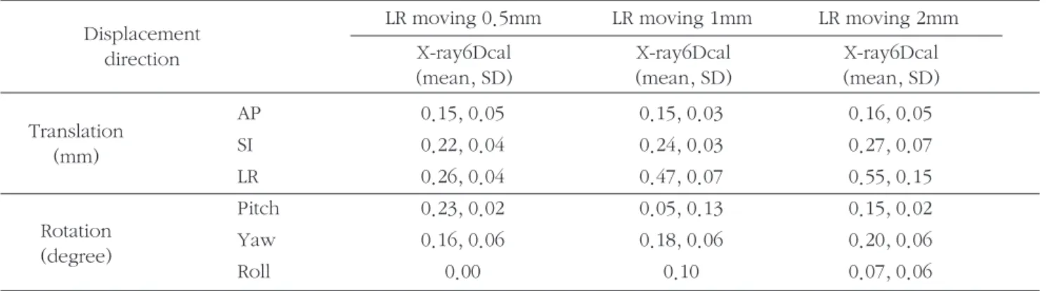
Table 1. Translation group(a, b, c) Rotation group(d, e, f) and divided ExacTrac 6.1 auto matching the average, standard deviation analysis. (Unit: mm, degree) (a) Translation group LR moving
LR moving 0.5mm LR moving 1mm LR moving 2mm X-ray6Dcal
(mean, SD)
X-ray6Dcal (mean, SD)
X-ray6Dcal (mean, SD) Displacement
direction
AP 0.15, 0.05 0.15, 0.03 0.16, 0.05
SI 0.22, 0.04 0.24, 0.03 0.27, 0.07
LR 0.26, 0.04 0.47, 0.07 0.55, 0.15
Pitch 0.23, 0.02 0.05, 0.13 0.15, 0.02
Yaw 0.16, 0.06 0.18, 0.06 0.20, 0.06
Roll 0.00 0.10 0.07, 0.06
Translation (mm)
Rotation (degree)
(b) Translation group AP moving
AP moving 0.5mm APmoving 1mm APmoving 2mm X-ray6Dcal
(mean, SD)
X-ray6Dcal (mean, SD)
X-ray6Dcal (mean, SD) Displacement
direction
AP 0.2, 0.06 0.43, 0.04 0.51, 0.06
SI 0.12, 0.03 0.19, 0.03 0.15, 0.05
LR 0.17, 0.06 0.16, 0.05 0.20, 0.07
Pitch 0.04, 0.25 0.19, 0.04 0.16, 0.01
Yaw 0.16, 0.06 0.17, 0.04 0.14, 0.01
Roll 0.15, 0.09 0.13, 0.06 0.18, 0.02
Translation (mm)
Rotation (degree)
여기서 VRI는 기준 등선량(reference isodose)의 체적이 고, PTV는 표적 체적(target volume)이다.
결 과
1. 6개 방향으로 couch 이동시 ExacTrac 6D couch 교정 값 분석
Translation group과 Rotation group으로 나누어 임의로 움직였을 때 ExacTrac 6.1 auto matching하여 획득된 6개 방향의 couch이동 수치의 평균, 표준편차 값을 분석하였다 [표 1]. Translation group에서 일정 값의 오차를 주었을 때 6D couch 교정 값은 6개 방향으로 최대 0.5mm로 이동했으 며 임의로 부여한 수치와 비슷한 오차 값을 보였으나 couch
의 움직임에 대한 일정한 규칙성은 파악하지 못하였다. 이 러한 결과 수치는 Se An Oh의 ExacTrac 6D 불확실성에 대 한 연구결과와 비슷하게 나타났다.6)Rotation group에서 교 정 값은 최소 0.2mm ,최대 23mm 까지 나타났으며, pitch 방향에서의 불규칙적인 오차가 가장 많이 발생한 것을 볼 수 있었다. couch이동이 6개 방향으로 일관된 규칙성이 없 이 움직였음을 알 수 있었다[그림 4].
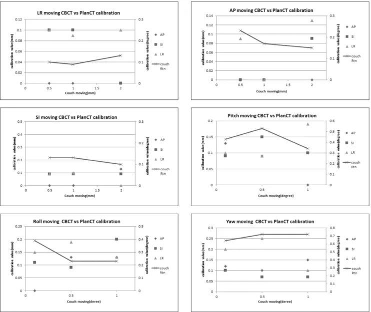
2. 치료좌표로 수정된 Phantom CBCT 와 Plan CT 횡단 면의 3D/3D matching 값 비교
ExacTrac 6D couch로 치료좌표가 수정된 Phantom을 사 방향 DRR이미지가 아닌 CBCT에서 획득된 삼차원 CT 이미 지와 Plan CT 횡단면 영상을 비교 분석하여 오차 값을 분석 하였다[표 2]. Translation group에서는 couch의 최대 X, Y,
Z 좌표 값이 0.1mm, couch Rtn(rotation)값은 0.1°이내로 측정되었다. Rotation group에서는 최대 X, Y, Z 좌표 값이 0.2mm, couch Rtn값은 0.7°로 오차 값이 Translation group의 값 보다 오차가 높음을 알 수 있었다[그림 5].
3. 3D/3D matching couch 오차 값에 따른 치료계획 분포 비교
3D/3D matching에서 획득한 couch 오차 값을 origin 치 료 좌표 couch 값에 반영하여 치료 계획 분포를 파악해 보 (d) Rotation group Pitch moving
Pitch moving 0.1° Pitch moving 0.5° Pitch moving 1°
X-ray6Dcal (mean, SD)
X-ray6Dcal (mean, SD)
X-ray6Dcal (mean, SD) Displacement
direction
AP 9.89, 0.59 12.96, 0.15 23.97, 1.70
SI 2.00, 0.11 0.41, 0.19 1.88, 0.14
LR 0.71, 0.05 0.39, 0.03 1.24, 0.07
Pitch 0.16, 0.06 0.3, 0.06 0.12, 0.03
Yaw 0.06, 0.01 0.07, 0.04 0.4, 0.39
Roll 1.04, 0.04 0.46, 0.01 1.2, 0.07
Translation (mm)
Rotation (degree)
(c) Translation group SI moving
SI moving 0.5mm SI moving 1mm SI moving 2mm X-ray6Dcal
(mean, SD)
X-ray6Dcal (mean, SD)
X-ray6Dcal (mean, SD) Displacement
direction
AP 0.13, 0.03 0.19, 0.07 0.22, 0.06
SI 0.26, 0.08 0.35, 0.10 0.19, 0.07
LR 0.15, 0.06 0.19, 0.06 0.25, 0.03
Pitch 0.15, 0.31 0.17, 0.06 0.15, 0.04
Yaw 0.07, 0.17 0.13, 0.06 0.18, 0.07
Roll 0.00 0.03, 0.06 0.03, 0.03
Translation (mm)
Rotation (degree)
(e) Rotation group Roll moving
Roll moving 0.1° Roll moving 0.5° Roll moving 1°
X-ray6Dcal (mean, SD)
X-ray6Dcal (mean, SD)
X-ray6Dcal (mean, SD) Displacement
direction
AP 0.54, 0.48 0.40, 0.47 0.51, 0.32
SI 0.64, 0.11 1.30, 0.48 2.23, 0.80
LR 0.58, 0.43 2.49, 1.42 3.74, 2.03
Pitch 0.24, 0.07 0.34, 0.11 0.04, 0.02
Yaw 0.36, 0.08 0.44, 0.02 0.76, 0.21
Roll 0.19, 0.04 0.07, 0.03 0.07, 0.02
Translation (mm)
Rotation (degree)
(f) Rotation group Yaw moving
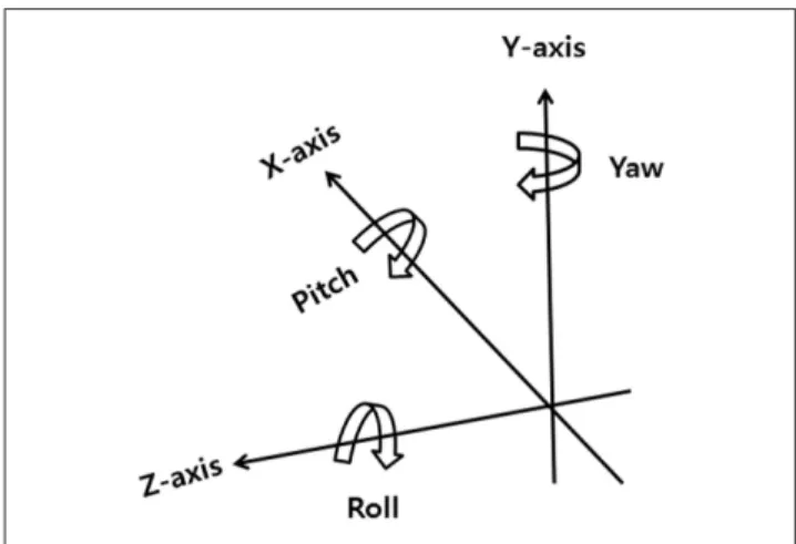
6D: 6 degree-of-freedom, CBCT: cone-beam computed tomography, SD: standard deviation, cal: calibration LR: left- right, SI: superior-inferior, AP: anterior-posterior, Pitch: rotation around LR direction, Roll: rotation around SI direction, Yaw: rotation around AP direction.
Yaw moving 0.5° Yaw moving 1° Yaw moving 2°
X-ray6Dcal (mean, SD)
X-ray6Dcal (mean, SD)
X-ray6Dcal (mean, SD) Displacement
direction
AP 0.50, 0.07 1.22, 0.11 1.20, 0.06
SI 1.38, 0.18 0.14, 2.10 1.26, 0.16
LR 0.75, 0.15 0.34, 0.14 0.62, 0.09
Pitch 0.22, 0.04 0.76, 0.1 1.77, 0.13
Yaw 0.20, 0.24 0.22, 0.02 0.29, 0.04
Roll 0.06, 0.15 0.07, 0.01 0.06, 0.02
Translation (mm)
Rotation (degree)
았다. 단편적인 couch 이동 값을 조정하여 선량의 균질성을 파악하는데 다소 무리가 되어보였지만, 본 논문에서는 부정 확한 IGRT가 선량분포에 미치는 영향을 알아볼 필요가 있 다고 생각이 되어서 치료계획을 다시 작성해 보았다. 종양 조직에 대한 선량분포 일치성 지표 값인 HI, CI로 비교했다.
정상조직에 대한 선량분포는 origin, Translation, Rotation group에서 선량 제한치 조건에 만족하였으며, Rotation group에서 처방선량 95%영역에서의 선량 분포 값이 다른 두 개의 치료계획 보다 저 선량 분포영역이 많음을 알 수 있 었다[그림6]. PTV HI, CI값 또한 Rotation group에서 균일 도와 일치성지수가 낮음을 알 수 있었다[표 3].
결론 및 고찰
방사선 치료기술의 발전에 따라 정밀하고 정확한 방사선 치료기법들에 대한 연구가 지속되고 있다. 방사선 선질의 종류의 다양화, 고도화된 치료 장비의 개발로 방사치료의 새로운 영역들이 개척되어지고 있다. 이러한 개발의 한 영 역으로 환자자세의 재현성을 높여 줄 수 있는 여러 방법들 이 모색되어지고 있으며, 환자자세의 불확실성에 대한 결과 로 정상조직의 과 선량 조사와 종양조직의 저 선량 조사를 최소 시킬 수 있는 다양한 영상 유도 방사선치료기법들이 다양이 개발되고 있다.7) 본 논문에서는 방사선치료과정에 서 안전하면서 빠르게 환자의 치료 자세를 정확하게 교정하 려는 장비로 개발된 BrainLab사의 ExacTrac 6D couch에
대해서 연구해 보았다. 단편적인 사 방향 X-ray 이미지의 조합으로 영상유도의 정확하게 구현하고 교정하는지에 대 해서 6개 방향을 부여하여 교정을 시행한 후 CBCT 이미지 를 획득하여 삼차원적인 분석과정을 통해서 정확도를 평가 해보았다.8) 환자의 움직임에 대한 변수를 최소하기 위하여 Brain Rando Phantom에 임의 종양을 만들어 실험하였으며, 6개 방향을 couch의 움직임에 따라 단편적인 Translation group, couch에 각도가 들어가는 Rotation group으로 나누 어 Exactrac 6D couch영상 유도 교정 값을 얻었다[그림 7].
임의의 위치, 각도 변화를 주었을 때 6D couch는 6개 방향 으로 상호 복합적인 방법으로 움직여서 Phantom의 위치를 맞추었다.9)6D couch 움직임에 대한 우선순위와 가중치 부 여 방식은 파악하지 못하였으며 ExacTrac 고유의 algorithm 으로 영상 유도됨을 알 수 있었다.10)
ExacTrac으로 교정된 Phantom에 CBCT 이미지를 획득 한 후 Plan CT 횡단면 이미지와 3D/3D matching 결과에서 는 Rotation group에서 couch 변화의 각도가 커짐에 따라 Translation group보다 비교적 오차 값이 커지는 것을 알 수 있었다.11) 치료 좌표의 변화가 없는 origin과 Translation, Rotation group에서 3D/3D matching 최대 couch 오차 값 에 대한 치료계획 분포 비교에서 Rotation group에서 종양 조직에 대한 저 선량분포영역이 많음을 알 수 있었다.. The Physics of Radiation Therapy(Faiz M. Khan),12)에서 권고하 는 couch Rtn 오차 범위 0.5°초과하는 값이 나온 Yaw방향 2°오차 에서 특히 저 선량 분포가 늘어났다. 즉 환자의 종 축 체위가 couch와 평행이 되지 않고 뒤틀림의 각이 커짐에
Fig 5. 3D / 3D matching value compared with the Phantom CBCT and treatment to modify the coordinates of the Plan CT
Fig 6.
In treatment planning dose distribution of PTV 95% compared three groups DVH
따라 ExacTrac의 교정 불확실도가 높음을 알 수 있었다.13,14) 고 선량, 고 선량률, 세기변조 방사선치료와 방사선 재 치료 환자의 정상조직 선량 제한치를 벗어나는 선량 분포 값은 치명적인 부작용을 초래 할 수 있음으로 ExacTrac 과 CBCT 를 모두 이용하여 더욱 정확성 있게 치료를 진행해야 된 다 고 사료된다.
ExacTrac 6D 영상유도방식은 기존의 어느 방식보다 빠 르게 환자의 위치를 교정해 주는 장점이 있었지만 여섯 방 향으로 임의 조작하여 정확도를 평가해본 결과 couch의 각 이 크게 있는 변화에서는 완벽한 정확도를 확보하기에는 무 리가 있어 보였다.15)실제 임상에서 환자 체위의 방향이 치 료 좌표보다 Rotation변화가 크게 예상되어진다고 여겨지 Fig 6. Directions randomly assigned to six adjustment
values in the schematic 6D couch
Table 2. 3D / 3D matching value comparison of Plan CT and CBCT and a modified Phantom in the Translation group(a, b, c), Rotation group(d, e, f) to coordinate treatment. (Unit: mm, degree) (a) Translation group LR moving
LR moving 0.5mm LR moving 1mm LR moving 2mm CBCTac & Plan CT cal
(mean, SD)
CBCTac & Plan CT cal (mean, SD)
CBCTac & Plan CT cal (mean, SD) Displacement
direction
AP 0 0 0
SI 0, 0.1 0, 0.1 0
LR 0, 0.1 0.03, 0.06 0, 0.1
couch Rtn 0.1 0.03, 0.06 0.07, 0.06
Translation (mm) Rotation(degree)
(b) Translation group SI moving
SI moving 0.5mm SI moving 1mm SI moving 2mm CBCTac & Plan CT cal
(mean, SD)
CBCTac & Plan CT cal (mean, SD)
CBCTac & Plan CT cal (mean, SD) Displacement
direction
AP 0 0 0.07, 0.06
SI 0.03, 0.06 0.03, 0.06 0.03, 0.06
LR 0.03, 0.06 0.03, 0.07 0
couch Rtn 0.07, 0.06 0.07, 0.06 0, 0.1
Translation (mm) Rotation(degree)
(c) Translation group AP moving
AP moving 0.5mm AP moving 1mm AP moving 2mm CBCTac & Plan CT cal
(mean, SD)
CBCTac & Plan CT cal (mean, SD)
CBCTac & Plan CT cal (mean, SD) Displacement
direction
AP 0 0 0
SI 0 0 0.03, 0.06
LR 0.03, 0.06 0 0.07, 0.06
couch Rtn 0.17, 0.06 0, .17 0.03, 0.12
Translation (mm) Rotation(degree)
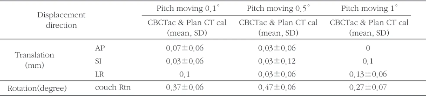
(d) Rotation group Pitch moving
Pitch moving 0.1° Pitch moving 0.5° Pitch moving 1°
CBCTac & Plan CT cal (mean, SD)
CBCTac & Plan CT cal (mean, SD)
CBCTac & Plan CT cal (mean, SD) Displacement
direction
AP 0.07±0.06 0.03±0.06 0
SI 0.03±0.06 0.03±0.12 0.1
LR 0.1 0.03±0.06 0.13±0.06
couch Rtn 0.37±0.06 0.47±0.06 0.27±0.07
Translation (mm) Rotation(degree)
(e) Rotation group Roll moving
Roll moving 0.1° Roll moving 0.5° Roll moving 1°
CBCTac & Plan CT cal (mean, SD)
CBCTac & Plan CT cal (mean, SD)
CBCTac & Plan CT cal (mean, SD) Displacement
direction
AP 0 0.07, 0.06 0.07, 0.06
SI 0., 0.11 0.03, 0.06 0.1, 0.1
LR 0.03, 0.12 0.13, 0.06 0.07, 0.06
couch Rtn 0.33, 0.06 0.17, 0.06 0.17, 0.06 Translation
(mm) Rotation(degree)
(f) Rotation group Yaw moving
CBCTac = CBCT Accurate calibration
Yaw moving 0.1° Yaw moving 0.5° Yaw moving 1°
CBCTac & Plan CT cal (mean, SD)
CBCTac & Plan CT cal (mean, SD)
CBCTac & Plan CT cal (mean, SD) Displacement
direction
AP 0.06, 0.06 0.1 0.09, 0.06
SI 0.1 0.01, 0.06 0.01, 0.06
LR 0.14, 0.06 0.19, 0.06 0.1
couch Rtn 0.62, 0.02 0.69, 0.03 0.7, 0.02
Translation (mm) Rotation(degree)
Table 3. Comparative PTV HI, CI value of the origin, Translation, Rotation groups.
Parameter origin plan Translation group plan Rotation group plan
HI 1.16 1.33 1.45
CI 1.0 1.01 1.03
PTV
거나 ExacTrac 시행하였을 때 Pitch, Roll, Yaw 값의 오차 가 크게 나타나면 CBCT 영상유도를 재 시행하여 검증해야 된다고 여겨진다.16)
연구의 제한점으로는 연구의 제한점으로써는 ExacTrac program 에서 Plan CT를 Loading 하여 ExacTrac 교정 값 과 비교 분석을 시도하였으나 빈번한 loading 에러로 추가 연구의 어려움이 있었습니다. 또한 실제 치료실에서 Rotation group의 ExacTrac 교정 값이 크게 나타났을 경우 환자 자세를 다시 조정하여 re IGRT 할 지 아니면 추가적으 로 CBCT 교정을 하여 치료를 할 것인지에 대한 고민이 필 요할 것으로 사료 됩니다.
참고문헌
1. Van Herk M: Different styles of image-guided radiotherapy. Semin Radiat Oncol 2007;17:258-67
2. Thierry Gevaert, Dirk Verellen, Koen Tournel: Setup accuracy of the novalis exactrac 6dof system for frameless radiosurgery. Int. J. Radiation Oncology Biol.
Phys 2012;88(5):1627-1635
3. Jian Yue Jin, Fang-Fang Yin, Stephen, Tenn, Paul: Use of the BrianLAB EXACTRAC X-ray 6D System in image- guided radiotherapy. Medical Dosimetry 2008;
33(2):124-134
4. Nedzi LA, Kooy HM, Alexander E 3rd, Svensson GK, Loeffler JS : Dynamic field shaping for stereotactic radiosurgery a modeling study. Int J Radiat Oncol Biol Phys 1993; 25:859-869
5. Dzyubak O, Kincaid R, Hertanto A, Hu Y.-C, Pham H, Rimner A: Evaluation of tumor localiza-tion in respiration motion-corrected cone-beam CT:
Prospective study in lung. Medical physics 2014;41(10):1018-1019
6. Se An Oh, Ji Woon Yea2, Min Kyu Kang, Jae Won Park, Sung Kyu Kim: Analysis of the Setup Uncertainty
and Margin of the Daily ExacTrac 6D Image Guide System for Patients with Brain Tumors. PLoS ONE 2016;11(3): e0151709doi:10.1371/journal.pone.0151709
7. Miyabe Y, Sawada A, Takayama K, Kaneko S, Mizowaki T, Kokubo M: Positioning accuracy of a new image-guided radiotherapy system. Medical physics 2011;38(5):2535-2541
8. Stanley, Papanikolaou, Gutirez: Development of image quality assurance mea-sures of the ExacTrac localization system using commercially available image evaluation software and hardware for image-guided radiotherapy. Journal of Applied Clinical Medical Physics 2014; 15(6)
9. 최우근, 박수연, 박도근, 송기원: CBCT와 EXACTRAC을 이용한 Spine SBRT의 유용성 평가. 대한방사선치료학회 지 2013; 25(2)167-172
10. Jin JY, Yin FF, Tenn SE, Medin PM, Solberg TD: Use of the brainlab ExacTrac X-ray 6D system in image- guided radiotherapy. Med Dosim 2008;33:124-34
11. Boda-Heggemann J, Walter C, Rahn A: Repositioning accuracy of two different mask systems-3D revisited:
comparison using true 3D/3D matching with cone- beam CT. Int J Radiat Oncol Biol Phys 2006;66:1568- 75
12. Faiz M. Khan: The Physics of Radiation Therapy. 5th ed. Wolters Kluwer health. Philadelphia, PA, 2015:387-389
13. Takakura T, Mizowaki T, Nakata M, et al. The geometric accuracy of frameless stereotactic radiosurgery using a 6D robotic couch system. Phys Med Biol 2010;55:1-10
14. Verellen D, Soete G, Erbel S, et al. Comment on
“Image-guided patient positioning: If one cannot
correct for rotational offsets in external-beam radiotherapy setup, how should rotational offsets be managed?. Med Phys 2007;34:4064-4065
15. Jinli Ma, Zheng Chang, Zhiheng Wang: ExacTrac X- ray 6 degree-of-freedom image-guidance for intracranial non-invasive stereotactic radiotherapy:
Comparison with kilo-voltage cone-beam CT.
Radiotherapy and Oncology 2009;93:602-608
16. Zheng Chang, Zhiheng Wang, Jinli Maa,b, Jennifer:
6D image guidance for spinal non-invasive stereotactic body radiation therapy: Comparison between ExacTrac X-ray 6D with kilo-voltage cone- beam CT. Radiotherapy and Oncology 2010;95:116- 121
To verify the accuracy of the image guided radiotherapy using ExacTrac 6D couch, the error values in six directions are randomly assigned and corrected and then the corrected values were compared with CBCT image to check the accurateness of ExacTrac.
The therapy coordination values in the Rando head Phantom were moved in the directions of X, Y and Z as the translation group and they were moved in the directions of pitch, roll and yaw as the rotation group. The corrected values were moved in 6 directions with the combined and mutual reactions. The Z corrected value ranges from 1mm to 23mm. In the analysis of errors between CBCT image of the phantom which is corrected with therapy coordinate and 3D/3D matching error value, the rotation group showed higher error value than the translation group. In the distribution of dose for the error value of the therapy coordinate corrected with CBCT, the restricted value of dosage for the normal organs in two groups meet the prescription dose. In terms of PHI and PCI values which are the dose homogeneity of the cancerous tissue, the rotation group showed a little higher in the low dose distribution range.
This study is designed to verify the accuracy of ExacTrac 6D couch using CBCT. It showed that in terms of the error value in the simple movement, it showed the comparatively accurate correction capability but in the movement when the angle is put in the couch, it showed the inaccurate correction values. So, if the body of the patient is likely to have a lot of changes in the direction of rotation or there is a lot of errors in the pitch, roll and yaw in ExacTrac correction, it is better to conduct the CBCT guided image to correct the therapy coordinate in order to minimize any side effects.
Park Ho Chun, Kim Hyo Jung, Kim Jong Deok, Ji Dong Hwa, Song Ju Young
Evaluation of accuracy in the ExacTrac 6D image induced radiotherapy using CBCT
Department of Radiation Oncology, Chonnam National University Hwasun Hospital Department of Radiation Oncology, Chonnam National University School, Gwangju, Korea
Abstract
Keyword :ExacTrac 6D couch, image guided radiotherapy, CBCT, pitch, roll and yaw