of January 13, 2016.
This information is current as
B Activation κ
by Macrophages Requires NF-
Inducible Nitric Oxide Synthase Expression -Potentiated 2
Secretory Phospholipase A
Jung-Hye Kim and Koing-Bo Kwun
Yoon-Jeong Lee, Hyeun-Wook Chang, Soo-Jung Lee, Suk-Hwan Baek, Taeg Kyu Kwon, Jun-Hee Lim,
http://www.jimmunol.org/content/164/12/6359 doi: 10.4049/jimmunol.164.12.6359
2000; 164:6359-6365; ; J Immunol
References
http://www.jimmunol.org/content/164/12/6359.full#ref-list-1
, 21 of which you can access for free at:
cites 43 articles This article
Subscriptions
http://jimmunol.org/subscriptions
is online at:
The Journal of Immunology Information about subscribing to
Permissions
http://www.aai.org/ji/copyright.html Submit copyright permission requests at:
Email Alerts
http://jimmunol.org/cgi/alerts/etoc
Receive free email-alerts when new articles cite this article. Sign up at:
Print ISSN: 0022-1767 Online ISSN: 1550-6606.
Immunologists All rights reserved.
Copyright © 2000 by The American Association of 9650 Rockville Pike, Bethesda, MD 20814-3994.
The American Association of Immunologists, Inc.,
is published twice each month by The Journal of Immunology
at Keimyung Univ Med Lib on January 13, 2016http://www.jimmunol.org/Downloaded from at Keimyung Univ Med Lib on January 13, 2016http://www.jimmunol.org/Downloaded from
2
Oxide Synthase Expression by Macrophages Requires NF-B Activation1
Suk-Hwan Baek,* Taeg Kyu Kwon,§ Jun-Hee Lim,* Yoon-Jeong Lee,* Hyeun-Wook Chang,† Soo-Jung Lee,‡ Jung-Hye Kim,* Koing-Bo Kwun‡2
The effect of secretory group II phospholipase A2 (sPLA2) on the expression of the inducible NO synthase (iNOS) and the production of NO by macrophages was investigated. sPLA2by itself barely stimulated nitrite production and iNOS expression in Raw264.7 cells. However, in combination with LPS, the effects were synergistic. This potentiation was shown for sPLA2enzymes from sPLA2-transfected stable cells or for purified sPLA2from human synovial fluid. The effect of PLA2 on iNOS induction appears to be specific for the secretory type of PLA2. LPS-stimulated activation of iNOS was inhibited by the well-known selective inhibitors of sPLA2such as 12-epi-scalaradial and-bromophenacyl bromide. In contrast, the cytosolic PLA2-specific inhibitors methyl arachidonyl fluorophosphate and arachidonyltrifluoromethyl ketone did not affect LPS-induced nitrite production and iNOS expression. Moreover, when we transfected cDNA-encoding type II sPLA2, we observed that the sPLA2-transfected cells produced two times more nitrites than the empty vector or cytosolic PLA2-transfected cells. The sPLA2-potentiated iNOS ex- pression was associated with the activation of NF-B. We found that the NF-B inhibitor pyrrolidinedithiocarbamate prevented nitrite production, iNOS induction, and mRNA accumulation by sPLA2plus LPS in Raw264.7 cells. Furthermore, EMSA analysis of the activation of the NF-B involved in iNOS induction demonstrated that pyrrolidinedithiocarbamate prevented the NF-B binding by sPLA2plus LPS. Our findings indicated that sPLA2, in the presence of LPS, is a potent activator of macrophages. It stimulates iNOS expression and nitrite production by a mechanism that requires the activation of NF-B. The Journal of Immunology, 2000, 164: 6359 – 6365.
M acrophage activation is a key component of the im- mune response. Several proinflammatory cytokines and bacterial products such as LPS participate actively in this process (1–3). Besides activating macrophages, LPS in- duces the synthesis of additional cytokines such as TNF-␣, IL-1, and IL-6, which leads to amplification of the original response (4, 5). Activated macrophages release NO, which is an important bac- tericidal and cytostatic gas (1–2, 6). However, massive production of this mediator can have detrimental effects on the host organism, such as occurs during septic shock or multiple organ failure (7, 8).
For this reason, the study of the mechanism of the actions of the inflammatory cytokines and drugs has attracted strong interest (7–
10). NO is the product of conversion ofL-arginine toL-citrulline, which is catalyzed by the enzyme NO synthase (NOS)3(11). Three
isoforms of NOS have been cloned and characterized: endothelial NOS, neuronal NOS, and inducible NOS (iNOS) (12, 13). NO, produced in low levels by the endothelial and neuronal NOS iso- forms, functions as a signaling molecule in several biological pro- cesses including the regulation of vascular tone and neuronal sig- naling (13–15). NO, produced in large quantities following induction of iNOS by cytokines and LPS, can have cytotoxic or cytostatic effects on macrophage (2). iNOS is expressed in various cell types, which include vascular smooth muscle cells, hepato- cytes, astrocytes, and macrophages and is induced in response to proinflammatory cytokines or bacterial LPS (16 –19).
NF-B appears to play a primary role in the transcriptional reg- ulation of the iNOS gene in macrophages (20, 21). In unstimulated cells, NF-B is present as an inactive heterodimer of p50/p65 sub- units bound to the NF-B inhibitor protein IB. Upon stimulation, IB becomes phosphorylated on specific serine residues. This tar- gets IB for degradation in an ubiquitin-dependent process (22).
Antioxidant inhibitors of NF-B activation, pyrrolidinedithiocar- bamate (PDTC) and diethyldithiocarbamic acid, prevent the induc- tion of iNOS expression and nitrite production by LPS in Raw264.7 cells, indicating that NF-B participates in the LPS- induced iNOS expression (21–23).
The details of the signal transduction cascade involved in the induction of iNOS in response to LPS are an active area of inves- tigation. Although LPS-induced iNOS induction in macrophages has been reported previously (20, 21, 23), the molecular events involved in this process are not yet fully understood. Many reports have suggested a potential role for phospholipase A2 (PLA2) in LPS-mediated iNOS induction. Secretory PLA2(sPLA2) is a lipo- lytic enzyme that catalyzes the hydrolysis of the acyl ester bond at the sn-2 position of phospholipids. sPLA2is thought to be an im- portant inflammatory agent because it is induced by inflammatory
Departments of *Biochemistry and Molecular Biology,†Pharmacy, and‡Surgery, College of Medicine, Yeungnam University, Taegu, South Korea; and§Department of Immunology, School of Medicine, Keimyung University, Taegu, South Korea.
Received for publication November 5, 1999. Accepted for publication March 30, 2000.
The costs of publication of this article were defrayed in part by the payment of page charges. This article must therefore be hereby marked advertisement in accordance with 18 U.S.C. Section 1734 solely to indicate this fact.
1This work was supported by Korea Research Foundation Grant KRF99-041- F00043.
2Address correspondence and reprint requests to Dr. Koing-Bo Kwun, Department of Surgery, College of Medicine, Yeungnam University, 317-1 Daemyung-Dong, Taegu 705-717, South Korea. E-mail address: [email protected]
3Abbreviations used in this paper: NOS, NO synthase; PLA2, phospholipase A2; sPLA2, secretory type II PLA2; cPLA2, cytosolic PLA2; iNOS, inducible NOS;
PDTC, pyrrolidinedithiocarbamate; MAFP, methyl arachidonyl fluorophosphate; AA- COCF3,arachidonyltrifluoromethyl ketone; [3H]AA, [5,6,89,11,12,14,15-3H]arachi- donic acid; PI 3-K, phosphatidylinositol 3-kinase; PLD, phospholipase D; ERK, ex- tracellular regulated kinase; IB, inhibitory B;L-NMMA,L-N-monomethylarginine;
-BPB, -bromophenacyl bromide.
Copyright © 2000 by The American Association of Immunologists 0022-1767/00/$02.00
at Keimyung Univ Med Lib on January 13, 2016http://www.jimmunol.org/Downloaded from
cytokines such as IL-1 and TNF-␣ and its activation can lead to the release of arachidonic acid and subsequent production of var- ious other proinflammatory mediators such as PGs, leukotrienes, and platelet-activating factors (24 –26). sPLA2is also suspected to play an important role in sepsis. Recent studies of patients with sepsis revealed a strong correlation between the plasma levels of sPLA2and sepsis. sPLA2plasma levels were significantly higher in patients who died of sepsis than in those who survived the illness (27–29). Nevertheless, the biological role of sPLA2in sep- tic shock remains unclear. More recently, several research groups have shown that PLA2regulates the cytokine production of mac- rophages and phagocytosis (29, 30). Furthermore, a PLA2inhibitor could simultaneously reduce NO production and superoxide gen- eration in a certain cell type (31). However, PLA2- and especially sPLA2-mediated NO production by macrophages is still not suf- ficiently understood.
The purpose of this study is to determine whether the activation of macrophages by sPLA2is linked to iNOS expression and nitrite production and if these events are dependent on NF-B activation.
We found that sPLA2in combination with LPS was a potent ac- tivator of murine macrophages and stimulated iNOS expression and nitrite production. The role of the PLA2 isoforms in LPS- stimulated nitrite production and iNOS expression was further eluci- dated by the use of type-specific inhibitors. In addition, we demon- strated that the sPLA2-potentiated iNOS expression is associated with the activation of NF-B. Our studies provide direct evidence that sPLA2is one of the effective molecules that mediates NO production of macrophages and that it does so in a NF-B-dependent mechanism.
Materials and Methods
Reagents
Type II sPLA2enzyme was obtained from the cDNA transfectants or purified from human synovial fluid as previously described (24).
[␣-32P]dCTP, [␥-32P]ATP, and enhanced chemiluminescence reagents were purchased from Amersham (Buckinghamshire, U.K.). RPMI 1640 and PBS were obtained from Life Technologies (Grand Island, NY). FCS was purchased from HyClone (Logan, UT). Rabbit polyclonal iNOS Ab and anti-rabbit IgG peroxidase-conjugated secondary Ab were purchased from Santa Cruz Biotechnology (Santa Cruz, CA). LPS (from Escherichia coli 0111:B4, gamma irradiated) and PDTC were obtained from Sigma (St.
Louis, MO). PLA2inhibitors methyl arachidonyl fluorphosphate (MAFP), arachidonyltrifluoromethyl ketone (AACOCF3), and 12-epi-scalaradial were purchased from Biomol (Plymouth Meeting, PA) and dissolved in DMSO before addition to cell cultures or enzyme assays; final concentra- tions of DMSO were 0.1% or less. Controls using DMSO alone were run in all cases.
Cell culture
The macrophage cell line Raw264.7 was obtained from the American Type Culture Collection (Manassas, VA). The cells were cultured in RPMI 1640 supplemented with 2 mML-glutamine, 100 U/ml penicillin, 100g/ml streptomycin, and 10% FCS. The cells were grown at 37°C, 5% CO2in fully humidified air and subcultured twice weekly. Cells were seeded on 12-well plates at 5⫻ 105cells/well or 6-well plates at 1⫻ 106cells/well.
The cells were stimulated for various lengths of time ranging from 1 to 24 h in the presence of LPS with or without inhibitors. LPS was diluted with culture medium to a final concentration of 1g/ml.
PLA2activity assay and measurement of [5,6,8,9,11,13,14,15-
3H]arachidonic acid ([3H]AA) release
PLA2activity of purified enzymes or transfectants supernatants was mea- sured as acylhydrolysis of 1-palmitoyl-2-[1-14C]linoleoylL-3-phosphati- dylethanolamine as previously described (24 –26). The samples were in- cubated with the enzyme and substrate for 10 min at 37°C. Results are calculated as cpm or dpm free fatty acid hydrolyzed. For [3H]AA release experiments, cells labeled with [3H]AA (1 Ci/ml) were used, and the incubations were performed in the presence or absence of cytosolic PLA2 (cPLA2) inhibitors. The supernatants were removed, cleared of detached cells by centrifugation, and assayed for radioactivity by liquid scintillation counting.
NO assay
Synthesis of NO was determined by assaying culture supernatants for ni- trite, the stable reaction product of NO with molecular oxygen. Briefly, 100
l of culture supernatant was allowed to react with 100 l of Griess re- agent (1% sulfanilamide, 0.1% naphthylethylenediamine dihydrochloride, and 2.5% phosphoric acid) at room temperature for 10 min. The OD of the assay sample was measured spectrophotometrically at 570 nm. Fresh cul- ture medium served as the blank in all experiments. Nitrite concentration was calculated from a standard curve derived from the reaction of NaNO2 under assay conditions.
Western blot analysis
Raw264.7 cells were plated in six-well plates (1⫻ 106cells/well) and treated with LPS for 18 h. The cells were washed with cold PBS, scraped off, and pelleted at 700⫻ g at 4°C. The cell pellets were resuspended in lysis buffer (50 mM Tris-HCl, pH 8.0, 5 mM EDTA, 150 mM NaCl, 0.5%
Nonidet P-40, 1 mM PMSF, 1g/ml aprotinin, 1 g/ml pepstatin, and 1
g/ml leupeptin) and centrifuged. Supernatants were saved as the whole- cell lysates. The proteins (20g) were separated by 8% reducing SDS- PAGE and transferred in 20% methanol, 25 mM Tris, and 192 mM glycine to a nitrocellulose membrane. The nitrocellulose membrane was blocked with 5% nonfat dry milk in TTBS (25 mM Tris-HCl, 150 mM NaCl, and 0.2% Tween-20), and subsequently incubated with anti-iNOS Ab for 4 h.
The membrane was then washed and incubated for 1 h with a secondary Ab conjugated to HRP. The membrane was then washed and developed using an enhanced chemiluminescence system.
Northern blot analysis
Raw264.7 cells (1⫻ 106cells) were cultured for 6 h at 37°C with the indicated concentrations of sPLA2and/or LPS. The cells were then washed three times with PBS containing 2% BSA, and RNA was isolated using the RNeasy kit (Quiagen, Chatsworth, CA). Then, 2-g aliquots of total RNA were denatured and fractionated by gel electrophoresis using a 1% agarose gel containing 2.2 M formaldehyde. RNA was transferred by capillary action with 20⫻ SSC (3 M NaCl, 0.3 M sodium citrate, pH 7.0) to a nylon membranes (Amersham). The blots were incubated with specific DNA probes for iNOS or GAPDH, which had been labeled with [␣-32P]dCTP by random priming using the Prime-a-Gene kit from Promega (Madison, WI).
The iNOS DNA probe corresponds to bases 1– 800 of the rat iNOS-coding region. The GAPDH probe was used as an internal control for RNA loading.
Transfection assay
Mouse type II sPLA2cDNA was subcloned into the mammalian expression vector pCDNA3.1 (Invitrogen, Carlsbad, CA). cDNA carrying or empty vector was transfected into human embryonic kidney 293 cells using the Lipofectamine reagent (Life Technologies, Grand Island, NY) according to the manufacturer’s instructions. Then, 2g of plasmid was mixed with 1
l of Lipofectamine in 200 l of Opti-MEM medium (Life Technologies) for 15 min and then added to cells that had grown to 40 – 60% confluence in 6-well plates. After incubation for 5 h, the medium was replaced with fresh culture medium. After an overnight incubation, the medium as re- placed again with fresh culture medium and culturing continued. For anal- ysis of transient expression, the cells were harvested 3 days after transfec- tion and used immediately. To obtain stable transfectants, cells transfected with cDNA were cloned by serial dilution in 96-well plates in a culture medium containing 700g/ml G418. After continued subculturing for 4 wk, wells representing a single colony were selected, and the expression of sPLA2was confirmed by measuring PLA2activity released into the super- natants. The cells were pellets and lysed in lysis buffer containing protease inhibitors. The lysates were then analyzed by Western blot analysis with anti-iNOS Ab.
Nuclear extracts
Raw264.7 cells (1⫻ 106cells) were incubated with sPLA2or LPS for 30 min as indicated. Cells were harvested in PBS containing 2% serum, washed twice with PBS, and resuspended in 400 l of buffer (10 mM HEPES, pH 7.9, 5 mM MgCl2, 10 mM KCl, 1 mM ZnCl2, 0.2 mM EGTA, 1 mM Na3VO4, 10 mM NaF, 0.5 mM DTT, 0.5 mM PMSF, 1g/ml leupeptin, 1g/ml aprotinin, and 1 g/ml pepstatin A). After the cells were incubated on ice for 10 min and then lysed by the addition of 50l of 10%
Nonidet P-40 (1.1% final concentration), the nuclei were harvested by cen- trifugation. The nuclear pellets were resuspended in 60l of extraction buffer (10 mM HEPES, pH 7.9, 5 mM MgCl2, 300 mM NaCl, 1 mM ZnCl2, 0.2 mM EGTA, 25% glycerol, 1 mM Na3VO4, 10 mM NaF, 0.5 mM DTT,
6360 sPLA2AND LPS ACT IN SYNERGY IN iNOS EXPRESSION
at Keimyung Univ Med Lib on January 13, 2016http://www.jimmunol.org/Downloaded from
0.5 mM PMSF, 1g/ml leupeptin, 1 g/ml aprotinin, and 1 g/ml pep- statin A) and incubated for 15 min on ice. Nuclear debris was removed by centrifugation (13,000 rpm for 10 min), and the nuclear protein extract was used for gel-shift analysis. Protein concentration was determined by the Bradford method.
EMSA
Gel-shift analysis of nuclear extracts was performed using oligonucleotides containing the consensus sequence for NF-B (5⬘-AGT TGA GGG GAC TTT CCC AGG-3⬘; Santa Cruz Biotechnology) end labeled with [␥-32P]ATP using T4 polynucleotide kinase (Promega). Typical binding reactions consisted of 10g of nuclear extract, 1 ng DNA probe, 2 g/ml poly[d(I-C)] in a buffer containing 20 mM HEPES, pH 7.9, 50 mM NaCl, 1 mM DTT, 1 mM EDTA, and 5% glycerol and were incubated at 30°C for 20 min. Binding reactions were separated on 6% Tris-glycine nondenatur- ing polyacrylamide gels in a 2⫻ Tris-glycine buffer system. The gels were transferred to Whatman paper (Tewksbury, MA), dried, and subjected to autoradiography.
Results
The effect of sPLA2on nitrite production and iNOS mRNA and protein expression in Raw264.7 cells
LPS by itself activates mouse macrophages to express iNOS and produce NO. To investigate whether sPLA2could induce NO pro- duction in the Raw264.7 cells, we monitored nitrite concentrations in the culture media of cells stimulated with a sPLA2-enriched super- natant. The sPLA2enzyme was obtained from sPLA2-cDNA-trans- fected cells as described in Materials and Methods. After appropriate selections, several transfectants stably expressing substantial levels of sPLA2had been isolated. While sPLA2activity was barely detectable in parental 293 cells (135 dpm), it was strongly detected in the sPLA2- cDNA transfectants (15,500 dpm). As shown in Fig. 1A, after 18 h of incubating Raw264.7 cells with the sPLA2(0 – 400 ng/ml), we saw little effect on nitrite production. However, sPLA2 in combination with LPS (1g/ml) stimulated nitrite production ⬎20-fold. The fact that this NO production could be inhibited withL-N-monomethylargi- nine (L-NMMA), a competitive inhibitor of NOS activity, suggests that the sPLA2-potentiated nitrite production in the LPS-stimulated Raw264.7 cells is dependent on the NOS-mediated arginine metabo- lism. Fig. 1B shows that sPLA2potentiated the production of nitrite in LPS-stimulated Raw264.7 cells in a dose-dependent manner. There was an agreement between the synthesis of nitrite and the level of iNOS. sPLA2itself did not cause induction of iNOS protein in these cells. Higher amounts of iNOS were expressed when the cells were treated with LPS. However, iNOS expression drastically increased in response to treatment with a combination of sPLA2and LPS (Fig.
1C). The effect of sPLA2, in the presence of LPS, on iNOS mRNA accumulation in Raw264.7 cells was examined by Northern blot anal- ysis. sPLA2and LPS both stimulated the expression of iNOS mRNA following a 6-h exposure. However, the combination of sPLA2and LPS stimulated in iNOS mRNA accumulation with synergy (Fig.
1D). GAPDH, used as a control, was detected in all samples.
As Fig. 2A shows, low doses of LPS induced nitrite production only to a small extend. However, when sPLA2was present in the presence of LPS, a dose-dependent increase of nitrite accumulation was seen in response to increasing amounts of LPS. This potenti- ation of NO synthesis was evident in cells treated with LPS and sPLA2in combination. The dose-dependence curve for LPS shows that saturation in the presence of sPLA2was obtained in LPS con- centrations above 100 ng/ml. Although LPS also stimulated iNOS expression, this effect was potentiated by sPLA2(Fig. 2B). Taken together, the results in Fig. 1 demonstrated that sPLA2stimulates iNOS expression and nitrite production and that sPLA2potentiates the LPS effect on Raw264.7 cells.
To confirm this sPLA2effect, we measured stimulation of nitrite production and iNOS expression using type II sPLA2purified from human synovial fluid. The effects achieved with the purified sPLA2
were similar to those obtained with the overexpressed sPLA2 in terms of nitrite production in Raw264.7 cells and is shown in Fig.
3A. When we added the type II sPLA2enzyme (0 – 800 ng/ml) to the cells, nitrite production was induced, but the level was very low (Fig. 3A, inset). However, this effect increased by purified FIGURE 1. Effects of sPLA2and LPS on nitrite production, protein expression, and iNOS mRNA accumulation in Raw264.7 cells. A, Raw264.7 cells (1 ⫻ 106cells/3 ml) were incubated for 18 h with the indicated concentrations of sPLA2, LPS (1 g/ml), andL-NMMA (0.5 mM). B, Cells were incubated with the indicated concentrations of sPLA2 in the presence or in the absence of LPS (1g/ml). Nitrite production in the culture supernatant was determined as described in Materials and Methods. C, Raw264.7 cells (1⫻ 106cells/3 ml) were incubated for 18 h with sPLA2(400 ng/ml) and LPS (1g/ml) concomitantly. The cells were isolated, and the expression of iNOS was determined by Western blot analysis as described in Materials and Methods. D, Raw264.7 cells (1⫻ 107cells/10 ml) were cultured for 6 h in the presence of sPLA2(400 ng/ml) and LPS (1g/ml). Total RNA was isolated and probed for iNOS and GAPDH by Northern blot analysis as described in Materials and Methods.
The results for nitrite are average values ⫾ SE from five independent experiments. The iNOS protein and mRNA data are representative of three and two independent experiments, respectively.
at Keimyung Univ Med Lib on January 13, 2016http://www.jimmunol.org/Downloaded from
sPLA2 with synergism in a dose-dependent manner (Fig. 3B).
Therefore, these results indicated that sPLA2is capable of stimu- lating Raw264.7 cells to produce of NO.
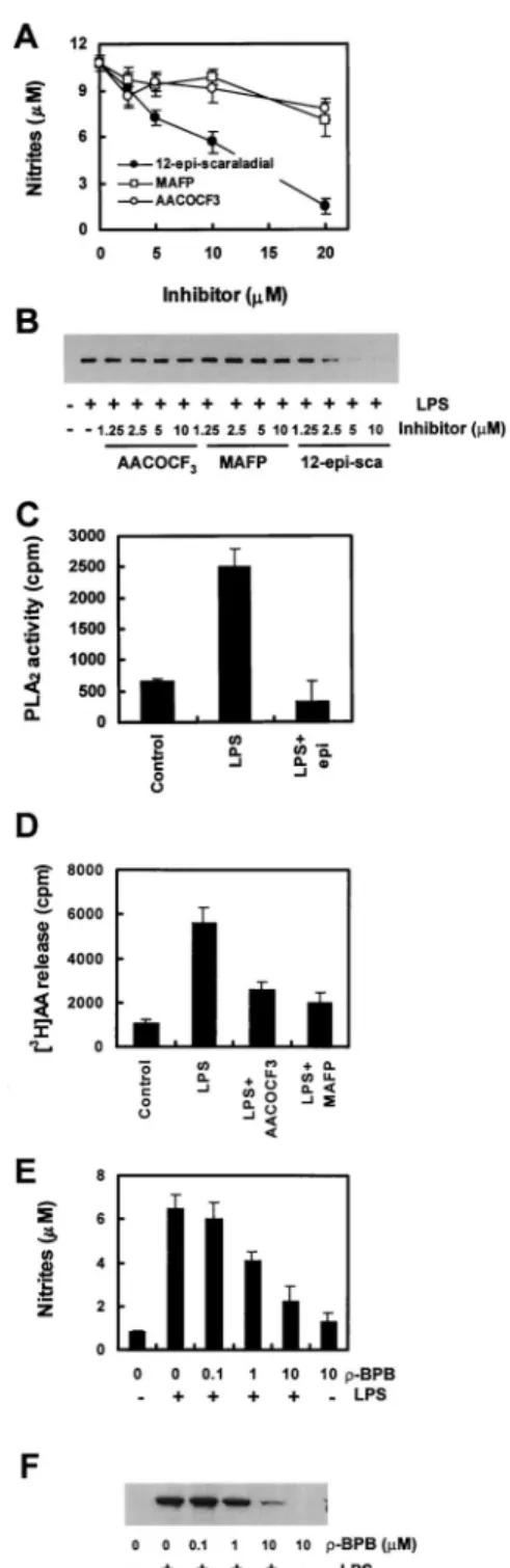
Effect of inhibitors of PLA2on LPS-induced nitrite production and iNOS expression in Raw264.7 cells
Because we have found that sPLA2raises the production of nitrite by Raw264.7 cells, we wanted to confirm the specificity of the PLA2 type. Therefore, we stimulated the cells with LPS in the presence of selective cPLA2inhibitors, synthetic arachidonic acid analogue MAFP or AACOCF3, and a specific sPLA2 inhibitor, 12-epi-scalaradial. AACOCF3and MAFP inhibit cPLA2-mediated phospholipid hydrolysis by binding tightly to the enzyme. 12-epi- scalaradial causes irreversible inhibition of sPLA2 by forming a Schiff’s base with a lysine residue on the surface of the enzyme (32). Nitrite production (Fig. 4A) as well as sPLA2activity (Fig.
4C) in response to LPS was inhibited in the presence of 12-epi- FIGURE 2. Dose-dependent induction of nitrite production and iNOS expression by sPLA2in LPS-stimulated Raw264.7 cells. Raw264.7 cells were incubated with the indicated concentrations of LPS and either culture medium or sPLA2for 18 h at 37°C. Nitrite production (A) was determined in the culture supernatant, and the level of iNOS protein (B) was deter- mined by Western bolt analysis as described in Materials and Methods.
Comparable results were obtained in three separate experiments.
FIGURE 3. Effect of purified sPLA2on nitrite production and iNOS ex- pression in LPS-stimulated Raw264.7 cells. Raw264.7 cells were incubated with the indicated concentrations of purified sPLA2in the presence or absence (A, inset) of LPS (1g/ml). Nitrite production in the culture supernatant was determined (A). Expression of iNOS (B) was determined by Western blot analysis as described in Materials and Methods. The results for nitrite are average values⫾ SE from three independent experiments, and iNOS expres- sion data are representative of three independent experiments.
FIGURE 4. Effects of PLA2inhibitors on nitrite production, iNOS ex- pression, and PLA2activity by Raw264.7 cells. Raw264.7 cells were pre- treated with the indicated concentrations of MAFP, AACOCF3, or 12-epi- sclaradial for 1 h followed by incubation with 1g/ml LPS for 18 h. Nitrite production (A) was determined in the culture supernatant. iNOS expression (B) was determined by Western blot analysis as described in Materials and Methods. C, The cells, pretreated with or without 10M 12-epi-sclaradial for 30 min, were challenged with LPS for 18 h. Afterward, supernatants were collected and assayed for PLA2activity. D, [3H]AA release was as- sayed in LPS-treated cells. The cells were treated with 20M MAFP or 20
M AACOCF3or neither for 30 min before the addition of LPS (1g/ml) for 18 h. The released [3H]AA was determined as described in Materials and Methods. The cells were pretreated with the indicated concentrations of-BPB for 1 h followed by incubation with LPS for 18 h. Nitrite pro- duction (E) was determined in the culture supernatant, and expression of iNOS (F) was determined by Western blot analysis as described in Mate- rials and Methods. The results for nitrite and PLA2activities are average values⫾ SE from three independent experiments, and iNOS expressions are representative of three independent experiments.
6362 sPLA2AND LPS ACT IN SYNERGY IN iNOS EXPRESSION
at Keimyung Univ Med Lib on January 13, 2016http://www.jimmunol.org/Downloaded from
scalaradial in a dose-dependent manner and was completely inhib- ited at a 20-M concentration of the inhibitor. However, cPLA2
inhibitors MAFP and AACOCF3at a high concentration (20M) had little effect on the LPS stimulation of the cells (Fig. 4A), al- though cPLA2activities were decreased to almost control levels by both cPLA2inhibitors (Fig. 4D). To evaluate whether this was due to inhibition of iNOS expression, we monitored LPS-induced iNOS levels using immunoblot analysis. LPS-mediated iNOS ex- pression was reduced in cells pretreated with 12-epi-scalaradial, while in the presence of MAFP or AACOCF3did not inhibit the LPS response (Fig. 4B). To strengthen this conclusion, we tested the effect of-bromophenacyl bromide (-BPB), the other struc- turally unrelated specific sPLA2inhibitor, on nitrite production and iNOS expression as well as sPLA2activity.-BPB also strongly inhibited nitrite production and iNOS expression (Fig. 4, E and F) concomitantly with sPLA2activity (data not shown). These results suggest that the LPS-induced activation of iNOS is indeed the specific effect of sPLA2. To confirm sPLA2 specificity for the iNOS induction, cDNAs encoding mouse cPLA2and sPLA2were separately subcloned into mammalian expression vector (pcDNA 3.1) and used to transiently transfect Raw264.7 cells. We then measured PLA2activity and the amount of nitrites produced. The activity of PLA2 increased about 4-fold in both transfectants as compare to empty vector transfectants. The production of nitrite was not detectable in untransfected Raw264.7 cells, while the ni- trite production by the sPLA2transfectants was about 2-fold over the empty vector or cPLA2transfectants. Furthermore, expression of iNOS was also increased in the sPLA2transfectants compared with others transfectants, although empty vector and cPLA2trans- fectants both slightly induced iNOS expression (Fig. 5).
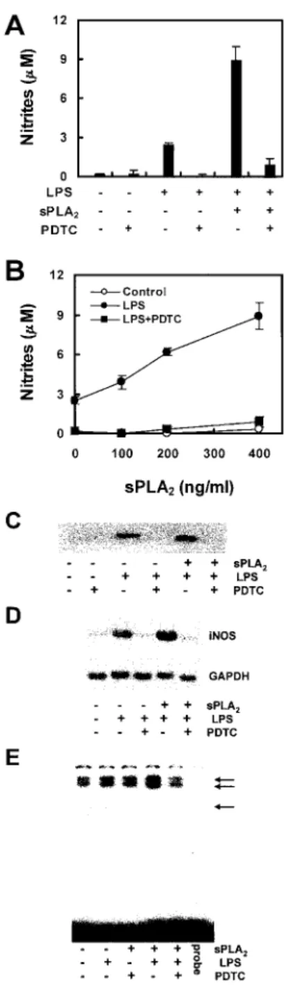
The role of NF-B in sPLA2-potentiated nitrite production, iNOS expression, and iNOS mRNA accumulation
Because sPLA2 potentiated the production of NO in Raw264.7 cells, we wanted to see whether sPLA2could be involved in the LPS-mediated activation of NF-B. One of the signaling mole- cules participating in the LPS-induction of iNOS expression is the transcriptional regulator NF-B. The antioxidant, PDTC, a potent inhibitor of NF-B activation, prevented LPS-induced iNOS ex- pression in Raw264.7 cells. To determine whether NF-B partic- ipated in sPLA2-potentiated nitrite production and iNOS expres- sion, the cells were pretreated for 1 h with 100M PDTC. After that, sPLA2or LPS and sPLA2plus LPS were added. The cells were then cultured for an additional 18 h. As seen in Fig. 6, A and B, the PDTC pretreatment prevented all LPS- and sPLA2 plus LPS-induced nitrite production. Consistent with its inhibitory ef-
fects on nitrite production, PDTC also inhibited LPS- and sPLA2
plus LPS-induced iNOS protein expression (Fig. 6C). We exam- ined iNOS mRNA accumulation in macrophages treated with FIGURE 5. Effect of transiently expressed sPLA2on nitrite production
and iNOS expression of transfected Raw264.7 cells. A, Raw264.7 cells were incubated with Lipofectamine complexes with sPLA2, cPLA2cDNA/
pCDNA 3.1, or empty vector for 72 h. After 72 h, nitrite production the medium was measured. B, Cell lysates from Raw264.7 cells transfected with the above cDNA were subjected to Western blot analysis with anti- iNOS Ab. The results for nitrite are average values⫾ SE from five inde- pendent experiments. iNOS expressions are representative of five indepen- dent experiments.
FIGURE 6. Effect of PDTC on nitrite production, iNOS expression, and mRNA accumulation in Raw264.7 cells. Raw264.7 cells were pretreated for 30 min with 100 M PDTC and then incubated for 18 h with the indicated concentrations of sPLA2in the presence of LPS (1g/ml). Nitrite production (A and B) in culture supernatant was determined. Expression of iNOS (C) was determined by Western blot analysis as described in Mate- rials and Methods. D, The cells were pretreated for 30 min with 100M PDTC and then incubated for 6 h with LPS (1g/ml) or LPS plus sPLA2
(400 ng/ml) as indicated. Total RNA was isolated and probed for iNOS and GAPDH by Northern blot analysis as described in Materials and Methods.
E, The cells were pretreated with 100 mM PDTC for 30 min before incu- bation with LPS or LPS plus sPLA2. Then, nuclear extracts were prepared.
NF-B-specific DNA-protein binding activity in nuclear extracts was de- termined by EMSA as described in Materials and Methods. The results for nitrite are average values⫾ SE from three independent experiments. iNOS protein data are representative of three independent experiments. iNOS mRNA and EMSA data are representative of two independent experiments.
at Keimyung Univ Med Lib on January 13, 2016http://www.jimmunol.org/Downloaded from
PDTC by Northern blot analysis. Fig. 6D show iNOS mRNA ac- cumulation in Raw264.7 cells stimulated with LPS and sPLA2plus LPS. However, in the cells pretreated with the NF-B inhibitor PDTC, the iNOS mRNA accumulation, even after LPS or sPLA2
plus LPS treatment, dropped to basal levels. In addition, we used EMSA to investigate the involvement of NF-B in the induction of iNOS. Raw264.7 cells were stimulated for 30 min with LPS or LPS plus sPLA2. In nuclear extracts of unstimulated macrophages, two faint DNA-protein complexes were identified, the intensity of which increased following exposure of the cells to LPS. However, the intensity of bands is markedly increased on the treated cells with LPS plus sPLA2. In addition, after treatment of the cells for 30 min with PDTC, the LPS plus sPLA2-induced activation of NF-B-specific DNA-protein complex formation was inhibited (Fig. 6E). These results suggest that the sPLA2plus LPS stimula- tion of iNOS mRNA transcription is dependent on NF-B participation.
Discussion
Among the macrophage responses to LPS exposure is the expres- sion of iNOS and increased production of NO (2, 6). Recent in- vestigations have shown evidence that sPLA2 enzymes may be important participants in the activation of macrophages by LPS (20 –23, 33). It has also been reported that sPLA2 enhances the response of leukocytes to LPS, which suggests a direct interaction of sPLA2 with LPS (34). Rupprecht et al. have shown and sug- gested cross talk between sPLA2and iNOS in rat renal mesangial cells (35). Recently, Tsukahara et al. reported that the PLA2in- hibitor quinacrine inhibited iNOS expression in alveolar macro- phages and reduced lung injury in acute pancreatitis. They sug- gested that PLA2 mediates NO production (36). Still, the mechanism by which PLA2 stimulates iNOS expression is unknown.
In our current study, we examined the effect of sPLA2on mac- rophage activation and the mechanism by which sPLA2activates iNOS. Treatment of Raw264.7 cells with LPS stimulated iNOS expression and nitrite production. Alone, sPLA2 also stimulated iNOS expression in Raw264.7 cells, but only slightly. However, in combination with LPS, sPLA2raised iNOS expression and nitrite production to high levels. The effect of PLA2on iNOS expression appears to be PLA2type specific. While sPLA2inhibitors, 12-epi- scalaradial and-BPB, inhibited LPS-induced iNOS expression in the cells, cPLA2inhibitors, MAFP or AACOCF3, did not inhibit nitrite production and iNOS expression. In addition, when cDNAs encoding either sPLA2 or cPLA2 were transfected into the cell, sPLA2 transfectants stimulated nitrite production significantly more than cPLA2or empty vector transfectants. In the process of responding to LPS, an early crucial step is the nuclear translocation of NF-B, which in turn induces the transcriptional activation of genes for various inflammatory cytokines. The sPLA2-potentiated iNOS expression in Raw264.7 cells also required the activation of NF-B. Our studies have shown that LPS- or LPS plus sPLA2- induced iNOS expression, mRNA accumulation, and nitrite pro- duction can be prevent by treatment of the cells with the NF-B inhibitor PDTC. In addition, PDTC inhibited LPS plus sPLA2- induced NF-B-specific DNA-protein binding and IB␣ degrada- tion by Raw264.7 cells (data not shown). These results suggest that sPLA2may participate in the iNOS induction, which then leads to the functional activation of NF-B.
Our study showed that sPLA2induces iNOS expression and NO generation of macrophages, which contribute to sepsis. These con- clusions are based on the observations of the direct effect of potent inhibitors with high selectivity against either sPLA2or cPLA2. The
role of sPLA2in endotoxic shock has been widely studied (27, 28).
Both activity and protein levels of this enzyme are enhanced in the serum of patients with endotoxic shock, and both increase after the production of proinflammatory cytokines including TNF-␣ and IL- 1. It is also well known that circulating PLA2causes tissue injury such as damage to the alveolar surfactant or, by reacting with cell membranes, releases inflammatory mediators such as eicosanoids and platelet-activating factor. Therefore, we hypothesize that in- creased levels of PLA2, especially type II, raise iNOS expression in macrophages and thus mediate sepsis or inflammation. This speculation is supported by several studies. Kurose et al. reported that an increased production of NO in rat Kupffer cells was pro- ceeded by activated NF-B, and the PLA2 inhibitor quinacrine significantly attenuated the increase in NF-B and NO production (37). Furthermore, in a study of an animal model of inflammation, when the rat air pouch was stimulated with zymosan, the levels of nitrites, sPLA2in exudates, and NOS activity in polymorphonu- clear leukocytes and monocytes increased (38).
At present, we have preliminary data concerning the upstream targets of iNOS. Some reports have shown a possible role for tyrosine kinase and phosphatidylinositol 3-kinase (PI 3K) in the process of macrophage activation and LPS-induced iNOS induc- tion (39 – 41). Inhibition of PI 3K by LY294002 results in down- regulation of iNOS expression, mainly through a mechanism that involves activation of NF-B. Furthermore, Chen et al. reported that LPS activates phospholipase D (PLD) via tyrosine phosphor- ylation by protein kinase C and NF-B activation, iNOS expres- sion, and finally NO release (42). We are currently examining the effects of signaling molecules such as tyrosine kinase, PI 3K, PLD, and extracellular regulated kinase (ERK) on sPLA2-potentiated iNOS expression in Raw264.7 cells. The PI 3K inhibitor LY294002 and the PLD inhibitor 1-butanol attenuate the sPLA2- potentiated effects as well as LPS effects, whereas the mitogen- activated protein kinase/ERK (MEK)/ERK inhibitor PD98059, which abrogates MEK/ERK activation by LPS, has little effect on iNOS expression (data not shown). The results suggest that the signal transduction pathways leading to iNOS and to MEK/ERK activation diverge downstream of PI 3K and PLD activation. This is in contrast to the situation in epithelial cell invasion by Listeria monocytogenes, in which both PI 3K and ERK are activated (43).
sPLA2 is a proinflammatory mediator found to be highly ele- vated both in the circulation and locally in tissues and in associ- ation with a number of pathologic conditions such as sepsis. The main proinflammatory effect of sPLA2is thought to be the gener- ation of arachidonic acid as a precursor for eicosanoids. Our results suggest a potentially new role for sPLA2, namely in the potentia- tion of iNOS and NF-B-regulated expression of genes involved in LPS signal transduction. The sPLA2- or LPS-mediated increase in NF-B and the cellular consequences should be of interest in the search for inhibitor compounds for the treatment of inflammatory conditions.
References
1. MacMicking, J., Q. W. Xie, and C. Nathan, 1997. Nitric oxide and macrophage function. Annu. Rev. Immunol. 15:323.
2. Nathan, C. 1992. Nitric oxide as a secretory product of mammalian cells. FASEB J. 6:3051.
3. Terenzi, F., M. J. Diaz-Guerra, M. Casado, S. Hortelano, S. Leoni, and L. Bosca.
1995. Bacterial lipopeptides induce nitric oxide synthase and promote apoptosis through nitric oxide-independent pathways in rat macrophages. J. Biol. Chem.
270:6017.
4. Cohen, J., and M. P. Glauser. 1991. Septic shock: treatment. Lancet 338:736.
5. Bone, R. C. 1991. The pathogenesis of sepsis. Ann. Intern. Med. 115:457.
6. Lorsbach, R. B., W. J. Murphy, C. J. Lowenstein, S. H. Snyder, and S. W. Russell. 1993. Expression of the nitric oxide synthase gene in mouse macrophages activated for tumor cell killing: molecular basis for the synergy between interferon-␥ and lipopolysaccharide. J. Biol. Chem. 268:1908.
6364 sPLA2AND LPS ACT IN SYNERGY IN iNOS EXPRESSION
at Keimyung Univ Med Lib on January 13, 2016http://www.jimmunol.org/Downloaded from
7. Nathan, C. 1997. Inducible nitric oxide synthase: what difference does it make?
J. Clin. Invest. 100:2417.
8. Brennan, F. M., and M. Feldmann. 1996. Cytokines in autoimmunity. Curr. Opin.
Immunol. 8:872.
9. Jiang, C., A. T. Ting, and B. Seed. 1998. PPAR-␥ agonists inhibit production of monocyte inflammatory cytokines. Nature 391:82.
10. Lopez-Collazo, E., S. Hortelano, A. Rojas, and L. Bosca. 1998. Triggering of peritoneal macrophages with IFN-␣/ attenuates the expression of inducible ni- tric oxide synthase through a decrease in NF-B activation. J. Immunol. 160:
2889.
11. Griffith, O. W., and D. J. Stuehr. 1995. Nitric oxide synthases: properties and catalytic mechanism. Annu. Rev. Physiol. 5:707.
12. Nathan, C., and Q. W. Xie. 1994. Regulation of biosynthesis of nitric oxide.
J. Biol. Chem. 269:13725.
13. Schmidt, H. H. 1992. NO䡠, CO and 䡠OH: endogenous soluble guanylyl cyclase- activating factors. FEBS Lett. 307:102.
14. Moncada, S., R. M. Palmer, and E. A. Higgs, 1991. Nitric oxide: physiology, pathophysiology, and pharmacology. Pharmacol. Rev. 43:109.
15. Snyder, S. H., and D. S. Bredt. 1992. Biological roles of nitric oxide. Sci. Am.
266:68.
16. Nunokawa, Y., N. Ishida, and S. Tanaka. 1993. Cloning of inducible nitric oxide synthase in rat vascular smooth muscle cells. Biochem. Biophys. Res. Commun.
191:89.
17. Lyons, C. R., G. J. Orloff, and J. M. Cunningham. 1992. Molecular cloning and functional expression of an inducible nitric oxide synthase from a murine mac- rophage cell line. J. Biol. Chem. 267:6370.
18. Geller, D. A., C. J. Lowenstein, R. A. Shapiro, A. K. Nussler, M. Di Silvio, S. C. Wang, D. K. Nakayama, R. L. Simmons, S. H. Snyder, and T. R. Billiar.
1993. Molecular cloning and expression of inducible nitric oxide synthase from human hepatocytes. Proc. Natl. Acad. Sci. USA 90:3491.
19. Galea, E., D. L. Feinstein, and D. J. Reis. 1992. Induction of calcium-independent nitric oxide synthase activity in primary rat glial cultures. Proc. Natl. Acad. Sci.
USA 89:10945.
20. Kim, H., H. S. Lee, K. T. Chang, T. H. Ko, K. J. Baek, and N. S. Kwon. 1995.
Chloromethyl ketones block induction of nitric oxide synthase in murine mac- rophages by preventing activation of nuclear factor-B. J. Immunol. 154:4741.
21. Heitmeier, M. R., A. L. Scarim, and J. A. Corbett. 1998. Double-stranded RNA- induced inducible nitric-oxide synthase expression and interleukin-1 release by murine macrophages requires NF-B activation. J. Biol. Chem. 273:15301.
22. Baeuerle, P. A., and T. Henkel. 1994. Function and activation of NF-B in the immune system. Annu. Rev. Immunol. 12:141.
23. Hu, Y., P. L. Fisette, L. C. Denlinger, A. G. Guadarrama, J. A. Sommer, R. A. Proctor, and P. J. Bertics. 1998. Purinergic receptor modulation of lipo- polysaccharide signaling and inducible nitric-oxide synthase expression in RAW264.7 macrophages. J. Biol. Chem. 273:27170.
24. Vadas, P., J. Browning, J. Edelson, and W. Pruzanski. 1993. Extracellular phos- pholipase A2expression and inflammation: the relationship with associated dis- ease states. J. Lipid Mediat. 8:1.
25. Crowl, R. M., T. J. Stoller, R. R. Conroy, and C. R. Stoner. 1991. Induction of phospholipase A2gene expression in human hepatoma cells by mediators of the acute phase response. J. Biol. Chem. 266:2647.
26. Kudo, I., M. Murakami, S. Hara, and K. Inoue. 1993. Mammalian non-pancreatic phospholipases A2. Biochim. Biophys. Acta 1170:217.
27. Endo, S., K. Inada, H. Nakae, T. Takakuwa, Y. Yamada, T. Suzuki, S. Taniguchi, M. Yoshida, M. Ogawa, and H. Teraoka. 1995. Plasma levels of type II phos-
pholipase A2and cytokines in patients with sepsis. Res. Commun. Mol. Pathol.
Pharmacol. 90:413.
28. Guidet, B., O. Piot, J. Masliah, V. Barakett, E. Maury, G. Bereziat, and G. Offenstadt. 1996. Secretory non-pancreatic phopholipase A2in severe sepsis:
relation to endotoxin, cytokines and thromboxane B2. Infection 24:103.
29. Bomalaski, J. S., T. Ford, A. P. Hudson, and M. A. Clark. 1995. Phospholipase A2-activating protein induces the synthesis of IL-1 and TNF in human mono- cytes. J. Immunol. 154:4027.
30. Weiss, J., M. Inada, M., Elsbach, and R. M. Crowl. 1994. Structural determinants of the action against Escherichia coli of a human inflammatory fluid phospho- lipase A2in concert with polymorphonuclear leukocytes. J. Biol. Chem. 269:
26331.
31. Gunasekar, P. G., A. G. Kanthasamy, J. L. Borowitz, and G. E. Isom. 1995.
NMDA receptor activation produces concurrent generation of nitric oxide and reactive oxygen species: implication for cell death. J. Neurochem. 65:2016.
32. Potts, B. C., D. J. Faulkner, and R. S. Jacobs. 1992. Phospholipase A2inhibitors from marine organisms. J. Nat. Prod. 55:1701.
33. Arbibe, L., D. Vial, I. Rosinski-Chupin, N. Havet, M. Huerre, B. B. Vargaftig, and L. Touqui. 1997. Endotoxin induces expression of type II phospholipase A2 in macrophages during acute lung injury in guinea pigs: involvement of TNF-␣ in lipopolysaccharide-induced type II phospholipase A2synthesis. J. Immunol.
159:391.
34. Cai, T. Q., N. Thieblemont, B. Wong, R. Thieringer, B. P. Kennedy, and S. D. Wright. 1999. Enhancement of leukocyte response to lipopolysaccharide by secretory group IIA phospholipase A2. J. Leukocyte Biol. 65:750.
35. Rupprecht, G., K. Scholz, K. F. Beck, H. Geiger, J. Pfeilschifter, and M. Kaszkin.
1999. Cross-talk between group IIA-phospholipase A2and inducible NO-syn- thase in rat renal mesangial cells. Br. J. Pharmacol. 127:51.
36. Tsukahara, Y., T. Morisaki, Y. Horita, M. Torisu, and M. Tanaka. 1999. Phos- pholipase A2mediates nitric oxide production by alveolar macrophages and acute lung injury in pancreatitis. Ann. Surg. 229:385.
37. Kurose, I., H. Saito, S. Miura, H. Ebinuma, H. Higuchi, N. Watanabe, S. Zeki, T. Nakamura, M. Takaishi, and H. Ishii. 1997. CD18/ICAM-1-dependent oxida- tive NF-B activation leading to nitric oxide production in rat Kupffer cells cocultured with syngeneic hepatoma cells. J. Clin. Invest. 99:867.
38. Paya, M., P. Garcia Pastor, J. Coloma, and M. J. Alcaraz. 1997. Nitric oxide synthase and cyclo-oxygenase pathways in the inflammatory response induced by zymosan in the rat air pouch. Br. J. Pharmacol. 120:1445.
39. Procyk, K. J., P. Kovarik, A. von Gabain, and M. Baccarini. 1999. Salmonella typhimurium and lipopolysaccharide stimulate extracellularly regulated kinase activation in macrophages by a mechanism involving phosphatidylinositol 3-ki- nase and phospholipase D as novel intermediates. Infect. Immun. 67:1011.
40. Diaz-Guerra, M. J., A. Castrillo, P. Martin-Sanz, and L. Bosca. 1999. Negative regulation by phosphatidylinositol 3-kinase of inducible nitric oxide synthase expression in macrophages. J. Immunol. 162:6184.
41. Salh, B., R. Wagey, A. Marotta, J. S. Tao, and S. Pelech. 1998. Activation of phosphatidylinositol 3-kinase, protein kinase B, and p70 S6 kinases in lipopo- lysaccharide-stimulated Raw264. 7 cells: differential effects of rapamycin, Ly294002, and wortmannin on nitric oxide production. J. Immunol. 161:6947.
42. Chen, C. C., J. K. Wang, W. C. Chen, and S. B. Lin. 1998. Protein kinase C mediates lipopolysaccharide-induced nitric-oxide synthase expression in primary astrocytes. J. Biol. Chem. 273:19424.
43. Ireton, K., B. Payrastre, H. Chap, W. Ogawa, H. Sakaue, M. Kasuga, and P. Cossart. 1996. A role for phosphoinositide 3-kinase in bacterial invasion. Sci- ence 274:780.
at Keimyung Univ Med Lib on January 13, 2016http://www.jimmunol.org/Downloaded from