ABSTRACT
Bioresorbable vascular scaffold (BRS) is an innovative device that provides structural support and drug release to prevent early recoil or restenosis, and then degrades into nontoxic compounds to avoid late complications related with metallic drug-eluting stents (DESs).
BRS has several putative advantages. However, recent randomized trials and registry studies raised clinical concerns about the safety and efficacy of first generation BRS. In addition, the general guidance for the optimal practice with BRS has not been suggested due to limited long-term clinical data in Korea. To address the safety and efficacy of BRS, we reviewed the clinical evidence of BRS implantation, and suggested the appropriate criteria for patient and lesion selection, scaffold implantation technique, and management.
Keywords: Coronary disease; Bioresorbable vascular scaffold; Stents; Thrombosis
Review Article
Jung-Min Ahn , MD1, Duk-Woo Park, MD1, Sung Jin Hong , MD2, Young Keun Ahn, MD3, Joo-Yong Hahn , MD4, Won-Jang Kim, MD5, Soon Jun Hong, MD6, Chang-Wook Nam , MD7, Do-Yoon Kang, MD1, Seung-Yul Lee , MD8, Woo Jung Chun, MD9, Jung Ho Heo, MD10,
Deok-Kyu Cho, MD11, Jin Won Kim, MD12, Sung-Ho Her, MD13, Sang Wook Kim, MD14, Sang-Yong Yoo, MD15, Myeong-Ki Hong , MD2, Seung-Jea Tahk, MD16,
Kee-Sik Kim, MD17, Moo Hyun Kim, MD18, Yangsoo Jang , MD2, and Seung-Jung Park , MD, PhD1
1Heart Institute, University of Ulsan College of Medicine, Asan Medical Center, Seoul, Korea
2Division of Cardiology, Severance Cardiovascular Hospital, Yonsei University College of Medicine, Seoul, Korea
3Division of Cardiology, Department of Medicine, Chonnam National University Hospital, Gwangju, Korea
4 Division of Cardiology, Department of Medicine, Samsung Medical Center, Sungkyunkwan University School of Medicine, Seoul, Korea
5Department of Cardiology, CHA Bundang Medical Center, CHA University, Seongnam, Korea
6Department of Cardiology, Cardiovascular Center, Korea University Anam Hospital, Seoul, Korea
7Department of Medicine, Keimyung University College of Medicine, Daegu, Korea
8Department of Internal Medicine, Wonkwang University Sanbon Hospital, Sanbon, Korea
9 Division of Cardiology, Department of Internal Medicine, Sungkyunkwan University Samsung Changwon Hospital, Changwon, Korea
10Division of Cardiology, Department of Internal Medicine, Kosin University Gospel Hospital, Busan, Korea
11Department of Cardiology, Myongji Hospital, Goyang, Korea
12Cardiovascular Center, Korea University Guro Hospital, Seoul, Korea
13Division of Cardiology, The Catholic University of Korea, Daejeon St. Mary's Hospital, Daejeon, Korea
14Department of Cardiology, Chung-Ang University Hospital, Seoul, Korea
15Cardiovascular Center, GangNeung Asan Hospital, Gangneung, Korea
16Division of Cardiology, Ajou University Medical Center, Suwon, Korea
17 Division of Cardiology, Department of Internal Medicine, Daegu Catholic University Medical Center, Daegu, Korea
18Department of Cardiology, Dong-A University Medical Center, Busan, Korea
Bioresorbable Vascular Scaffold Korean Expert Panel Report
Received: Aug 31, 2017 Accepted: Sep 12, 2017 Correspondence to Seung-Jung Park, MD, PhD
Heart Institute, University of Ulsan College of Medicine, Asan Medical Center, 88, Olympic- ro 43-gil, Songpa-gu, Seoul 05505, Korea.
E-mail: [email protected] Copyright © 2017. The Korean Society of Cardiology
This is an Open Access article distributed under the terms of the Creative Commons Attribution Non-Commercial License (https://
creativecommons.org/licenses/by-nc/4.0) which permits unrestricted noncommercial use, distribution, and reproduction in any medium, provided the original work is properly cited.
ORCID iDs Jung-Min Ahn
https://orcid.org/0000-0003-4031-391X Sung Jin Hong
https://orcid.org/0000-0003-4893-039X Joo-Yong Hahn
https://orcid.org/0000-0002-4412-377X Chang-Wook Nam
https://orcid.org/0000-0002-3370-5774 Seung-Yul Lee
https://orcid.org/0000-0002-9039-9806 Myeong-Ki Hong
https://orcid.org/0000-0002-2090-2031 Yangsoo Jang
https://orcid.org/0000-0002-2169-3112 Seung-Jung Park
https://orcid.org/0000-0002-9187-5405
Conflict of Interest
The authors have no financial conflicts of interest.
INTRODUCTION
Bioresorbable vascular scaffold (BRS) is an innovative device that provides structural support and drug release to prevent early recoil or restenosis, and then degrades into nontoxic compounds to avoid late complications related with metallic drug-eluting stents (DESs).
BRS has several putative advantages including early restoration of physiological processes, superior conformability, beneficial edge-vascular response and suppression of late-stent malapposition.1) In addition, 5-years follow-up of BRS showed late lumen enlargement and restoration of vasomotor response and suggested a possible plaque stabilizing effect.2-4) However, recent randomized trials and registry studies raised clinical concerns about the safety and efficacy of first generation BRS. They showed that higher rate of procedural related myocardial infarction (MI), and scaffold thrombosis compared with metallic DES.5)6) Thus, at March 2017, the US Food and Drug Administration (FDA) warned physicians that treating patients with first generation BRS.
BRS has been commercially available in Korea since January 2016, and as of August 2017, about 2,800 BRSs were implanted. However, the general guidance for the optimal practice with BRS has not been suggested due to limited long-term clinical data in Korea. Therefore, at 4th August 2017, 18 Korean heart centers combined efforts to address clinical issues raised by previous studies. We reviewed the clinical evidence of BRS implantation, and suggested the appropriate criteria for patient and lesion selection, scaffold implantation technique, and management.
The scope of this document is limited to the Absorb Bioresorbable Vascular Scaffold (Absorb BRS; Abbott Vascular, Abbott Park, IL, USA), which is the only available BRS in Korea.
DEVICE DESCRIPTION
The Absorb BRS (Abbott Vascular) consists of a 157-μm-thick bioresorbable poly (L-lactide) scaffold with a 7-μm-thick bioresorbable poly (D,L-lactide) coating, which elutes everolimus (7). About 80% of the drug elutes in the first 30 days, and the remainder elutes over 120 days.
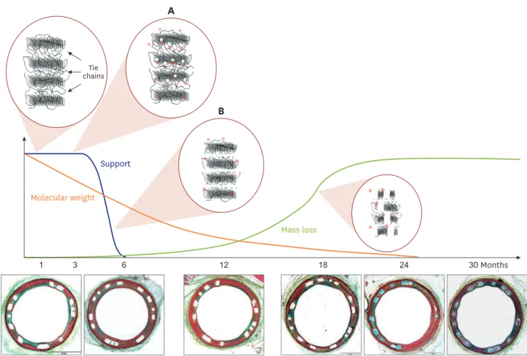
Scaffold are fully bioresorbable, with complete bioresorption expected by approximately 24 to 36 months. The initial reduction in molecular weight, the decrease in radial support occurs at approximately equal to 6 months, and finally the loss in mass starts at 12 months (Figure 1).8) To compared with metallic DESs, BRS has less acute gain, and smaller lumen area. In addition, it has a limited expansion capability. Experimental in-vitro study indicated the fracture threshold was +1.0 mm. Overexpansion beyond this threshold can lead to strut disconnections and focal loss of mechanical support.9)
CURRENT EVIDENCE
Early pilot trials
The ABSORB cohort A and B experiences provided objective characterization of BRS resorption and coronary healing process in humans.10)11) Serial intravascular imaging studies showed that strut resorption and vascular healing after BRS implantation were associated with late lumen enlargement.2) The formation of a neointima layer after BRS resorption suggested to seal or “cap” the necrotic core plaque, and to prevent plaque rupture in the future.3) In addition, the restoration of vasomotor response to stimuli was demonstrated by 5 years.4) Early promising results called for clinical comparison studies with standard metallic DES.
Randomized trials
ABSORB II is the first randomized controlled trial to compare Absorb BRS with everolimus- eluting stent (EES) in 501 patients.1) The primary endpoint was angiographic vasomotor reactivity after nitrate injection and angiographic late luminal loss. At 1 year, first new or worsening angina was lower with BRS, although clinical outcomes were similar between groups. However, at 3-year follow-up, the vasomotor reactivity, angina status, and exercise capacity were not different. In addition, the late luminal loss was larger in the BRS group.
The rate of a device-oriented composite endpoint (DOCE) was significantly higher in the BRS group, mainly driven by target vessel MI. Definite or probable device thrombosis was also significantly higher in the BRS group (Figure 2).5)
ABSORB III is the first large-scale, multicenter, randomized trial for US FDA regulatory approval.7) This study enrolled 2,008 patients with relatively simple coronary lesions. This study demonstrated that the BRS group was non-inferior to the EES group in the respective to target lesion failure (TLF) at 1-year. However, the 2-year results presented at American College of the Cardiology (ACC) 2017 that the rates of TLF were significantly higher in the BRS group due to the increased risk of target vessel MI in the BRS group. However, ABSORB China and ABSORB Japan at EuroPCR 2017 showed that BRS had comparable safety and efficacy to EESs at 3 years.
Mass loss Molecular weight
1 3 6 12 18 24 30
chainsTie
Months Support
A
B
Figure 1. Scaffold biodegradation. (A) Hydrolysis randomly cleaves amorphous tie chains, leading to a decrease in molecular weight without altering radial strength. (B) When enough tie chains are broken, the device begins losing radial strength. After 2–3 years, BRS was fully bioresorbed.
BRS = bioresorbable vascular scaffold.
Recently, the Amsterdam Investigator-initiateD Absorb Strategy (AIDA) study reported early because of the higher incidence of device thrombosis in the BRS group.6) This study enrolled 1,845 patients with more complex lesion subset than ABSORB III trial in the context of routine clinical practice: acute coronary syndrome (ACS) was 54% and small vessel disease was about 20%. In addition, postdilatation (74%) was still underused. Although target vessel failure (TVF) was not significantly different, device thrombosis, and subsequently target vessel myocadial infarction were significantly higher in the BRS group. Table 1 summarized currently available randomized trials.
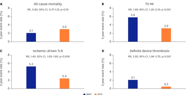
Update meta-analysis of 2-year outcomes from 7 randomized trials showed that BRS had higher 2-year risk of the DOCE than EES.12) This difference was mainly derived from target vessel MI and ischemic-driven target lesion revascularization (TLR). In addition, device thrombosis was significantly higher in the BRS group. However, cardiac mortality was not different between groups (Figure 3).
A B C
D E F
Figure 2. A case of acute scaffold thrombosis. (A) Baseline coronary angiography, (B) after Absorb 3.0×23 mm BRS implantation, (C) follow-up coronary angiography due to chest pain with ST elevation after 7 hours, (D) OCT showing acute scaffold thrombosis with underexpansion and malapposition, (E) final angiography after high-pressure balloon dilatation, and (F) final OCT image showing well-apposed scaffold.
BRS = bioresorbable vascular scaffold; OCT = optical coherent tomography.
Table 1. Summary of randomized trials with the Absorb BRS Clinical trial No. of patients
(BRS:DES) Primary endpoint Primary outcome DOCE rate
(BRS vs. DES) Scaffold thrombosis rate (BRS vs. DES)
ABSORB II1) 501 (335:166) Vasomotor reactivity/angiographic
lumen loss at 3 years 0.47 mm vs. 0.56 mm (p=0.49)/
0.37 mm vs. 0.25 mm (pnon-inferiority=0.78) 5% vs. 3% (p=0.35) 0.9% vs. 0% (p=0.55) ABSORB III7) 2,008 (1,322:686) Target-lesion failure at 1 year 7.8% vs. 6.1% (p=0.16, pnon-inferiority=0.007) Same as primary outcome 1.5% vs. 0.7% (p=0.13) ABSORB Japan42) 400 (266:134) Target-lesion failure at 1 year 4.2% vs. 3.8% (pnon-inferiority<0.0001) Same as primary outcome 1.5% vs. 1.5% (p>0.99) ABSORB China43) 480 (241:239) In-segment lumen loss at 1 year 0.19 mm vs. 0.13 mm (p=0.01) 3.4% vs. 4.2% (p=0.62) 0.4% vs. 0% (p>0.99) EVERBIO II64) 240 (80:160) Late lumen loss at 9 months 0.28±0.39 mm
vs. 0.25±0.36 mm (p=0.30) 12% vs. 9% (p=0.6) 1.3% vs. 0%
TROFI II65) 191 (95:96) Healing score at 6 months 1.74 vs. 2.80 (pnon-inferiority<0.001) 1.1% vs. 0% 1.1% vs. 0%
AIDA6) 1,845 (924:921) Target-vessel failure at 2 years 11.7% vs. 10.7% (p=0.43) 10.3% vs. 8.9% (p=0.31) 3.5% vs. 0.9% (p<0.001) AIDA = Amsterdam Investigator-initiated Absorb Strategy; BRS = bioresorbable vascular scaffold; DES = drug-eluting stent; DOCE = device-oriented composite endpoint; EVERBIO = Comparison of Everolimus- and Biolimus-Eluting Coronary Stents with Everolimus-Eluting Bioresorbable Vascular Scaffold; TROFI II
= Comparison of the ABSORB Everolimus Eluting Bioresorbable Vascular Scaffold System With a Drug-Eluting Metal Stent (Xience™) in Acute ST-Elevation Myocardial Infarction.
Registry studies
The Gauging coronary Healing with bioresorbable Scaffolding plaTforms in EUrope (GHOST- EU) registry evaluated the performance of the Absorb BRS in a real-world practice from 10 European heart centers.13) The incidence of TLF was 2.2% at 30 days and 4.4% at 6 months.
However, definite or probable scaffold thrombosis was 1.5% at 30 days and 2.1% at 6 months.
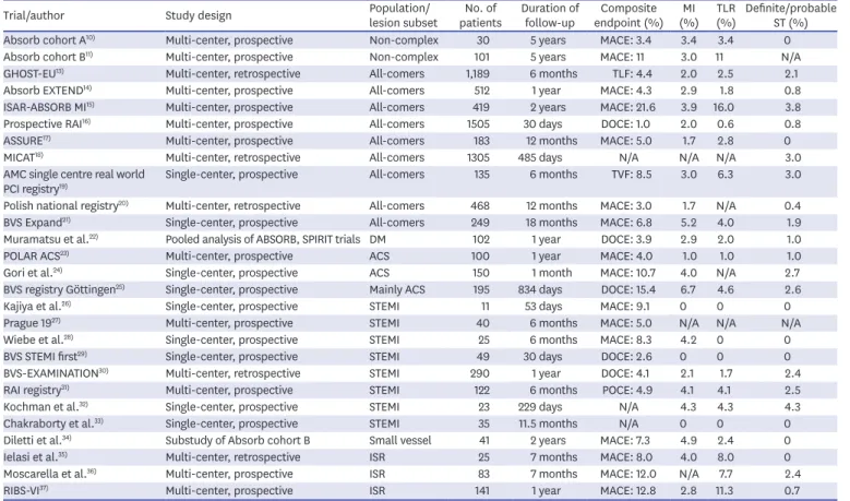
ABSORB expand registry reported that 12-month clinical outcomes in the first 512 patients.14) At one year, the ischemia-driven TVF was 4.9%. The cumulative rate of definite and probable scaffold thrombosis for this population was 0.8%. A Prospective, Randomized Trial of Bioresorbable Vascular Scaffold Versus Everolimus Eluting Stent in Patients Undergoing Coronary Stenting for Myocardial Infarction (ISAR-ABSORB MI) registry enrolled more complex population with diabetes (31.5%), ACS (39.0%), and bifurcation (13.1%).15) At 2 years, the composite of death, MI, or TLR occurred in 21.6%. Definite scaffold thrombosis occurred in 3.8%. This study showed the higher event rates than expected, which raised concerns about the daily use of BRS. The Registro Absorb Italiano (RAI) registry enrolled 1,505 patients (22.4% diabetes, 59.6% ACS) from 23 Italian heart centers16). All lesions were predilated and 96.8% lesions were post-dilated after BRS implantation. At 30 days, TLR occurred in 0.6% and definite or probable scaffold thrombosis occurred in 0.8%. This registry suggested that when accurate BRS implantation technique was used, an unrestricted BRS use would be safe and effective.17-21) In addition, BRS was evaluated in the complex patients and lesions subset including diabetes,22) ACS,23-25) MI,26-33) small vessel,34) and in-stent restenosis.35-37) However, the interpretation of such studies should be careful considering the biased nature of registry studies. Table 2 summarized currently available registry studies.
B
2 year event rate (%)
RR, 0.80; 95% CI, 0.57–1.12; p=0.19
2.1 3.0
5.8
3.2 RR, 1.68; 95% CI, 1.29–2.19; p<0.001
All-cause mortality TV-MI
Ischemic-driven TLR Definite device thrombosis
8 6 4
0 2
A
2 year event rate (%)
8 6 4
0 2
D
2 year event rate (%)
RR, 1.40; 95% CI, 1.09–1.80; p=0.009 5.3
2.4 2.1
0.5 RR, 3.95; 95% CI, 1.96–5.72; p<0.001 8
6 4
0 2
C
2 year event rate (%)
8 6 4
0 2
BRS EES
Figure 3. Meta-analysis from 7 randomized trials: 2 year outcomes.
BRS = bioresorbable vascular scaffold; CI = confidence interval; EES = everolimus-eluting stent; RR = relative risk; TLR = target lesion revascularization; TV-MI = target vessel myocardial infarction.
RISK FACTORS FOR SCAFFOLD THROMBOSIS
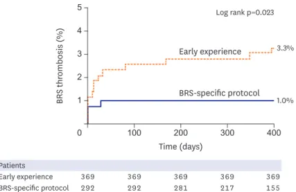
Risk factors for scaffold thrombosis were multifactorial in the combination of the device, procedural, and patient factors. Mainz IntraCoronAry daTabase (MICAT) registry enrolled 42 scaffold thrombosis. Multivariate analysis showed that ostial lesions and impaired left ventricular ejection fraction were independent predictors of scaffold thrombosis. In addition, early discontinuation of dual antiplatelet therapy (DAPT) was frequent in those patients.
Most striking finding was that a BRS-specific implantation strategy reduced 12-months scaffold thrombosis rate from 3.3% to 1.0% (Figure 4).18)
Recent meta-analysis from ABSORB trials reported that diabetes and preprocedural reference vessel diameter (<2.25 mm vs. ≥2.25 mm) were independent predictors for definite or probable device thrombosis.12)
Table 2. Summary of registry studies with the Absorb BRS
Trial/author Study design Population/
lesion subset No. of
patients Duration of
follow-up Composite endpoint (%) MI
(%) TLR
(%) Definite/probable ST (%)
Absorb cohort A10) Multi-center, prospective Non-complex 30 5 years MACE: 3.4 3.4 3.4 0
Absorb cohort B11) Multi-center, prospective Non-complex 101 5 years MACE: 11 3.0 11 N/A
GHOST-EU13) Multi-center, retrospective All-comers 1,189 6 months TLF: 4.4 2.0 2.5 2.1
Absorb EXTEND14) Multi-center, prospective All-comers 512 1 year MACE: 4.3 2.9 1.8 0.8
ISAR-ABSORB MI15) Multi-center, prospective All-comers 419 2 years MACE: 21.6 3.9 16.0 3.8
Prospective RAI16) Multi-center, prospective All-comers 1505 30 days DOCE: 1.0 2.0 0.6 0.8
ASSURE17) Multi-center, prospective All-comers 183 12 months MACE: 5.0 1.7 2.8 0
MICAT18) Multi-center, retrospective All-comers 1305 485 days N/A N/A N/A 3.0
AMC single centre real world
PCI registry19) Single-center, prospective All-comers 135 6 months TVF: 8.5 3.0 6.3 3.0
Polish national registry20) Multi-center, retrospective All-comers 468 12 months MACE: 3.0 1.7 N/A 0.4
BVS Expand21) Single-center, prospective All-comers 249 18 months MACE: 6.8 5.2 4.0 1.9
Muramatsu et al.22) Pooled analysis of ABSORB, SPIRIT trials DM 102 1 year DOCE: 3.9 2.9 2.0 1.0
POLAR ACS23) Multi-center, prospective ACS 100 1 year MACE: 4.0 1.0 1.0 1.0
Gori et al.24) Single-center, prospective ACS 150 1 month MACE: 10.7 4.0 N/A 2.7
BVS registry Göttingen25) Single-center, prospective Mainly ACS 195 834 days DOCE: 15.4 6.7 4.6 2.6
Kajiya et al.26) Single-center, prospective STEMI 11 53 days MACE: 9.1 0 0 0
Prague 1927) Multi-center, prospective STEMI 40 6 months MACE: 5.0 N/A N/A N/A
Wiebe et al.28) Single-center, prospective STEMI 25 6 months MACE: 8.3 4.2 0 0
BVS STEMI first29) Single-center, prospective STEMI 49 30 days DOCE: 2.6 0 0 0
BVS-EXAMINATION30) Multi-center, retrospective STEMI 290 1 year DOCE: 4.1 2.1 1.7 2.4
RAI registry31) Multi-center, prospective STEMI 122 6 months POCE: 4.9 4.1 4.1 2.5
Kochman et al.32) Single-center, prospective STEMI 23 229 days N/A 4.3 4.3 4.3
Chakraborty et al.33) Single-center, prospective STEMI 35 11.5 months N/A 0 0 0
Diletti et al.34) Substudy of Absorb cohort B Small vessel 41 2 years MACE: 7.3 4.9 2.4 0
Ielasi et al.35) Multi-center, retrospective ISR 25 7 months MACE: 8.0 4.0 8.0 0
Moscarella et al.36) Multi-center, prospective ISR 83 7 months MACE: 12.0 N/A 7.7 2.4
RIBS-VI37) Multi-center, prospective ISR 141 1 year MACE: 12.8 2.8 11.3 0.7
ACS = acute coronary syndrome; AMC = Academic Medical Center; ASSURE = An Absorb post-marketing surveillance registry to monitor the everolimus-eluting bioresorbable vascular scaffold in patients with coronary artery disease; BRS (BVS) = bioresorbable vascular scaffold; DM = diabetes mellitus; DOCE = device- oriented composite endpoint; GHOST-EU = Gauging coronary Healing with bioresorbable Scaffolding plaTforms in EUrope; ISAR-ABSORB MI = A Prospective, Randomized Trial of Bioresorbable Vascular Scaffold Versus Everolimus Eluting Stent in Patients Undergoing Coronary Stenting for Myocardial Infarction; ISR
= in-stent restenosis; MACE = major cardiac adverse event; MI = myocardial infarction; MICAT = Mainz IntraCoronAry daTabase; N/A = not applicable; PCI
= percutaneous coronary intervention; POCE = patient-oriented composite endpoint; POLAR ACS = POLishAbsorb Registry for ACS Patients; RAI = Registro Absorb Italiano; RIBS-VI = Restenosis Intra-stent: drug eluting Balloon vs. everolimus-eluting Stent-VI; SPIRIT = A Clinical Evaluation of the XIENCE V Everolimus Eluting Coronary Stent System; ST = stent thrombosis; STEMI = ST-segment elevation myocardial infarction; TLF = target lesion failure; TLR = target lesion revascularization; TVF = target vessel failure.
KOREAN EXPERIENCE
The Absorb BRS has been used since October 2015 in Korea. As of August 2017, a total of 2,833 BRSs were implanted. Among those, only 9 scaffold thrombosis were reported in 9 patients.
All patients presented with ACS (5 acute MI, 4 unstable angina). BRSs were implanted under the intracoronary imaging guidance in all cases. Postdilatation was performed in 8 patients.
Scaffold thrombosis occurs in 2 cases within 24 hours and in 7 cases between 1 day and 30 days.
No late scaffold thrombosis was reported. Possible mechanisms of scaffold thrombosis are early continuation of DAPT in 5 cases, underexpansion in 4 cases, and scaffold malapposition in 1 case. All patients were successfully treated without events (Table 3).
Interventional Cardiology Research In-cooperation Society Fractional Flow Reserve Bioresorbable Vascular Scaffold (IRIS BVS) registry is the ongoing prospective, multicenter
BRS thrombosis (%)
Time (days)
Early experience
BRS-specific protocol
100 200 300 400
4 3 2 1
0
Log rank p=0.023
3.3%
1.0%
5
Patients
Early experience 369 369 369 369 369
BRS-specific protocol 292 292 281 217 155
Figure 4. BRS specific implantation protocol. BRS specific implantation protocol was associated with about 70%
reduction of scaffold thrombosis.
BRS = bioresorbable vascular scaffold.
Table 3. BRS thrombosis from Korean population
Characteristic Case 1 Case 2 Case 3 Case 4 Case 5 Case 6 Case 7 Case 8 Case 9
Sex/age M/54 M/45 M/67 M/87 F/75 F/68 M/58 M/73 M/74
Diagnosis UA NSTEMI NSTEMI STEMI UA UA NSTEMI STEMI ACS
Location m LAD p LAD m LAD m LAD p LAD p-m LAD m LAD m LAD m LAD
BRS (mm) 3.5×28 2.5×18 2.5×28 3.0×18 3.0×18 3.5×28 3.5×28 3.0×23 3.0×18
Image guidance IVUS OCT OCT OCT OCT IVUS/OCT IVUS/OCT None IVUS
Post-dilatation Done Done Done Not done
(complete Scaffold apposition)
Done Done Done Done
(3.5×12 mm) Done (3.0×12 mm)
Days post-procedure 12 days 14 days 20 days 8 days 7 days 5 hours 13 days 7 hours 2 months
DAPT Aspirin,
Clopidogrel Aspirin, Ticagrelor Aspirin,
Ticagrelor Aspirin,
Clopidogrel Aspirin,
Clopidogrel Aspirin,
Clopidogrel Aspirin,
Ticagrelor Aspirin,
Clopidogrel Aspirin, clopidogrel D/C for 3 days D/C for 2 days D/C for 3 days D/C for 5 hours D/C for 3 days
Possible mechanism DAPT D/C DAPT D/C DAPT D/C Under-
expansion Under-
expansion Jailed diagonal
branch DAPT D/C Scaffold
malapposition DAPT D/C Underexpansion Underexpansion
Outcomes Survival Survival Survival Survival Survival Survival Survival Survival Survival
ACS = acute coronary syndrome; BRS = bioresorbable vascular scaffold; DAPT = dual antiplatelet therapy; D/C = discontinuation; IUVS = intravascular ultrasound;
LAD = left anterior descending; m = mid; NSTEMI = non-ST-segment elevation myocardial infarction; OCT = optical coherent tomography; p = proximal; ST = stent thrombosis; STEMI = ST-segment elevation myocardial infarction; UA = unstable angina.
registry to enroll all patients who underwent the Absorb BRS implantation from 15 Korean heart centers. Preliminary data of early 352 patients was presented at Transcatheter Cardiovascular Therapeutics Angioplasty Summit (TCTAP) 2017. All procedures were performed under the intracoronary imaging guidance. Predilatation was performed in 94%;
postdilation with non-compliant balloon in 99%. Mean scaffold diameter was 3.5±1.9 mm.
Final non-compliant balloon pressure was 19.5±5.3 mm. Final balloon diameter was 3.6±0.3 mm. Mean balloon to artery ratio was 1.22±0.30. Mean inflation time was 25±12 seconds. At 1 year, no scaffold thrombosis occurred. Only 2 TLFs related with periprocedural MI occurred (Table 4). Compared with other studies, the IRIS BVS registry had the higher rate of imaging guidance, and pre- and postdilation. This could be a plausible reason of favorable BRS outcomes with very low rate of scaffold thrombosis in Korea, although preliminary.
INDICATIONS FOR BRS IMPLANTATION
Patient selection
As current BRSs resolve 2–3 years after implantation, improvement in outcome comparing with metallic DESs is expected in long-term over those periods. Therefore, ideal BRS candidate is a relatively young with a good life expectancy (>5 years). On the other hand, the use of BRS in patients with limited life expectancy with multiple comorbidities, and at high bleeding risk was not supported.38)
Lesion selection
BRS can be implanted in non-complex lesions including de novo lesions with diameter of 2.25–4.0 mm on on-site quantitative coronary angiography (QCA), relative short lesions, and stable angina presentation. The culprit lesion of ST segment elevated MI, bifurcation lesion treating with 2 scaffolds, and aorto-ostial lesion, extreme tortuous vessel and small vessel (<2.25 mm) were less favorable lesions for BRS implantation. Table 5 summarized the BRS favorable patient and lesion characteristics.38)
BRS specific implantation techniques
Optimal sizing and BRS implantation technique is of paramount importance for achieving favorable long-term outcomes. Operators should understand that thick scaffold struts and their plastic nature results in a lower lumen gain and a smaller post-implanted minimal lumen Table 4. IRIS BVS registry: 1 year outcome (n=352)
Variable No. of patients (%)
Device-oriented endpoint
TVF 2 (0.45)
Cardiac death 0 (0.0)
MI 2 (0.45)
Periprocedural MI 2 (0.45)
Spontaneous MI 0 (0.0)
Target-vessel revascularization 0 (0.0)
Scaffold thrombosis 0 (0.0)
Patient-oriented endpoint
Death from any cause 0 (0.0)
Cardiac death 0 (0.0)
Non-cardiac death 0 (0.0)
Stroke 0 (0.0)
IRIS BVS = Interventional Cardiology Research In-cooperation Society Fractional Flow Reserve Bioresorbable Vascular Scaffold; MI = myocardial infarction; TVF = target vessel failure.
diameter compared with conventional metallic DES. Importantly, report that early scaffold thrombosis occurs at a time when most patients received DAPT suggested that scaffold thrombosis would be related to procedure related factors.5) In addition, in randomized trials and registry studies, the optimal techniques for BRS implantation was underused. The systemic introduction of a BRS-specific protocol which has come to be known as preparation, sizing, and postdilatation (PSP) are associated with an up to 70% decrease in scaffold thrombosis to rates similar to those reported in metallic DESs (Figure 4).18) We suggest the “effective” PSP methods for BRS implantation (Table 6, Figure 5).38)39)
Step 1. lesion preparation with predilatation
The lesions should be prepared using adequate predilatation with semi or non-compliant balloon (1:1 vessel to balloon ratio). At the same time, operator should be cautious to avoid severe dissection over the BRS covered zone. Particularly for calcified lesions, aggressive lesion preparation is mandatory due to the relatively lack of sufficient radial force in BRS.
Scoring/cutting balloon or rotational atherectomy can be used with lower threshold.40) If predilatation balloons cannot fully expand lesions, BRS implantation should be avoided.
Step 2. sizing and implantation
Intracoronary imaging including intravascular ultrasound or optical coherent tomography is a useful tool to assess pre-intervention lesion characteristics and optimize stent implantation.41) However, even in clinical trial setting, intracoronary imaging was
significantly underused.7)42)43) In addition, angiography guidance with visual estimation may be inaccurate.44)
Table 5. BRS favorable patient and lesion characteristics
• A relatively young with a good life expectancy (>5 years)
• De novo lesions
• Diameter 2.35–4.0 mm on QCA
• Maximum length 28 mm
• One BRS scaffold overlap
• Stable or silent ischemia
BRS = bioresorbable vascular scaffold; QCA = quantitative coronary angiography.
Table 6. Effective PSP Prepare lesion
• Use a non-compliant balloon (1:1 balloon to vessel ratio)
• Encourage scoring/cutting balloon or rotational atherectomy in calcified lesions
• Avoid BRS implantation in the lesion not achieving full balloon expansion Sizing
• Use intracoronary imaging to select adequate device size
• Otherwise, use on-line QCA with automatic calibration to select device size
• Select device size relying on the proximal Dmax on on-line QCA (example)
Postdilatation
• Use a non-compliant balloon with 0.5 mm bigger size than scaffold with high inflation pressure (16–25 atm)
• Target balloon to artery ratio of >1.2 (or balloon to device ratio of >1.15)
• Maintain target pressure for at least 30 seconds
BRS = bioresorbable vascular scaffold; Dmax = maximal lumen diameter; PSP = preparation, sizing, and postdilatation; QCA = quantitative coronary angiography.
QCA proximal Dmax (mm) BRS size (mm) Final balloon diameter (mm)
2.5 2.5 3.1–3.2
3.0 3.0 3.6–3.7
3.5 3.5 4.1–4.2
Instead, online QCA with automatic calibration offers reliable assessment of vessel sizing without additional cost.45)46) We suggest that BRS sizing relies on proximal and distal maximal lumen diameter (Dmax) at the level of intended BRS implantation zone after nitroglycerin administration.47) Except in case of extreme vessel tapering, the scaffold selection should match the proximal Dmax in proximal device landing zone. Considering Dmax from online QCA is about 0.5 mm smaller than vessel diameter from intracoronary imaging,48) this sizing method with additional bigger non-compliant balloon inflation can well-negotiate the risk between underexpansion of proximal edge and dissection of distal edge. In addition, this strategy can limit excessive scaffold/artery ratio, and may decrease thrombogenicity, and neointimal thickness.49)50) BRS should cover normal looking segment at either edge of lesion. Scaffold deployment should be performed slowly with long-duration. In general, high-pressure inflation with delivery balloon was not recommended. When implanting multiple BRS, minimal overlapping techniques to minimize scaffold thickness are suggested such as “marker-to marker” (up to 1 mm of overlap) or “scaffold-to-scaffold” (no overlap) technique.8) Step 3. postdilatation with a non-compliant balloon
Postdilitation is also very important during BRS implantation. In previous registry, all acute or subacute BRS thrombosis occurred in severe underexpanded scaffold.51) Postdilatation uses a non-compliant balloon with 0.5 mm bigger size than scaffold device with high inflation pressure (16–25 atm). Recent data showed that higher balloon to artery ratio (>1.2, or balloon to device ratio >1.15) was associated with expansive vessel wall remodeling.52) Target pressure should be maintained for at least 30 seconds, because a significant larger lumen diameter is obtained with a longer inflation time.53)
G B
C D
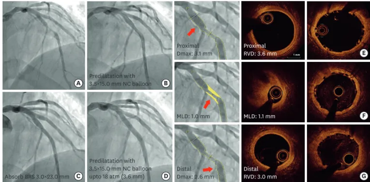
E
A Predilatation with 3.5×15.0 mm NC balloon
Predilatation with 3.5×15.0 mm NC balloon upto 18 atm (3.6 mm) Absorb BRS 3.0×23.0 mm
Proximal
Dmax: 3.1 mm Proximal
RVD: 3.6 mm
MLD: 1.0 mm MLD: 1.1 mm F
Distal
Dmax: 2.6 mm Distal
RVD: 3.0 mm
Figure 5. A case of “Effective” PSP. Fifty-seven years old man admitted due to effort related chest pain. Coronary angiography showed tight stenosis of LAD artery (A). On-line QCA showed that proximal Dmax was 3.1 mm and distal Dmax was 2.6 mm (E-G). Predilatation was performed using 3.5×1.5 mm non-compliant balloon (B). Absorb BRS 3.0×23 mm was implanted (C). Postdilatation was done by 3.5×15 mm non-compliant balloon up to 18 atm (final balloon diameter: 3.6 mm). Final optimal coherent tomography showed well expanded and apposed scaffold without acute complications.
BRS = bioresorbable vascular scaffold; Dmax = maximal lumen diameter; LAD = left anterior descending; MLD = mean lung dose; NC = non-compliant; PSP = preparation, sizing, and postdilatation; QCA = quantitative coronary angiography; RVD = reference vessel diameter.
TREATMENT FOR BRS FAILURE (SCAFFOLD THROMBOSIS AND IN-SCAFFOLD RESTENOSIS)
Understanding the fundamental pathophysiological mechanism underlying BRS failure is of key importance to guide proper subsequent treatment.54) Therefore, intracoronary imaging study is highly recommended in cases of BRS failure.55) Multiple treatment strategy for treating BRS failure was proposed including DES, plane balloon angioplasty, drug- coated balloon, or BRS.55)56) Mechanical causes can be treated first with balloon angioplasty with non-compliant balloon. In-scaffold restenosis due to neointimal hyperplasia can be treated by a drug-coated balloon. If mechanical factors cannot be corrected by balloon angioplasty, DES implantation can be considered. In addition, if BRS failure occurs 6 months after implantation, DES or BRS implantation can be considered because after 6 months disintegration of the scaffold begin and additional radial strength is necessary.55)
HYBRID PERCUTANEOUS CORONARY INTERVENTION
Due to the clinical and mechanical limitations of current generation BRS, complex coronary lesions are frequent unsuitable for pure BRS implantation. To minimize the length of permanent metallic caging, and achieving optimal BRS result, hybrid approach in combination of BRS, DES, and drug-coated balloon was proposed.57) For the BRS less favorable lesion including large vessel, aorto-ostial lesion, side-branch of bifurcation, large size discrepancy, and small vessel, conventional DES was implanted. Drug-coated balloon can be used in small diffuse coronary artery disease.58) BRS was implanted only in BRS favorable lesions overlapping with DES. Aggressive post-dilatation should be performed at the overlapping site to minimize the risk of late malapposition of metallic DES after complete resorption of BRS.57)
ANTIPLATELET THERAPY AFTER BRS IMPLANTATION
For metallic DES, at least 6-month DAPT after PCI for stable ischemic heart disease, and 12-month for ACSs are recommended in American and European guidelines.59)60) However, optimal duration of DAPT for BRS remains to be evaluated. Randomized trials stated the use of DAPT for at least 1 year per protocol.6)7)42)43) Regarding several reports on early as well as late scaffold thrombosis,61) some physicians suggest the longer DAPT regimen (>12 months), and/or more potent agents (e.g., ticagrelor or prasugrel)62)63) or triple antiplatelet therapy, particularly in the early period after BRS implantation. In this context, patients who cannot tolerate a long-term DAPT or are at high risk of bleeding may not be ideal candidate for BRS implantation.
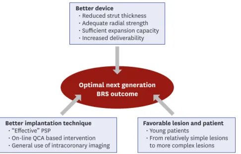
LESSONS FROM FIRST GENERATION ABSORB BRS
At 14th September 2017, Abbott Vascular announced a halt to sales of the Absorb BRS. The experience of the first generation BRS provides the valuable insight for the next generation (Figure 6). First, the new BRS needs to be mechanically stronger, have thinner struts, and available in a broad range of length and diameter. In addition, complete biodegradation occurs without significant inflammatory reaction within 1–2 years. Second, treating
physicians should realize that BRS profiles may differ significantly from conventional metallic DESs and should adopt specific BRS implantation technique for favorable outcomes.
Third, new BRS should be extensively tested in a stepwise fashion from relatively simple lesion to more complex lesion.
CONCLUSION
Although promising early reports, recent studies have raised concerns about the safety and efficacy of BRS compared with contemporary standard metallic DESs. However, we have experienced in the interventional technology field that the drawbacks of old device have greatly motivated technological innovation to solve previous problems. With rapid evolving technology of BRS under a number of current ongoing clinical tests, newer BRS overcoming current issues are expected in a near future.
REFERENCES
1. Serruys PW, Chevalier B, Dudek D, et al. A bioresorbable everolimus-eluting scaffold versus a metallic everolimus-eluting stent for ischaemic heart disease caused by de-novo native coronary artery lesions (ABSORB II): an interim 1-year analysis of clinical and procedural secondary outcomes from a randomised controlled trial. Lancet 2015;385:43-54.
PUBMED | CROSSREF
2. Karanasos A, Simsek C, Gnanadesigan M, et al. OCT assessment of the long-term vascular healing response 5 years after everolimus-eluting bioresorbable vascular scaffold. J Am Coll Cardiol 2014;64:2343-56.
PUBMED | CROSSREF
3. Brugaletta S, Radu MD, Garcia-Garcia HM, et al. Circumferential evaluation of the neointima by optical coherence tomography after ABSORB bioresorbable vascular scaffold implantation: can the scaffold cap the plaque? Atherosclerosis 2012;221:106-12.
PUBMED | CROSSREF
Better device
• Reduced strut thickness
• Adequate radial strength
• Sufficient expansion capacity
• Increased deliverability
Better implantation technique
• “Effective” PSP
• On-line QCA based intervention
• General use of intracoronary imaging
Favorable lesion and patient
• Young patients
• From relatively simple lesions to more complex lesions Optimal next generation
BRS outcome
Figure 6. Optimal BRS outcomes.
BRS = bioresorbable vascular scaffold; PSP = preparation, sizing, and postdilatation; QCA = quantitative coronary angiography.
4. Simsek C, Karanasos A, Magro M, et al. Long-term invasive follow-up of the everolimus-eluting bioresorbable vascular scaffold: five-year results of multiple invasive imaging modalities. EuroIntervention 2016;11:996-1003.
PUBMED
5. Serruys PW, Chevalier B, Sotomi Y, et al. Comparison of an everolimus-eluting bioresorbable scaffold with an everolimus-eluting metallic stent for the treatment of coronary artery stenosis (ABSORB II): a 3 year, randomised, controlled, single-blind, multicentre clinical trial. Lancet 2016;388:2479-91.
PUBMED | CROSSREF
6. Wykrzykowska JJ, Kraak RP, Hofma SH, et al. Bioresorbable scaffolds versus metallic stents in routine PCI. N Engl J Med 2017;376:2319-28.
PUBMED | CROSSREF
7. Ellis SG, Kereiakes DJ, Metzger DC, et al. Everolimus-eluting bioresorbable scaffolds for coronary artery disease. N Engl J Med 2015;373:1905-15.
PUBMED | CROSSREF
8. Tamburino C, Latib A, van Geuns RJ, et al. Contemporary practice and technical aspects in coronary intervention with bioresorbable scaffolds: a European perspective. EuroIntervention 2015;11:45-52.
PUBMED | CROSSREF
9. Foin N, Lee R, Bourantas C, et al. Bioresorbable vascular scaffold radial expansion and conformation compared to a metallic platform: insights from in vitro expansion in a coronary artery lesion model.
EuroIntervention 2016;12:834-44.
PUBMED | CROSSREF
10. Onuma Y, Dudek D, Thuesen L, et al. Five-year clinical and functional multislice computed tomography angiographic results after coronary implantation of the fully resorbable polymeric everolimus-eluting scaffold in patients with de novo coronary artery disease: the ABSORB cohort A trial. JACC Cardiovasc Interv 2013;6:999-1009.
PUBMED | CROSSREF
11. Serruys PW, Ormiston J, van Geuns RJ, et al. A polylactide bioresorbable scaffold eluting everolimus for treatment of coronary stenosis: 5-year follow-up. J Am Coll Cardiol 2016;67:766-76.
PUBMED | CROSSREF
12. Ali ZA, Serruys PW, Kimura T, et al. 2-year outcomes with the absorb bioresorbable scaffold for treatment of coronary artery disease: a systematic review and meta-analysis of seven randomised trials with an individual patient data substudy. Lancet 2017;390:760-72.
PUBMED | CROSSREF
13. Capodanno D, Gori T, Nef H, et al. Percutaneous coronary intervention with everolimus-eluting bioresorbable vascular scaffolds in routine clinical practice: early and midterm outcomes from the European multicentre GHOST-EU registry. EuroIntervention 2015;10:1144-53.
PUBMED | CROSSREF
14. Abizaid A, Ribamar Costa J Jr, Bartorelli AL, et al. The ABSORB EXTEND study: preliminary report of the twelve-month clinical outcomes in the first 512 patients enrolled. EuroIntervention 2015;10:1396-401.
PUBMED | CROSSREF
15. Wiebe J, Hoppmann P, Colleran R, et al. Long-term clinical outcomes of patients treated with everolimus- eluting bioresorbable stents in routine practice: 2-year results of the ISAR-ABSORB Registry. JACC Cardiovasc Interv 2017;10:1222-9.
PUBMED | CROSSREF
16. Cortese B, Ielasi A, Moscarella E, et al. Thirty-day outcomes after unrestricted implantation of bioresorbable vascular scaffold (from the prospective RAI Registry). Am J Cardiol 2017;119:1924-30.
PUBMED | CROSSREF
17. Wöhrle J, Naber C, Schmitz T, et al. Beyond the early stages: insights from the ASSURE registry on bioresorbable vascular scaffolds. EuroIntervention 2015;11:149-56.
PUBMED | CROSSREF
18. Puricel S, Cuculi F, Weissner M, et al. Bioresorbable coronary scaffold thrombosis: multicenter
comprehensive analysis of clinical presentation, mechanisms, and predictors. J Am Coll Cardiol 2016;67:921-31.
PUBMED | CROSSREF
19. Kraak RP, Hassell ME, Grundeken MJ, et al. Initial experience and clinical evaluation of the absorb bioresorbable vascular scaffold (BVS) in real-world practice: the AMC Single Centre Real World PCI Registry. EuroIntervention 2015;10:1160-8.
PUBMED | CROSSREF
20. Rzeszutko Ł, Siudak Z, Tokarek T, et al. Twelve months clinical outcome after bioresorbable vascular scaffold implantation in patients with stable angina and acute coronary syndrome. Data from the Polish National Registry. Postepy Kardiol Interwencyjnej 2016;12:108-15.
PUBMED | CROSSREF
21. Felix CM, Fam JM, Diletti R, et al. Mid- to long-term clinical outcomes of patients treated with the everolimus-eluting bioresorbable vascular scaffold: the BVS Expand Registry. JACC Cardiovasc Interv 2016;9:1652-63.
PUBMED | CROSSREF
22. Muramatsu T, Onuma Y, van Geuns RJ, et al. 1-year clinical outcomes of diabetic patients treated with everolimus-eluting bioresorbable vascular scaffolds: a pooled analysis of the ABSORB and the SPIRIT trials. JACC Cardiovasc Interv 2014;7:482-93.
PUBMED | CROSSREF
23. Dudek D, Rzeszutko Ł, Zasada W, et al. Bioresorbable vascular scaffolds in patients with acute coronary syndromes: the POLAR ACS study. Pol Arch Med Wewn 2014;124:669-77.
PUBMED | CROSSREF
24. Gori T, Schulz E, Hink U, et al. Early outcome after implantation of absorb bioresorbable drug-eluting scaffolds in patients with acute coronary syndromes. EuroIntervention 2014;9:1036-41.
PUBMED | CROSSREF
25. Hellenkamp K, Becker A, Gabriel YD, et al. Mid- to long-term outcome of patients treated with everolimus-eluting bioresorbable vascular scaffolds: data of the BVS registry Göttingen predominantly from ACS patients. Int J Cardiol 2017;234:58-63.
PUBMED | CROSSREF
26. Kajiya T, Liang M, Sharma RK, et al. Everolimus-eluting bioresorbable vascular scaffold (BVS) implantation in patients with ST-segment elevation myocardial infarction (STEMI). EuroIntervention 2013;9:501-4.
PUBMED | CROSSREF
27. Kočka V, Malý M, Toušek P, et al. Bioresorbable vascular scaffolds in acute ST-segment elevation myocardial infarction: a prospective multicentre study ‘Prague 19’. Eur Heart J 2014;35:787-94.
PUBMED | CROSSREF
28. Wiebe J, Möllmann H, Most A, et al. Short-term outcome of patients with ST-segment elevation myocardial infarction (STEMI) treated with an everolimus-eluting bioresorbable vascular scaffold. Clin Res Cardiol 2014;103:141-8.
PUBMED | CROSSREF
29. Diletti R, Karanasos A, Muramatsu T, et al. Everolimus-eluting bioresorbable vascular scaffolds for treatment of patients presenting with ST-segment elevation myocardial infarction: BVS STEMI first study.
Eur Heart J 2014;35:777-86.
PUBMED | CROSSREF
30. Brugaletta S, Gori T, Low AF, et al. Absorb bioresorbable vascular scaffold versus everolimus-eluting metallic stent in ST-segment elevation myocardial infarction: 1-year results of a propensity score matching comparison: the BVS-EXAMINATION Study (bioresorbable vascular scaffold-a clinical evaluation of everolimus eluting coronary stents in the treatment of patients with ST-segment elevation myocardial infarction). JACC Cardiovasc Interv 2015;8:189-97.
PUBMED | CROSSREF
31. Cortese B, Ielasi A, Romagnoli E, et al. Clinical comparison with short-term follow-up of bioresorbable vascular scaffold versus everolimus-eluting stent in primary percutaneous coronary interventions. Am J Cardiol 2015;116:705-10.
PUBMED | CROSSREF
32. Kochman J, Tomaniak M, Pietrasik A, et al. Bioresorbable everolimus-eluting vascular scaffold in patients with ST-segment elevation myocardial infarction: optical coherence tomography evaluation and clinical outcomes. Cardiol J 2015;22:315-22.
PUBMED | CROSSREF
33. Chakraborty R, Patra S, Banerjee S, et al. Outcome of everolimus eluting bioabsorbable vascular scaffold (BVS) compared to non BVS drug eluting stent in the management of ST-segment elevation myocardial infarction (STEMI) - a comparative study. Cardiovasc Revasc Med 2016;17:151-4.
PUBMED | CROSSREF
34. Diletti R, Farooq V, Girasis C, et al. Clinical and intravascular imaging outcomes at 1 and 2 years after implantation of absorb everolimus eluting bioresorbable vascular scaffolds in small vessels. Late lumen enlargement: does bioresorption matter with small vessel size? Insight from the ABSORB cohort B trial.
Heart 2013;99:98-105.
PUBMED | CROSSREF
35. Ielasi A, Latib A, Naganuma T, et al. Early results following everolimus-eluting bioresorbable vascular scaffold implantation for the treatment of in-stent restenosis. Int J Cardiol 2014;173:513-4.
PUBMED | CROSSREF
36. Moscarella E, Ielasi A, Granata F, et al. Long-term clinical outcomes after bioresorbable vascular scaffold implantation for the treatment of coronary in-stent restenosis: a multicenter Italian experience. Circ Cardiovasc Interv 2016;9:e003148.
PUBMED | CROSSREF
37. Alfonso F, Cuesta J, Pérez-Vizcayno MJ, et al. Bioresorbable vascular scaffolds for patients with in-stent restenosis: the RIBS VI Study. JACC Cardiovasc Interv 2017;10:1841-51.
PUBMED | CROSSREF
38. Everaert B, Wykrzykowska JJ, Koolen J, et al. Recommendations for the use of bioresorbable vascular scaffolds in percutaneous coronary interventions: 2017 revision. Neth Heart J 2017;25:419-28.
PUBMED | CROSSREF
39. Indolfi C, De Rosa S, Colombo A. Bioresorbable vascular scaffolds - basic concepts and clinical outcome.
Nat Rev Cardiol 2016;13:719-29.
PUBMED | CROSSREF
40. Miyazaki T, Latib A, Ruparelia N, et al. The use of a scoring balloon for optimal lesion preparation prior to bioresorbable scaffold implantation: a comparison with conventional balloon predilatation.
EuroIntervention 2016;11:e1580-8.
PUBMED | CROSSREF
41. Hibi K, Kimura K, Umemura S. Clinical utility and significance of intravascular ultrasound and optical coherence tomography in guiding percutaneous coronary interventions. Circ J 2015;79:24-33.
PUBMED | CROSSREF
42. Kimura T, Kozuma K, Tanabe K, et al. A randomized trial evaluating everolimus-eluting absorb bioresorbable scaffolds vs. everolimus-eluting metallic stents in patients with coronary artery disease:
ABSORB Japan. Eur Heart J 2015;36:3332-42.
PUBMED | CROSSREF
43. Gao R, Yang Y, Han Y, et al. Bioresorbable vascular scaffolds versus metallic stents in patients with coronary artery disease: ABSORB China Trial. J Am Coll Cardiol 2015;66:2298-309.
PUBMED | CROSSREF
44. Campbell PT, Mahmud E, Marshall JJ. Interoperator and intraoperator (in)accuracy of stent selection based on visual estimation. Catheter Cardiovasc Interv 2015;86:1177-83.
PUBMED | CROSSREF
45. Gomez-Lara J, Diletti R, Brugaletta S, et al. Angiographic maximal luminal diameter and appropriate deployment of the everolimus-eluting bioresorbable vascular scaffold as assessed by optical coherence tomography: an ABSORB cohort B trial sub-study. EuroIntervention 2012;8:214-24.
PUBMED | CROSSREF
46. Pinton FA, Falcão Bd, Mariani J JrKajita LJ, Filho AE, Lemos PA. Accuracy and precision of online quantitative coronary angiography with automatic calibration: a pilot study. Rev Bras Cardiol Invasiva 2015;23:58-60.
CROSSREF
47. Farooq V, Gomez-Lara J, Brugaletta S, et al. Proximal and distal maximal luminal diameters as a guide to appropriate deployment of the ABSORB everolimus-eluting bioresorbable vascular scaffold: a sub-study of the ABSORB Cohort B and the on-going ABSORB EXTEND Single Arm Study. Catheter Cardiovasc Interv 2012;79:880-8.
PUBMED | CROSSREF
48. Goto K, Mintz GS, Litherland C, et al. Lumen measurements from quantitative coronary angiography and IVUS: a PROSPECT Substudy. JACC Cardiovasc Imaging 2016;9:1011-3.
PUBMED | CROSSREF
49. Kawamoto H, Jabbour RJ, Tanaka A, Latib A, Colombo A. The bioresorbable scaffold: will oversizing affect outcomes? JACC Cardiovasc Interv 2016;9:299-300.
PUBMED | CROSSREF
50. Ishibashi Y, Nakatani S, Sotomi Y, et al. Relation between bioresorbable scaffold sizing using QCA-Dmax and clinical outcomes at 1 year in 1,232 patients from 3 study cohorts (ABSORB Cohort B, ABSORB EXTEND, and ABSORB II). JACC Cardiovasc Interv 2015;8:1715-26.
PUBMED | CROSSREF
51. Souteyrand G, Amabile N, Mangin L, et al. Mechanisms of stent thrombosis analysed by optical coherence tomography: insights from the national PESTO French registry. Eur Heart J 2016;37:1208-16.
PUBMED | CROSSREF
52. Serruys PW, Katagiri Y, Sotomi Y, et al. Arterial remodeling after bioresorbable scaffolds and metallic stents. J Am Coll Cardiol 2017;70:60-74.
PUBMED | CROSSREF