emergency room within 50 minutes after developing severe headache and lethargic mentality. Her blood pressure was 160/100 mm Hg on arrival; the infusion of nicardipine hydro- chloride reduced it to below 130 mm Hg before CT scanning.
An emergent unenhanced CT scan (SOMATOM Definition Flash, Siemens, Forchheim, Germany) showed diffuse SAH and subdural hematoma (Fig. 1A). The CTA was then per- formed to assess for an underlying aneurysm. To enhance the cerebral arteries, 120 mL nonionic contrast media was infused into the antecubital vein at a flow rate of 4 mL/s using a power injector. The delay between the infusion of contrast and the start of scanning was 40 seconds. The CTA source images were reformatted with the volume rendering method using a work- station (Wizard, Siemens, Forchheim, Germany). Aneurysms at ACoA and middle cerebral artery were demonstrated on the infused CT scan done 1.5 hours after admission (Fig. 1B). This patient suddenly fell into a deep stupor immediately after the CTA examination. Postcontrast CT scans taken after CTA re- vealed enlargement of SAH and new IVH in the lateral ventri- cle (Fig. 1C, D).
INTRODUCTION
Three-dimensional (3D) computed tomography angiography (CTA) is now utilized as the first imaging modality for detecting underlying lesions in patients with acute subarachnoid hemor- rhage (SAH)1). In spite of its increasing application, the reports of bleeding from aneurysms during CTA examination are few, so little information is available regarding the imaging findings for extravasation of ruptured cerebral aneurysms8,18). Furthermore, intraventricular hemorrhage (IVH) is commonly developed dur- ing rupture of anterior communicating artery (ACoA) aneu- rysm; however the CT picture for anatomical pathway of this condition has not been demonstrated2). The author describes a case of rerupturing ACoA aneurysm in which the blood is spilled through the lamina rostralis and the septum pellucidum into the ventricular system during CTA performed in the acute stage.
CASE REPORT
A 52-year-old woman with hypertension presented to the
Rupturing Anterior Communicating Artery Aneurysm during Computed Tomography Angiography :
Three-Dimensional Visualization of Bleeding into the Septum Pellucidum and the Lateral Ventricle
Ealmaan Kim, M.D., Ph.D.
Department of Neurosurgery, Dongsan Medical Center, Keimyung University School of Medicine, Daegu, Korea
Computed tomography angiography (CTA) is commonly used in setting of subarachnoid hemorrhage, but imaging features of aneurysm rupturing taking place at the time of scanning has rarely been described. The author reports a case of actively rebleeding aneurysm of the anterior communi- cating artery with intraventricular extravasation on the hyperacute CTA imaging. The rebleeding route, not into the third ventricle but into the lateral ventricles, can be visualized by real-time three-dimensional CT pictures. The hemorrhage broke the septum pellucidum and the lamina rostralis rather than the lamina terminalis.
Key Words : Anterior communicating artery aneurysm · Computed tomography · Computed tomography angiography · Lamina rostralis · Subarachnoid hemorrhage.
Case Report
•Received : January 15, 2014 •Revised : April 6, 2014 •Accepted : June 11, 2014
•Address for reprints : Ealmaan Kim, M.D., Ph.D.
Department of Neurosurgery, Dongsan Medical Center, Keimyung University School of Medicine, 56 Dalseong-ro, Jung-gu, Daegu 700-712, Korea Tel : +82-53-250-7823, Fax : +82-53-250-7356, E-mail : [email protected]
•This is an Open Access article distributed under the terms of the Creative Commons Attribution Non-Commercial License (http://creativecommons.org/licenses/by-nc/3.0) which permits unrestricted non-commercial use, distribution, and reproduction in any medium, provided the original work is properly cited.
J Korean Neurosurg Soc 55 (6) : 357-361, 2014 Copyright © 2014 The Korean Neurosurgical Society
rysm bleeding clearly delineated the interhemispheric cistern, the lamina terminalis, and the corpus callosum, similar to a ventriculographic manner.
The leaking trajectory of contrast medi- um, ejected from ACoA aneurysm, was 1) through the lamina rostralis above the lamina terminalis, 2) into the space between the septal leaves and 3) into the frontal horn of the lateral ventricle (Fig. 3E, F). The coronal CT scans dy- namically depicted the hemorrhage from ACoA aneurysm broke through the initial intraseptal hematoma and extended into the left frontal horn (Fig.
3G, H).
The authors were certain that the an- eurysm rerupture occurred while the routine CT scans for 3D angiography were being acquired. The patient was intubated for declining mental status and was urgently transferred to the op- erating room. The aneurysms were clipped simultaneously after hematoma evacuation. The surgeon did not find perforation of the lamina terminalis from direct hemorrhage of ACoA aneurysm. She re- gained consciousness and was discharged 3 weeks later after the event. This patient recovered neurologically including most of her cognitive functions, although with minimal memory deficit persisting at the 5-year follow-up.
DISCUSSION
Recently, brain CTA can be immediately performed after the recognition of SAH on precontrast CT in many institutions.
This expedites an early triage to the proper management and avoids delay between the diagnostic angiogram and early aneu- rysm repair24). Nevertheless, rebleeding from ruptured aneu- rysms is a still major cause of morbidity and death in the pa- tients presenting with spontaneous SAH13).
Rupture of aneurysms or other vascular malformations dur- ing conventional cerebral angiography has been frequently re- ported previously19). The published cases may reflect iatrogenic aneurysm rupture from forceful contrast infusion into the ca- rotid artery10,21). Such consideration does not apply to CTA, where the contrast agent is administered slowly into a peripher- al vein. There have been few studies in which the causal rela- tionship between performance of CT scan and rupture of the aneurysm has been discussed2,5,14). For this reason, the frequen- cy of aneurysm rupture during CTA has not been defined, but it is seems to be extremely rare. In the case reported here, the CTA showing active contrast leakage was performed within 3 hours of initial aneurysm rupture. Accordingly, it is likely that The results of brain CTA clearly visualized an ACoA aneu-
rysm from which the contrast medium was actively ejecting. An area consistent with contrast jet was identified around the aneu- rysm (Fig. 2A). The maximum intensity projection illustrated the diffuse curvilinear spill out of the contrast medium arising from the aneurysm dome, extended into the lateral ventricle (Fig. 2B). The author realized that this vessel-like extravasation represented the aneurysm rebleeding and the consequential contrast swirling at the time of imaging. The extravasations of blood were detected in the preexisting intracerebral hematomas on the raw data of the CTA, and they had the same high densi- ties as the internal carotid artery (Fig. 3A–D). On sagittal re- formed 3D CT images, the clots developed from the first aneu-
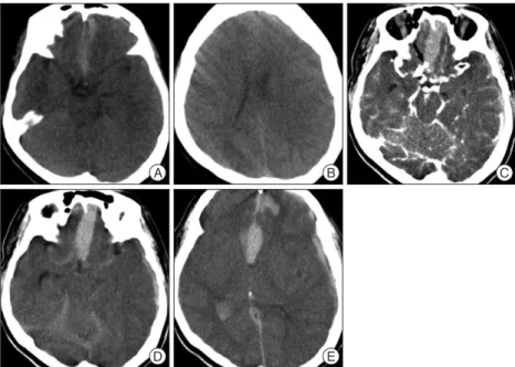
Fig. 1. A : Initial plain CT scan shows thin SAH and subdural hematoma. B : There is no cavum sep- tum pellucidum on the vertex cut. C : Source image of CTA reveals saccular aneurysms at the ante- rior communicating artery (arrow) and the left middle cerebral artery. D and E : Delayed images im- mediately after CTA show extensive SAH associated with intracerebral and intraventricular hemorrhages as well as blood within the septum pellucidum. CTA : computed tomography angiog- raphy, SAH : subarachnoid hemorrhage.
D A
E
B C
Fig. 2. A : Three-dimensional reconstruction of CTA depicts the spatial relationship between the aneurysm (arrow) and extravasated contrast medium (arrowhead). B : CTA with maximum intensity projection out- lines the entire region of extravasated fresh blood (arrowheads) spouting from the ruptured ACoA aneurysm (arrow). ACoA : anterior communicat- ing artery, CTA : computed tomography angiography.
B A
soon after, it was correctly interpreted as an aneurysm bursting.
The prior reports have described other patterns of a blood ex- travasation; cap sign, corkscrew sign, hematoma opacification sign, starburst appearance, and nebulous high attenuation4-6,16). This variability in imaging features seemed to depend on the following factors; site of rupture of the aneurysm, the location and dome direction of aneurysm, the configuration of the cis- ternal space, the presence of preexisting hematomas, amount of the bleeding, and the cerebrospinal fluid flow2,8). Those collec- tions of extraluminal contrast adjacent to ruptured aneurysm are needs to be differentiated from irregular blebs, calcification of the aneurysm and the artery, veins of variable thickness, and the cause of this bleeding from ACoA aneurysm was due to a
second spontaneous hemorrhage, which coincided with CT scanning.
Besides a spontaneous bleeding, direct effects of contrast mate- rial are supplementary factors for hemorrhage in reported rup- tured aneurysms during CTA10,25). The previously documented potentiality was that injuring endothelium and activating antico- agulant could initiate rebleeding from an aneurysm which is only coated by a platelet plug at an acute phase of SAHs11,12). More- over, some patients obviously are intolerable to contrast medi- um very well, as they usually vomited after injection of 30 mL (the total dose given was up to 200 mL)6,16). An increased blood pressure during emesis might be incriminated as a cause for in- tra-CTA rebleedings. In addition, the physical activity neces- sary for the examination procedure might be considered casual as well20). Therefore, more attention should be paid to deep se- dation and strict blood pressure control before and during CTA evaluation especially for at-risk patients of early rebleeding3,17). Practically, an antifibrinolytic therapy might be helpful for re- ducing the aneurysm rebleeding for the patients immediately transferred after the onset like the author’s case22).
As the rebleeding of aneurysm resuls in catastrophic conse- quences, it is imperative to understand the specific features of continuous bleeding on the CTA images. On 3D reformatted images of the current case, the contrast extravasation was de- picted to have a ribbon-like appearance that mimicked a vascu- lar structure, and it had a long trajectory traveling through the subarachnoid space, the preexistent hematoma, and the ventri- cles. Initially, it was misconstrued as a movement artifact, but
Fig. 3. A--D : Axial images of CTA demonstrate the caudorostral pathway of the extravasated contrast medium (arrowheads) into the preexisting he- matoma of lower density from the interhemispheric cistern into the septum pellucidum and into the frontal horn and body of the lateral ventricle. E and F : Sagittal CTA views illustrate the diffusion of the contrast medium (arrowheads) from the rebleeding ACoA aneurysm (arrow) through the lami- na rostralis (asterisk) into the septum pellucidum without penetrating into the lamina terminalis. G and H : Coronal reformatted CT scan clearly shows the ejected blood (arrowheads) which pass through the septum pellucidum up to the frontal horn of the left lateral ventricle. ACoA : anterior communi- cating artery, CTA : computed tomography angiography.
E A
F B
G C
H D
Fig. 4. Coronal T2-weighted magnetic resonance image shows the thin strip of white matter of the lamina rostralis (arrowhead). This separates the anterior interhemispheric cistern (lower arrow) from the space be- tween the leaves of the septum pellucidum (upper arrow) as well as the floor of the frontal horns in the mature brain.
extension into the lateral ventricle.
The previously reported patients with rebleeding around the time of CTA often had disturbances in consciousness and their prognosis was generally poor15,25). In contrast, this case showed a favorable recovery. For the selected patients with neurological responsiveness even after detection of extravasation on CTA, we should consider immediate and effective treatment.
CONCLUSION
The author reports on the diagnosis of an intra-CTA aneu- rysm rebleeding. This case provides a radiographic documenta- tion for the mechanism of IVH from a bleeding ACoA aneu- rysm through the lamina rostralis and the septum pellucidum.
References
1. Dehdashti AR, Rufenacht DA, Delavelle J, Reverdin A, de Tribolet N : Therapeutic decision and management of aneurysmal subarachnoid haemorrhage based on computed tomographic angiography. Br J Neu- rosurg 17 : 46-53, 2003
2. Desai S, Friedman JA, Hlavin J, Kash F : Actively bleeding intracranial aneurysm demonstrated by CT angiography. Clin Neurol Neurosurg 111 : 94-96, 2009
3. Fujii Y, Takeuchi S, Sasaki O, Minakawa T, Koike T, Tanaka R : Ultra- early rebleeding in spontaneous subarachnoid hemorrhage. J Neuro- surg 84 : 35-42, 1996
4. Gosselin MV, Vieco PT : Active hemorrhage of intracranial aneurysms : diagnosis by CT angiography. J Comput Assist Tomogr 21 : 22-24, 1997 5. Hashiguchi A, Mimata C, Ichimura H, Morioka M, Kuratsu J : Rebleed- ing of ruptured cerebral aneurysms during three-dimensional comput- ed tomographic angiography : report of two cases and literature review.
Neurosurg Rev 30 : 151-154, 2007
6. Holodny AI, Farkas J, Schlenk R, Maniker A : Demonstration of an ac- tively bleeding aneurysm by CT angiography. AJNR Am J Neuroradiol 24 : 962-964, 2003
7. Im SH, Oh CW, Hong SK, Kwon OK, Kim SH : CT angiography dem- onstration of the development of intraventricular hemorrhage during aneurysm rupture. Clin Neurol Neurosurg 109 : 299-301, 2007 8. Kathuria S, Deveikis JP, Westesson PL, Gandhi D : Improved diagnosis
of actively bleeding aneurysm on CT angiography using delayed CT images. Eur J Radiol 79 : 328-331, 2011
9. Kier EL, Truwit CL : The lamina rostralis : modification of concepts con- cerning the anatomy, embryology, and MR appearance of the rostrum of the corpus callosum. AJNR Am J Neuroradiol 18 : 715-722, 1997 10. Klisch J, Weyerbrock A, Spetzger U, Schumacher M : Active bleeding
from ruptured cerebral aneurysms during diagnostic angiography : emergency treatment. AJNR Am J Neuroradiol 24 : 2062-2065, 2003 11. Kuhn J, Vehlen C, Mennel HD, Mahkorn D, Bewermeyer H : Rupture
of an internal carotid artery aneurysm during angiography with leakage of contrast medium via an external ventricular drain. Neuroradiology 45 : 905-907, 2003
12. Kusumi M, Yamada M, Kitahara T, Endo M, Kan S, Iida H, et al. : Re- rupture of cerebral aneurysms during angiography--a retrospective study of 13 patients with subarachnoid hemorrhage. Acta Neurochir (Wien) 147 : 831-837, 2005
13. Larsen CC, Astrup J : Rebleeding after aneurysmal subarachnoid hem- orrhage : a literature review. World Neurosurg 79 : 307-312, 2013 14. Nagai M, Koizumi Y, Tsukue J, Watanabe E : A case of extravasation
from a cerebral aneurysm during 3-dimensional computed tomography intra-aneurysmal thrombus14,25).
An ACoA aneurysm is often accompanied by IVH in the very act of bleeding. In theory, retrograde entry of SAH into the ven- tricular system is possible, but an unlikely cause of significant IVH. Recently, a report cited the lamina terminalis as the proba- ble access for blood to the third ventricle; however the radio- graphic passage is not evidently from their CT images shown7). Investigators stated that the lamina terminalis does not make a strong resistance to hemorrhage from ACoA aneurysms, sug- gesting the lamina terminalis as an entry point from the inter- hemispheric fissure into the ventricular system4,16). However, the lamina terminalis is a solid membrane that requires sharp incision for anterior third ventriculostomy during surgery for tumors and aneurysms in this region. Furthermore, as con- firmed in the present case, surgeons mentioned that direct pen- etration of the lamina terminalis has not been seen at clipping surgery for the ruptured ACoA aneurysms1,24). In this report, 3D CT images clearly illustrated the anatomical pathway of hemorrhage from ACoA aneurysm via the interhemispheric subarachnoid space through the lamina rostralis into the sep- tum pellucidum and the frontal horn of the lateral ventricle.
Anatomically, the rostrum of the corpus callosum extends from the genu to the lamina terminalis, and consists of two sec- tions : the thick beaked segment and the thin lamina rostralis.
The lamina rostralis, the most rostral part of the corpus callo- sum, is visualized distinctly on spin-echo magnetic resonance images with 2 mm thickness9). The developing rostral corpus callosum from the rostral commissural plate seals the cavum septum pellucidum off from the anterior interhemispheric fis- sure during early fetal life20). Hence, the normally formed ca- vum septum pellucidum remains closed toward the anterior in- terhemispheric fissure. Because a cavum pellucidium regresses with maturation of the brain, the septum contains no open space in most human beings26). The lamina rostralis segment is superiorly continuous with the septum pellucidum and blends posteroinferiorly with the lamina terminalis just above the an- terior commissure. The junction of the lamina rostralis and lamina terminalis is embedded within the very thin layers of the embryonic lamina reuniens segment of the lamina termina- lis (Fig. 4).7,23) Consequently, in most individuals, the lamina rostralis secludes the anterior interhemispheric fissure from the imaginary interlaminar space within the septum pellucidum as well as the floor of the frontal horns. This filmy strip of nonmy- elinated part of the rostrum is likely to offer less resistance to pressure than the lamina terminalis8,24). For that reason, passing through the lamina rostralis can give direct access from the subarachnoid spaces of the anterior interhemispheric fissure to the frontal horn of the lateral ventricle. In this case, the contrast extravasated during CTA was seen to penetrate into the frontal horn via the lamina rostralis through the caval-septal hemato- ma developed from the initial bleeding. The septal leaves may act as a buffer, containing the clot until the pressure within the closed intraseptal space is over their resistance, with subsequent
rysmal pressure changes during angiography in coil embolization. Surg Neurol 48 : 451-457, 1997
22. Starke RM, Kim GH, Fernandez A, Komotar RJ, Hickman ZL, Otten ML, et al. : Impact of a protocol for acute antifibrinolytic therapy on an- eurysm rebleeding after subarachnoid hemorrhage. Stroke 39 : 2617- 2621, 2008
23. Tsuang FY, Su IC, Chen JY, Lee JE, Lai DM, Tu YK, et al. : Hyperacute cerebral aneurysm rerupture during CT angiography. J Neurosurg 116 : 1244-1250, 2012
24. Westerlaan HE, van Dijk JM, Jansen-van der Weide MC, de Groot JC, Groen RJ, Mooij JJ, et al. : Intracranial aneurysms in patients with sub- arachnoid hemorrhage : CT angiography as a primary examination tool for diagnosis--systematic review and meta-analysis. Radiology 258 : 134-145, 2011
25. Wu TC, Tsui YK, Chen TY, Lin CJ, Wu TC, Tzeng WS : Rebleeding of aneurysmal subarachnoid hemorrhage in computed tomography angi- ography : risk factor, rebleeding pattern, and outcome analysis. J Com- put Assist Tomogr 36 : 103-108, 2012
26. Yanaka K, Meguro K, Narushima K, Takano S, Doi M, Nose T : Basal perforating artery aneurysm within the cavum septi pellucidi. Case re- port. J Neurosurg 88 : 601-604, 1998
angiography. Surg Neurol 69 : 411-413, 2008
15. Naidech AM, Janjua N, Kreiter KT, Ostapkovich ND, Fitzsimmons BF, Parra A, et al. : Predictors and impact of aneurysm rebleeding after sub- arachnoid hemorrhage. Arch Neurol 62 : 410-416, 2005
16. Nakatsuka M, Mizuno S, Uchida A : Extravasation on three-dimension- al CT angiography in patients with acute subarachnoid hemorrhage and ruptured aneurysm. Neuroradiology 44 : 25-30, 2002
17. Ohkuma H, Tsurutani H, Suzuki S : Incidence and significance of early aneurysmal rebleeding before neurosurgical or neurological manage- ment. Stroke 32 : 1176-1180, 2001
18. Pérez-Núñez A, Alén JF, Ramos A, Millán JM : Aneurysm re-rupture during computed tomography angiography. Acta Radiol 47 : 419-421, 19. Saitoh H, Hayakawa K, Nishimura K, Okuno Y, Murayama C, Miyaza-2006
wa T, et al. : Intracarotid blood pressure changes during contrast medi- um injection. AJNR Am J Neuroradiol 17 : 51-54, 1996
20. Scholtes F, Signorelli F, Bojanowski MW : Rupture of anterior commu- nicating artery aneurysms during computed tomography angiography : description of the pathway for intraseptal and intraventricular hemor- rhage. J Neurosurg 115 : 617-620, 2011
21. Sorimachi T, Takeuchi S, Koike T, Minakawa T, Tanaka R : Intra-aneu-