Received: March 30, 2018 Revised: May 31, 2018 Accepted: June 5, 2018 Trauma and InJury
Correspondence to Kyoung Hoon Lim, M.D.
Department of Surgery, Kyungpook National University Hospital, 130 Dong- deok-ro, Jung-gu, Daegu 41944, Korea Tel: +82-53-200-5605
Fax: +82-53-420-0510 E-mail: [email protected]
http://www.jtraumainj.org Copyright © 2018 The Korean Society of Trauma
Endovascular Salvage for Traumatic midthoracic aortic rupture with left diaphragmatic Injury
Shin-Ah Son, M.D.
1, Tak-Hyuk Oh, M.D.
1, Gun-Jik Kim, M.D.
2, Deok Heon Lee, M.D., Ph.D.
2, Kyoung Hoon Lim, M.D.
31
Department of Thoracic and Cardiovascular Surgery, Kyungpook National University Hospital, Daegu, Korea
2
Department of Thoracic and Cardiovascular Surgery,
3Department of Surgery, School of Medicine, Kyungpook National University, Kyungpook National University Hospital, Daegu, Korea
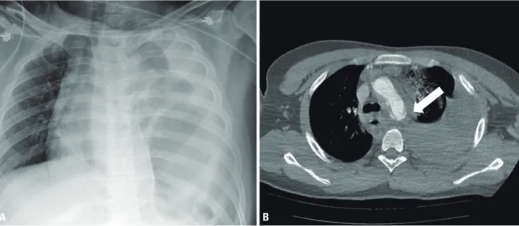
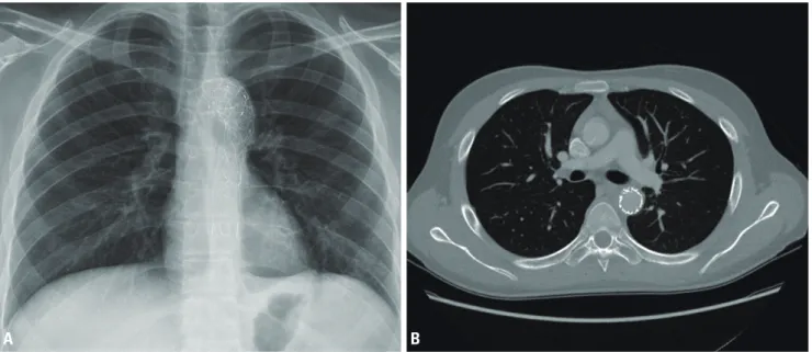
Patients with traumatic aortic rupture rarely reach the hospital alive. Even among those who arrive at the hospital alive, traumatic aortic rupture after high-speed motor vehicle accidents leads to a high in-hospital mortality rate and is associated with other major injuries. Here, we report a rare case of descending midthoracic aortic rupture with blunt diaphragmatic rupture. Successful management with emergency laparotomy after an immediate endovascular procedure resulted in a favorable prognosis in this case.
Keywords: Aortic rupture; Endovascular procedure; Multiple trauma
INTRODUCTION
The incidence of traumatic aortic injury (TAI) secondary to blunt trauma due to high-speed transportation accidents is rapidly increasing. TAI in accident survivors undoubtedly represents a direct threat to life and requires a fast-track diagnostic and therapeutic plan for a successful resuscitation.
CASE REPORT
A 19-year-old man with no prior medical history was struck by a motor vehicle while
riding his motorcycle, and was admitted to our trauma center with 119 emergency
Endovascular Salvage for Traumatic midthoracic aortic rupture with left diaphragmatic Injury
Shin-Ah Son, M.D.
1, Tak-Hyuk Oh, M.D.
1, Gun-Jik Kim, M.D.
2, Deok Heon Lee, M.D., Ph.D.
2, Kyoung Hoon Lim, M.D.
31
Department of Thoracic and Cardiovascular Surgery, Kyungpook National University Hospital, Daegu, Korea
2