∙ Received: October 15, 2007. Accepted: October 30, 2007.
∙ Corresponding author: Chang Ho Lee
Department of Nuclear Medicine, Severance Hospital, Yonsei University Health System, Seoul, 120-749, Korea
Tel: +82-2-2228-6050, Fax: +82-2-2227-7062 E-mail: nuclear@yuhs.ac
Chun Goo Kang, Jae Sam Kim, Chang Ho Lee
Department of Nuclear Medicine, Severance Hospital, Yonsei University Health System, Department of Diagnostic Radiology Severance Hospital, Yonsei University Health System*
Purpose: In FDG-PET/CT of breast cancer, a sensitivity was 80~96% and a specificity was 75~95%
commonly. It was valuable to identify a cancer in early stage been difficult in Mammography. Most of the PET/CT scans have been examined on supine position, so, the image of breast has been acquired by reconstructed whole body scan image. However, using prone position with a compensator, a shape of breast was reassembly shown to be real by gravity. Therefore, the purpose of this study was to evaluate diagnostic value of prone position in FDG PET-CT of breast cancer. Materials and Methods: 30 female patients with doubtful or positive breast cancer were examined. The PET-CT whole body scan was acquired at 60 minutes after 18F-FDG injection on Supine position. Then, regional breast spot scan was progressed on prone position using a compensator. Each image was evaluated by physicians blinded to patient’s data, and statistical analysis did through SUVs measured in PET-CT images. Results: In 27 of 30 patients, prone position was shown accurate discrimination and diagnostic value, but in another 3 patients had a lesion 1cm below, PET-CT couldn’t detect it, unlike MRI. Consequently, prone position distinguished a lesion better than Supine position, because of low degree of metamorphosis by gravity. The SUVs analysis of each position was significant (p value=0.004). Conclusion: In PET-CT of breast cancer, prone position could detect micrometastasis as well as primary lesion, better than supine position. Therefore, this study proposes that any technical change considered morphological feature like prone position can offer adequate and useful diagnostic information, together with complementary quantitative analysis. (Korean J Nucl Med Technol 2008;
12(1):44-48)
Key Words : PET-CT, Breast Cancer, Prone Position
서 론
유방암의 경우 여성의 악성종양 중에서 가장 이완율이 높 은 질환이며, 최근 증가하는 경향이 있다1). 발병 연령층이 비
교적 젊은 나이인 20대부터 고연령층까지 넓다2). 또한 조기 발견, 조기 치료에 의해 장기 생존이 가능한 암의 종류이다3). 발병 후 치료를 마친 경우에도 장기 경과를 거쳐 재발, 전이 를 초래하는 경우가 많다4). 전이의 경우 림프절 절이, 골전이, 간전이, 폐전이가 많으며, 골전이는 조골성(osteoblastic), 화 골성(myositis), 혼합성(mixed patterns) 등의 다양한 패턴을 나타낸다5,6).
이러한 유방암에서의 FDG를 이용한 PET-CT의 유용성과 다른 검사와의 관계(원발병소)는 예민도가 80~96%, 특이도
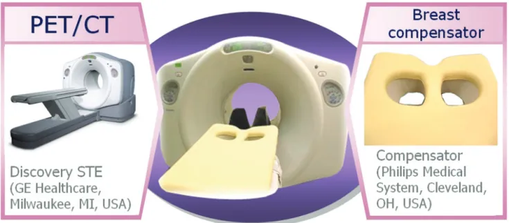
Fig. 1. Most of the PET-CT scans have been examined on supine position. Therefore, the image of breast has been acquired by reconstructed whole body scan image. However using breast compensator, ashape of breast was reassembly shown to be real by gravity. Consequently, we believe that diagnostic value of prone position scan method for breast cancer patients was prominent in PET-CT scan.
Fig. 2. A combined PET-CT inline system (Discovery STE, Milwaukee, Wis., USA) was used to acquire whole body 3-dimentional scans. Left side is PET-CT system. Right side breast of device (breast compensator). Close up of the breast cup openings. Center shows the position on scanner table.
가 75~95% 정도이며, 병변은 위음성(false negative), 위양성 (false positive)을 모두 가진다. 치밀유방 등으로 X-선 유방촬 영에 적합하지 않은 수진자에게 유용하며, 유방촬영 소견 상 판단이 어려운 수진자에게도 유용하다. 경우에 따라서는 섬 유낭성 변화(fibrocystic change)가 강하며, FNA (Fine Needle Aspiration)에서 음성인 환자의 진단에도 유용하다. 가슴확대 수술(breast surgically enhanced operation) 등의 시술을 받은 경우의 종양 검출에 유용하며, 검진으로서의 조기 암 검출도 기대할 수 있다.
림프절 전이의 평가의 경우 액와림프절의 전이의 검출률에
있어 예민도가 65~100%, 특이도가 66~100%로 높으며, 미세 전이(micrometastasis)는 검출할 수 없기 때문에 경계(sentinel) 림프절 생검이 필수적이다. 또한 흉골 림프절은 해부학적 영상에서 평가가 어려운 경우가 많으나 FDG를 이용한 PET- CT에서는 관찰이 가능하다.
대부분의 유방암 PET-CT 검사에서는 앙와위(supine) 자 세로 검사를 시행하며, 이러한 자세는 수진자의 측면에서는 유방이 중력에 의하여 왜곡된 상태에서 영상이 획득된다. 이 러한 문제점을 극복하기 위하여 최초 MRI에서 유방검사용 코일(breast coil)을 이용하여 prone position으로 검사하는 점
Fig. 3. All patients were examined in the supine position with whole body scan. But, breast lesional scan used to prone position with breast apparatus. The breast PET apparatus, was constructed to have the same geometry as the commercially available MRI coil with the breasts hanging freely under gravity.
을 착안하여, PET-CT 검사에서도 유방보정구(breast compen- sator)를 이용하여 prone position으로 검사를 진행하였다. 보 정구를 이용하여 prone position을 취하여 유방을 보다 원형 에 가깝게 표현함으로써 영상의 진단가치를 높일 수 있기에 유용성을 평가하였다(Fig. 1).
대상 및 방법
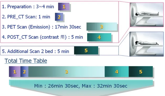
유방암으로 의심되거나 확진 된 여성 환자 20명(연령범위 : 17~60세, 평균연령 47.4세)을 대상으로 진행하였으며, 검사 는 환자에게 주사 전 500~1000 cc 정도의 충분한 수분을 섭취 하게 한 후 18F-FDG (8~12 mCi)를 말초혈관에 정맥 주사하 였으며, 측정한 선량을 모두 인체에 주입하기 위하여 3-way stop cock를 이용해 생리식염수 10 cc를 30초간 천천히 정맥 주사하였다. 환자는 주사 전 최소 6시간 이상 금식하였고, 주사 후에는 근육의 섭취가 증가됨을 막기 위해 40~50분간 움직임을 제재하였다. 주사 60분 후 전신 검사를 하였으며, 방출스캔(emission scan)을 한 후 투과스캔(transmission scan) 은 감쇠보정용 컴퓨터 단층촬영(CTAC)으로 획득하였다.
또한 유방에 대한 추가적인 검사는 2분30초/bed로 2 bed의 방출영상을 획득하였다(Fig. 2, 3).
결 과
유방암(진행성)을 가진 여성환자 20명에게 보정구를 이용 한 prone position에서 검사를 시행하였으며, 이중 17명의 환 자에게서 prone position이 진단 용이성과 정확한 판별률이 높았으며, 나머지 3명의 경우는 병소가 1 cm 이하의 크기로 인하여, 유방일반촬영, 초음파 또는 MRI에서는 나타났지만 PET-CT에서는 진단에 의미있는 관찰을 할 수 없었다. Su- pineposition과 비교하여 proneposition에서는 구조물의 중력 에 의한 변형수치가 낮고, 감별 영역이 넓어짐에 따라 구별이 곤란한 경우 명확하게 감별하는데 도움이 되었다(Fig. 4).
SUV를 측정한 결과 supine position과 prone position에서 유사한 SUV 측정치를 얻음으로써 자세에 의한 병변의 특성 에는 영향을 미치지 않았지만, 보다 명확한 병변의 위치를 확인하는데 prone position의 유용성을 확인할 수 있었다 (Table 1).
결 론
Supine자세를 통한 PET-CT 전신검사 후의 보정구를 이용 한 prone position으로 병변에 대한 추가검사(lesional scan)는
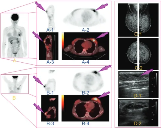
Fig. 4. FDG PET-CT scan in a patient with locally advanced breast cancer. (A) All patients were studied in the supine position.
(B) On the other hand, all patients retried in the prone position. (A, B) 3-dimentional images; A: supine position, B: prone position.
(A-1, A-2) Supine position in FDG PET; A-1: sagittal, A-2: axial. (A-3, A-4) Supine position in PET-CT fusion images; A-3:
sagittal, A-4: axial. Arrow indicated breast cancer lesion. (B-1, B-2) Prone position in FDG-PET; B-1: sagittal, B-2: axial. (B-3, B-4) Prone position in PET-CT fusion images; B-3: sagittal, B-4: axial. Arrow indicated breast cancer lesion too. (C-1, C-2) The same patient view of mammography images. (D-1, D-2) Ultrasound images in breast cancer lesion. (D-1) Arrow showed cancer lesion.
(D-2) Ultrasound of biopsy.
Table 1. Comparison of each SUVs.
두 자세의 비교가 모두 가능하였으며, 경우에 따라 결정적으 로 진단에 영향을 주었다. 두 가지 자세의 영상비교에 있어 prone position이 원발성(primary) 병변의 진단은 물론, 미세 전이(micrometastasis)의 검출이 가능하였으며, 분해능이 MRI
에 비해 낮은 단점을 보완하며 병변의 구별이 명확하게 나타 났다. 이러한 prone position을 기반으로 구조적인 문제를 정 량적 분석과 함께 극복한다면 병변의 진단에 있어 보다 유용 한 진단적 정보를 얻을 수 있으리라 사료된다.
, 유방을 중력에 의존하여 보다 원형에 가깝게 표현 함으로써 영상의 진단가치를 높일 수 있기에 그 유용성을 평 가하였다.
유방암으로 의심되거나 확진 된 여성 환자 30명을 대상으 로 하였으며, 18F-FDG 주사한 뒤 60분 후 supine position 으로 whole body PET-CT scan 시행 후, 보정구를 이용하여 prone position으로 병변에 대한 추가검사(lesional scan)를 시 행하였다. 각각 획득된 영상은 핵의학과 의사로부터 blind test를 하였고, PET-CT 영상의 SUV를 측정하여 분석하였다.
30명 중 27명의 환자에게서 prone자세가 진단 용이성과 정확한 판별률이 높았으며, 나머지 3명의 경우는 병소가 1 cm 이하의 크기로 인하여, MRI상에는 나타났지만 PET-CT에서 는 진단에 의미있는 관찰을 할 수 없었다. supine position과 비교하여 prone position은 구조물의 중력에 의한 변형수치 가 낮아, 병변의 구별이 곤란한 경우 명확하게 감별되었다.
각 자세에서 SUV분석은 유의수준 0.004로 유의하였다.
REFERENCES
1. Crippa F, et al. Prospective evaluation of fluorine-18-FDG-PET in pre-surgical staging of axilla in breast cancer. J Nucl Med.
39:4-8, 1998.
2. Moon DP, et al. Accuracy of whole-body fluorine-18-FDG PET for the detection of recurrent of metastatic breast cancer, J Nucl Med. 39:431-435, 1998.
3. Vranjesevic D, et al. Relationship between 18F-FDG uptake and breast density in women with normal breast tissue. J Nucl Med.
44:1238-1242, 2003.
4. Cook GJ, et al. Detection of bone metastases in breast cancer with FDG-PET. Differing metabolic activity in osteoblastic and osteolytic lesions. J Clin Oncol. 16:3375-3379, 1998.
5. Landheer ML, et al. Value of fluorodeoxyglucose positron emission tomography in women with breast cancer. Br J Surg.
92:1363-1367, 2005.
6. Tatsumi M, et al. Initial experience with FDG-PET/CT in the evaluation of breast cancer. Eur J Nucl Med Mol Imaging. 33:
254-262, 2006.