J Nucl Med Technol Vol. 14, No. 1, June 2010
147
∙ Received: March 23, 2010. Accepted: March 31, 2010.
∙ Corresponding author: Hyo Yeong Lee
Department of Nuclear Medicine, Pusan National University Hospital 305 Gudeok-Ro, Seo-gu, Pusan, 602-739, Korea
Tel: +82-51-240-7385, Fax: +82-51-241-5570 E-mail: [email protected]
Case Report
폐와 심근에서 전이성 석회화가 발견된 골 스캔부산대학교병원 핵의학과
송현석⋅이효영⋅윤종준⋅이화진⋅이무석⋅박세윤⋅정지욱
Metastatic Calcification Revealed by the Bone Scan at Both Lung and a Myocardium
Hyeon Seok Song, Hyo Yeong Lee, Jong Jun Yun, Hwa Jin Lee, Moo Seok Lee, Se Yun Park and Ji Uk Jeong
Department of Nuclear Medicine. Pusan National University Hospital
Introduction: A metastatic calcification is known for taking in bone scintigram medicine at metastatic calcification lesion due to abnormal distribution of the calcium and phosphorus. The one paper reports that a metastatic calcification occurs mainly at lung, stomach, kidney and myocardium. Index: The patient is seventy four years old man who is afflicted with clonic kidney disease, hypercalcemia, hypertension. Because of an ability of the multiple myeloma, we take a bone scan after intravenous injection 99mTc-DPD 25 mCi in three hours. We found out homogeneous 99mTc-DPD uptake at both lung and myocardium. Conclusions: Nothing unusual was found in other bone scan. We obtains a purity beyond 95 percent at 99mTc-DPD vial. In spite of no evidence about a myocardial infarction, the patient has a 99mTc-DPD uptake at both lung and myocardium.
(Korean J Nucl Med Technol 2010;14(1):147-148)
Key Words : Metastatic calcification, 99mTc-DPD, Clonic kidney disease, Hypercalcemia, Lung uptake, Myocardium uptake
서 론
전이성 석회화는 원발성 부갑상선 기능 항진증1)에서의 미만성 간질성 폐석회화와 폐 전이 없이 육종 또는 암종에 동반된 고칼슘 혈증의 경우 그리고 신부전증 환자에서 생기 는 것으로 알려져 있다. 전이성 석회화는 조직 검사를 시행 하여 확인할 수 있으나 석회화된 부위에 골 스캔 상 방사능의 섭취가 되는 예가 있다2). 이번 증례보고의 환자는 99mTc-DPD 를 이용한 골 스캔 상에서 양측 폐 부위와 심근에서의 섭취 가 나타나 이를 보고하는 바이다.
증 례
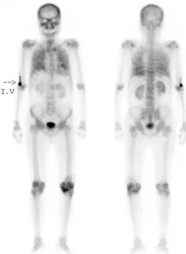
환자는 74세의 남자로 만성 신부전증에서 급성 신부전증 이 동반되어 있었으며, 고 칼슘혈증, 고혈압의 병력을 가지 고 있었고 다발성 골수종이 의심되어 타 병원에서 본원으로 검사 의뢰한 환자이다. 99mTc-DPD 25 mCi를 정맥 주사하고 약 3시간 뒤 전신 골 스캔을 실시하였다. 검사 후 판독 결과 왼쪽 무릎에서 증가된 방사선 의약품의 섭취가 관찰되었으 며, 양측 폐부위의 균일한 섭취 및 심근의 섭취가 나타났다 (Fig. 1, Fig. 2). 무릎은 관절의 퇴행 양상이며, 양측 폐와 심 근의 섭취는 전이성 석회화를 시사한다고 하였다.
고 찰
99mTc 인산염 유도체에 의한 골 스캔이 임상적으로 널리 이용된 이래 골격 외 연조직에 이 제제가 섭취되는 경우가 보고되고 있다3). 이 중에서 골 스캔 상 폐에 섭취를 보이는
핵의학기술 제14권 제1호 2010
148 Fig. 1. Metastatic calcification revealed by the whole body bone scan at both lung and a myocardium.
Fig. 2. LAO image at chest
원인으로서 전이성 석회화, 원발성 폐암, 폐에 대한 방사선 치료 후 상태 등이 흔한 것으로 알려져 있다. 본 환자에서 폐와 심근 부위에 섭취를 보인 것은 전이성 석회화 이외에 다른 원인의 증거를 찾을 수 없었다. 골 스캔 시 골 이외의 갑상선이나 위 부위에 방사선이 섭취되는 경우가 있다4). 이 에 대한 가장 흔한 원인은 방사성의약품의 불완전한 순도 때문에 유리 형태의 99mTc-DPD가 골격 외 연조직에 섭취되 는 경우이다. 그러나 이때 사용한 99mTc-DPD 바이알에서 순 도측정을 실시한 결과 95% 이상의 순도를 얻었으며 갑상선 및 구강 점막 부위의 방사능 증가 소견이 없었으며 동시에 동일 조건으로 골 스캔을 시행한 다른 환자에서 폐 및 위 부 위의 방사능 증가는 관찰되지 않았다. 환자는 상기의 병력이 있는 환자로 폐에 전이성 석회화가 관찰되며 임상적으로 심 근경색의 증거가 없음에도 불구하고 골 스캔 상에서 심근에 서 섭취가 나타났다.
REFERENCES
1. Itoh, k., et al. diffuse lung uptake on bone imaging in primary hyperparathyroidism before and after excision of parathyroid adenoma. clin. Nucl. Med 1979;4:382.
2. Richards, A.G. Metastatic calcification detected through scanning with 99mTc-polyphosphate. J. nucl. Med 1974;15:1057.
3. Brill, D.R. Radionuclide imaging of nonneoplastic soft tissue disorders. Semin. Nucl. Med 1981;11:277.
4. 이동수 외 4명. 전이성 석회화가 골 스캔에서 발견된 예. 대한 핵의학회지 1984;18:67-9.