※ This research was supported by Basic Science Research Program (2015-R1A2A2A04006136) and the Brain Research Program (grant number-2017-11-0625) through the National Research Foundation of Korea(NRF) funded by the Ministry of Science, ICT & Future Planning.
†Corresponding author:Sanghoon Han (Department of Psychology, Yonsei University) E-mail:[email protected]
TEL:02-2123-5436 FAX:02-2123-8330
Estimation of Reward Probability in the Fronto-parietal Functional Network: An fMRI Study
Yeonsoon Shin*․Hye-young Kim*․Seokyoung Min*․Sanghoon Han*
,**†
*Department of Psychology, Yonsei University
**Graduate Program in Cognitive Science, Yonsei University
Abstract
We investigated the neural representation of reward probability recognition and its neural connectivity with other regions of the brain. Using functional magnetic resonance imaging (fMRI), we used a simple guessing task with different probabilities of obtaining rewards across trials to assay local and global regions processing reward probability. The results of whole brain analysis demonstrated that lateral prefrontal cortex, inferior parietal lobe, and postcentral gyrus were activated during probability-based decision making. Specifically, the higher the expected value was, the more these regions were activated. Fronto-parietal connectivity, comprising inferior parietal regions and right lateral prefrontal cortex, conjointly engaged during high reward probability recognition compared to low reward condition, regardless of whether the reward information was extrinsically presented. Finally, the result of a regression analysis identified that cortico-subcortical connectivity was strengthened during the high reward anticipation for the subjects with higher cognitive impulsivity. Our findings demonstrate that interregional functional involvement is involved in valuation based on reward probability and that personality trait such as cognitive impulsivity plays a role in modulating the connectivity among different brain regions.
Key words: fMRI, Inferior Parietal Lobe, Lateral Prefrontal Cortex, Reward Probability Processing
1. Introduction
To choose optimal options, we need to take both the outcome value of the actions and the probability that leads an action to the desired outcome into consideration.
These two considerations are the constituents of “expected value”, an important concept both in economics and psychology (von Neumann and Morgenstern, 1944;
Rotter, 1972; Bandura, 1977; Kahneman and Tversky,
1984). Probability perception is especially critical since
when the actions are incorporated with incentives, the
probability determines the ultimate amount of reward
considering that the magnitude of reward is often given
in the context. Although several neuroimaging and
comparative animal studies have demonstrated that
lateral intraparietal or lateral prefrontal regions is
https://doi.org/10.14695/KJSOS.2017.20.4.101especially active when encoding the likelihood of gain during decision making (e.g., Platt & Glimcher, 1999;
Huettel et al., 2005; Camara et al., 2009; Liu et al., 2011;
Gerlach et al., 2014), less is debated on the brain networks that govern probability-based decision process.
The frontoparietal network, including the inferior parietal lobule (IPL) and lateral prefrontal cortex (PFC), is thought to be a likely candidate neural substrate for representing the reward probability. Prior neuroimaging studies on the neural representation of probability-related decision variables such as risk, uncertainty, and ambiguity, also suggests that this parietal region might be sensitive to the likelihood of receiving reward outcomes (Huettel et al., 2005; Weber & Huettel, 2008;
Peters & Büchel, 2009; d'Acremon et al., 2013; Liljeholm et al., 2013).
In addition to parietal cortex, lateral PFC might also be engaged in computation of the reward probability (Dreher et al., 2006; Tobler et al., 2007, 2008, 2009;
Labudda et al., 2008; Bach et al., 2009). For example, Labudda et al. (2008) showed that explicit information about the reward probability and magnitude induced activation in dorsolateral PFC and IPL regions.
More recently, some researchers investigating large- scale brain networks found that IPL and lateral PFC regions constitute a functionally connected network and may work in concert with each other (Dosenbach et al., 2007; Liu et al., 2011; Power et al., 2011; Vincent et al., 2008; Yeo et al., 2011; Cole et al., 2014; Gerlach et al., 2014). Although some studies found that both regions are co-activated in the recognition of reward probability (Huettel et al., 2005; Weber & Huettel, 2008;
Labudda et al., 2008), little is known about how these regions interact each other during probability-based decision-making.
In the present study using functional magnetic resonance imaging (fMRI), we used a simple guessing task with different probabilities of obtaining rewards across trials to assay local and global regions processing reward probability while the design keeps constant the
frequency and magnitude of the reward. The current study can explicate how human brain calculates and represents expected value based on probability.
Furthermore, it examines the functional connectivity through Psycho-Physiological Interaction (PPI) analyses, exploring neural substrates in valuation based on reward probability. Finally, although prior studies provide evidence that individual trait modulates the reward- related functioning of brain regions (e.g., Simon et al., 2010), how the variability of personality traits affects the functional connectivity among neural substrates in reward probability recognition and valuation has yet to be investigated. Therefore, personality trait such as cognitive impulsivity was expected to play a role in modulating the connectivity among different brain regions was investigated. For example, increased cortico-subcortical functional coupling was expected during high relative to low gain probability task for more impulsive people.
2. Methods
2.1. Participants
Twenty native English-speaking volunteers partici- pated. Participants were 11 females and 9 males, with a mean age of 24 years (age range 20-33 years). One subject was excluded from analysis for failure to complete the experiment. Informed consent was obtained in compliance with the Institutional Review Board of Duke University Medical Center. The participants were paid $20 for each hour of participation and additional rewards provided depending on their performance after experiment.
2.2. Experimental Tasks & Procedures
2.2.1. Monetary incentive guessing task
A simple incentive guessing task was examined to
investigate the regions involved in perceiving gain
Fig. 1. Schematic of trial design for (A) low reward anticipation condition and (B) high reward anticipation condition
probability. Subjects were presented with four boxes on
the screen indicating possible locations where a target (a ‘$’) could subsequently appear. There were two guessing conditions. First, for the “Pick target location”
condition, subjects received a reward only if they guessed correctly the one of four locations where the target would appear. Thus, the chance to obtain a reward (a dollar) was 25%. This is termed the low anticipation condition since subjects were more likely to miss obtaining the reward. The other condition was the high anticipation condition where instead the prompt read “Pick non- target location”. In this condition subjects were rewarded if they picked a location in which the target did not subsequently appear. Since the success rate was on average 75%, this constituted the condition in which there was high anticipation of reward. The final factor we manipulated was the feedback availability. On half the trials subjects received payout indication of their response correctness (red or green), on the other half of the trials the prompt (‘XXXX’) turned gray with no payout information. The purpose of the no-feedback condition was to see whether any existing neural responses are rooted on the effect of external feedback delivery rather than probability estimation process. Again, subjects were informed beforehand of the existence of no- feedback trials and were aware that they could still receive payouts regardless of whether the trial specific feedback was present.
The trial structure was depicted in Fig. 1 (Cue/Probe presentation for 3.5 s, immediate reward presentation following response with a blank .5 s) (SOA of 4s). There were two scan runs with a total of 80 guessing trials (40 low anticipation + 40 high anticipation conditions) in each test scan. Test trials were interspersed with 20 fixation trials as determined by an optimal stimulus/trial sequencing program (Wager & Nichols, 2003) to maximize the efficiency of estimating hemodynamic responses. The reward was always a dollar and there was no punishment for incorrect guesses. Subjects were informed that only one of the two runs would be selected
for payout at the completion of the experiment. The run selected for payment was determined by coin toss following the experiment and subjects received either the payout tallied from the selected scan run or a maximum of 40 dollars.
2.2.2 Post-scan Self-report Personality Questionnaire Survey
Following scanning and outside the scanner prior to payment, subjects completed the Barratt Impulsiveness Scale (11th Edition; BIS-11) (Barratt & Patton, 1983).
BIS-11 is a commonly used personality inventory mea-
suring trait impulsivity with 30 behavioral and
preference questions and the total scores range from
30 to 120, with the higher scores representing more
impulsiveness. Recently, it has been argued that
subscale scores representing separable subdomains may
be applicable to report subscale correlates (Stanford et
al., 2009; Reise et al., 2013). Therefore, among the
three sub-traits (motor, cognitive, and non-planning) of
the BIS-11 scale, we selected the cognitive subscale
to explore the modulation effect of impulsivity trait on
functional connectivity during reward-based decisions
given that the score is closely related to the tendency
to make quick decisions.
2.3. fMRI Data Acquisition
Scanning was performed on a 3T General Electric (Waukesha, WI) scanner using a standard head coil.
Functional data were acquired by using a gradient echo echo-planar pulse sequence (acquisition matrix 64×64, FOV = 240×240 mm, flip angle = 60°, TR = 2000, TE
= 31 msec, 34 axial slices parallel to the AC – PC plane with near-isotropic voxels of 3.75×3.75×3.8 mm, no gap, interleaved collection). Before functional data collection, four dummy volumes were discarded to allow for equilibration effects. Participants’ head motion was minimized by using foam padding. High-resolution T1- weighted anatomical images (3-D spoiled gradient recalled acquisition [SPGR], isomorphic voxel size = 1mm, 256×256 matrix, flip angle = 20°, TR = 22 ms, TE = 5.4 ms) were acquired for visualization.
2.4. fMRI Data Preprocessing & Analyses
Data were processed using SPM8 (Wellcome Department of Cognitive Neurology, London). Slice acquisition timing was corrected by resampling all slices in time relative to the middle slice collected, followed by rigid body motion correction across all scans.
Functional data were spatially normalized to a canonical echo-planar imaging (EPI) template using a 12-parameter affine and nonlinear cosine transformation, with volumes then resampled into 2 mm cubes and spatially smoothed with an 8-mm fullwidth at half-maximum isotropic Gaussian kernel. Each scanning session was rescaled such that the mean global signal was 100 across the volumes. For the analyses, volumes were treated as a temporally correlated time series and modeled by convolving a canonical hemodynamic response function (HRF) and its temporal derivative with a delta function marking each trial onset. The resulting functions were used as covariates in a general linear model, along with a basis set of cosine functions that were used to high-pass filter the data and a covariate representing session effects.
The least squares parameter estimates of the best-fitting synthetic HRF for each condition of interest (averaged across scans) were used in pair-wise contrasts and stored as a separate image for each subject. These different images were then tested against the null hypothesis of no difference between contrast conditions using one- tailed t tests. The data were statistically analyzed treating subjects as a random effect. The threshold for whole- brain GLM statistical significance was set at p < .05, corrected for multiple comparisons with the family-wise error (FWE) estimation through Monte Carlo simulation of cluster-extent thresholding (Slotnick et al., 2003), unless otherwise stated.
Functional regions of interest (ROIs) were extracted using the MarsBar Toolbox (Brett et al., 2002) using coordinates obtained from the SPM contrast maps.
Percent signal averages, using peristimulus time averaging, and parameter estimates for each condition were obtained for the significant voxels within an 8-mm radius of each of the SPM-identified maxima and further analyzed using off-line statistical software. Although prior research mentioned in the Introduction did not particularly emphasize the hemispheric asymmetry of the reward- related decision process in the brain, our current findings demonstrated mostly right hemispheric involvement of frontoparietal regions during reward probability. Among the functionally defined ROIs, the current analysis further investigated the right ventrolateral prefrontal regions and ventral-anterior inferior parietal lobule.
2.5. Psychophysiological interaction (PPI) analysis
PPI analyses were computed to investigate how
activities in brain regions covary with the regions of
interest (‘seed region’) along task conditions (Friston et
al., 1997). Peak voxels of anterior IPL and frontal regions
activated in a main contrast (i.e., high versus low
probability condition) were used as seed regions for PPI
analysis. The model of psychophysiological interaction
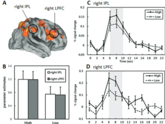
Fig. 2. fMRI results and ROI extraction. (A) Lateral view for brain areas showing significant activation on high versus low reward anticipation and ROIs (right IPL, right LPFC), (B) graphs representing the parameter estimates of each reward condition in the ROIs, (C), (D) time courses showing greater hemodynamic responses during high versus low conditions in the right IPL and the right LPFC, respectively
between deconvolved neural activity of a seed region (e.g., inferior parietal lobule) and the task conditions was generated for each subject. Each subject’s hemodynamic response parameters then were estimated based on these PPI models, generating beta parameters representing the degree to which the hemodynamic responses (HDR) to high versus low probability task condition in each voxel correlates with the seed regions’ HDR to the task.
Second-level random-effects analysis was used to find generally activated region across subjects.
Also, another second-level analysis of PPI contrast images with Barratt Impulsivity Scale, the 11th edition, cognitive subscale (Barratt & Patton, 1983) as regressor was performed in a simple regression analysis. The resulting parameters identify brain regions which show greater connectivity with the seed regions as a function of the BIS 11 cognitive subscores.
3. Results
3.1. Behavioral Results
Participants were not more likely to take less time during high probability condition compared to low probability condition (863 ms ± 226 versus 857 ms ± 251, mean ± SEM respectively, t (18) =.75, n.s.). On average, the participants received 75% on the high probability condition and 23% on the low probability trials (mean total reward; 30$ and 9$ for high versus low conditions, respectively).
3.2. fMRI Results
3.2.1 Whole brain general linear model analysis In order to compare the general probability sensitivity, high versus low probability contrast analysis collapsing feedback conditions (Feedback, No-Feedback) was conducted. As seen in Fig. 2A and Table 1, significantly greater activation was observed during high compared
to low probability conditions in right lateral PFC, Inferior Parietal Lobule (IPL), precuneus, postcentral, caudate, demonstrating that frontoparietal regions are relatively more recruited during probability- based decision-making process when outcome value is expected higher. To further scrutinize the difference between the high versus low conditions, we conducted ROI analyses on frontoparietal regions that showed significant effect of probability (right IPL, x=62, y=-22, z=37; right inferior frontal region, x=50, y=32, z=17).
Within the regions, each ROI exhibited significant mean parameter effects (beta value) in the high anticipation condition than in the low anticipation condition (right IPL: t (18) = 4.714; p < .001; right LPFC:
t (18) = 4.531, p < .001; Fig. 2B). Also, the averaged amplitude of fMRI hemodynamic responses peaked at approximately 6 ~ 10 sec showed significant difference between high probability and low probability conditions (right IPL: t (18) = 3.232, p < .005; right LPFC: t (18)
= 4.019, p < .001; Fig. 2C and 2D, respectively). In
sum, these results demonstrate that frontoparietal regions
are actively engaged in the recognition of probability or
expected value.
Regions Lat. BA
x y z z-score Superior/Medial Fontal Gyrus R 6/8 3 25 50 3.47
3 2 59 2.97
9 7 51 2.78
Superior/Middle Frontal Gyrus R 6 33 -1 59 3.35
9 27 38 32 2.66
9/10 33 58 8 2.72
Middle/Inferior Frontal Gyrus* R 46 50 32 17 3.28
9 48 12 25 3.14
Middle Frontal Gyrus/Precentral Gyrus R 6 45 -1 51 3.31 Inferior Frontal Gyrus/Precentral Gyrus R 9/44 59 14 18 2.91 Supramarginal Gyrus/IPL R 40 48 -40 33 3.75 Precuneus/Postcentral Gyrus L 7 -6 -53 62 3.33 Postcentral Gyrus/IPL* R 1/2/3/40 62 -22 37 3.19 Precuneus/Superior Parietal Lobule R 7 21 -71 52 2.84 Superior Parietal Lobule/IPL R 7/40 30 -54 54 2.74 Middle/Superior Temporal Gyrus L 21/22 -62 -47 6 2.88 Superior Temporal Gyrus R 19/22 39 -30 0 2.8
Caudate R 12 -9 19 3.19
Cingulate Gyrus/Caudate L -18 -11 24 2.8
Insula R 13 33 -20 22 3.07
Parahippocampal Gyrus R 19/30 12 -45 -4 2.74 Thalamus/Parahippocampal Gyrus R 27 6 -30 5 3.74
Thalamus R 12 -26 15 3.39
6 -12 9 2.7
Lingual Gyrus L 18 -12 -77 -13 3.4
Cuneus/Middle Occipital Gyrus R 17/18 9 -88 13 3.37
Fusiform Gyrus L 19 -33 -66 -14 2.91
* Regions selected for ROI analyses
Table 1. Whole brain activation table (overall high vs low contrast)
3.2.2 PPI analysis - interregional functional coupling We examined functional connectivity during high compared to low probability condition to reveal frontoparietal involvement for gain probability re- cognition. Fig. 2A above shows two seed regions including the right inferior parietal lobule and right lateral prefrontal cortex. PPI analyses identified the brain regions covarying with the seed regions during high (compared to low) gain probability task condition (See Table 2).
During high probability condition, there was a great deal of functional connectivity in right lateral PFC seed
region (rlPFC; x, y, z = 50, 32, 17) with various regions
including bilateral parietal, right posterior temporal,
postcentral, and occipital regions. Especially activity of
this seed was also coupled with that of right IPL (x,
y, z = 62, -22, 37) which was the other ROI seed,
consisting a conjoint frontoparietal network for pro-
bability recognition. The analysis also identified other
regions such as frontopolar (x, y, z = -3, 49, 11), bilateral
precentral gyrus (L: x, y, z = -45, 8, 41, R:x, y, z =
39, -2, 46), postcentral gyrus (x, y, z = -53, -22, 37)
and subcortical regions such as striatum (x, y, z = 6,
Regions Lat. BA x y z z-score Seed 1. Right ventrolateral PFC [50, 32, 17]
Medial Frontal Gyrus/ Frontopolar L 10 -3 49 11 3.01 Middle Frontal Gyrus/ Precentral Gyrus L 6 -45 7 41 2.97
R 6 39 -2 46 3.69
48 -16 40 3.66
45 -8 42 3.23
50 -2 39 2.87
Postcentral Gyrus L 2 -53 -22 37 3.12
3 -39 -19 45 2.84
Inferior Parietal Lobule/Postcentral Gyrus L 40 -48 -34 45 3.22
R 40 59 -25 37 3.68
56 -23 20 3.66
48 -31 48 3.62
48 -34 33 3.38
62 -41 23 3.30
2 48 -20 30 2.81
Precuneus L 7 -15 -54 57 2.85
R 7 18 -60 49 3.43
33 -66 32 2.95
Superior Temporal Gyrus R 13 45 -47 16 3.36
22 65 -38 15 3.15
Middle Temporal Gyrus L 37 -53 -65 4 2.81
R 37 48 -65 7 4.45
39 48 -56 9 4.39
Cingulate L 29/30 -9 -50 11 3.28
Putamen 18 10 -7 2.92
Caudate head R 6 2 -4 3.37
Fusiform R 37 36 -54 -12 2.84
Occipital Lobe L 17 -6 -91 0 3.82
R 17 9 -91 3 2.75
Seed 2. Right Postcentral Gyrus [62, -22, 37]
Middle Frontal Gyrus L 6 -42 4 44 5.27 46 -45 13 43 4.11 Ventrolateral Prefrontal Cortex* R 6/8 50 35 14 3.00 Inferior Frontal Gyrus* R 9 59 12 25 3.13 Precentral Gyrus* L 4 -27 -25 48 2.79
R 4 33 -22 45 2.82
33 -25 53 2.88 Postcentral Gyrus/ Inferior Parietal Lobule R 2/40 59 -25 37 3.13 Inferior Parietal Lobule* L 40 -48 -40 41 2.82
R 40 50 -31 45 2.71
Superior Parietal Lobule/Precuneus R 7 39 -63 49 3.32 18 -69 47 2.85 19 30 -72 37 3.28 Middle Temporal Gyrus R 39 39 -61 24 3.48 Cingulate Gyrus* L 31 -3 -37 36 2.85 23 0 -26 27 2.88
Occipital Lobe R 27 -69 30 3.09
Lat. = Laterality, * lax threshold (p < .005/5 extent voxels) adopted for exploration purpose
Table 2. Psycho-physiological interaction (high vs low contrast)2, -4) as well (Table 2).
PPI analysis using the right IPL (x, y, z = 62, -22, 37) seed also showed functional connectivity with various
frontal and parietal regions. When employed with more
lax threshold (p < .005, 5 contiguous voxels) for
exploring the reciprocal frontoparietal connectivity,
bilateral middle frontal gyrus (MFG, L: x, y, z = -42, 4, 44, R: x, y, z = 50, 35, 14), precuneus (x, y, z = 39, -63, 49), inferior frontal gyrus (x, y, z = 59, 12, 25), and left IPL (x, y, z = -48, -40, 41) showed functional coupling with the right IPL seed region.
3.2.3 PPI analysis – Feedback vs No-Feedback contrast
The current analyses revealed that the functional couplings corroborate frontoparietal network that is hired for high probability versus low probability regarding incentive task. However, it could be argued that this result is not solely rooted on the probability effect, but also affected by feedback processing, which was not separated in the overall high versus low probability contrast condition modeled as a psychological variable in PPI analyses. Therefore, we conducted additional PPI analysis under feedback compared to no-feedback condition to directly test if the existing functional coupling is modulated by overall feedback relative to no-feedback condition. The same PPI method above was used except the psychological variable modeled using overall Feedback versus No-Feedback contrast. More impor- tantly, the frontoparietal network that exhibited signifi- cant pattern of functional connectivity during high compared to low probability condition was no more activated during feedback compared to no-feedback condition even at very liberal standard, P < .01. Thus, we can conclude that the reciprocal frontoparietal network is especially hired when processing probability regardless of the existence of feedback presence.
3.2.4 Personality differences in PPI strength during incentive probability perception
There have been ample previous studies demonstrating that personality traits affect neurobiological activity (Cohen et al., 2009; Krebs et al., 2009). We examined not only neurobiological response of discrete brain region but the strength of interregional psychophysiological interaction as personality traits differ. The seed region
of interest was right IPL (see above) which constitutes frontoparietal network for probability recognition.
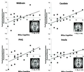
Here, with more impulsive personality, higher cortico- subcortical interaction was hypothesized. To ascertain whether the functional coupling of discrete brain regions is modulated by this personality trait, we performed simple regression analysis on connectivity strength with BIS 11 cognitive subscale. The regression analysis identified brain regions that showed better functional coupling with IPL seed region during high versus low probability condition for subjects with cognitively more impulsive personality (See Fig. 3). Right IPL showed significantly greater functional coupling under high versus low probability condition with various brain regions including midbrain, caudate, parahippocampal gyrus, and insula as cognitive impulsivity score increases.
Table 3 shows all regions that showed greater functional coupling with IPL for more impulsive personality. The results ensure that impulsive personality trait modulates cortico-subcortical functional connectivity while per- forming incentive tasks with different reward pro- babilities.
Fig. 3. Representative subcortical brain regions showing stronger functional coupling with IPL seed during high versus low probability recognition modulated by BIS 11 cognitive impulsivity personality scores. Clock-wise from
the top left corner; Midbrain, Caudate, Insula, Parahippocampal gyrus (PHG) correlation.
Regions Lat. BA
x y z z-score
Putamen L -24 5 1 4.16
-27 -15 2 3.39
R 24 2 6 4.15
27 5 -7 3.67
24 -15 12 3.21
30 -12 4 3.07
Midbrain R 9 -15 12 3.43
12 -16 -6 3.19 0 -29 -13 3.17
Caudate L -18 2 11 4.1
Parahippocampal Gyrus/Amygdala L 34 -33 1 -14 4.02
R 21 -4 -12 3.06
Hippocampus L -33 -27 -5 3.21
Insula L -30 14 5 3.98
-39 -21 12 3.3
R 13 48 -12 17 3.66
45 -23 17 3.47
39 -1 6 3.15
39 11 3 2.97
33 -21 14 2.93 Inferior Frontal Gyrus L 47 -33 13 -17 3.42 -27 19 -10 2.98
Precentral Gyrus L -6 -24 68 3.32
R 53 3 16 3.11
Precentral/Paracentral Lobule R 6 30 -15 60 3.19 0 -33 63 2.99 Middle Frontal Gyrus R 46 36 32 24 4.07
10 -30 55 6 2.73
Cingulate Gyrus L 24 -12 -11 37 3.61
-12 4 36 3.51
Table 3. BIS11 simple regression with PPI estimates (IPL seed)
4. Conclusions
We investigated the neural representation of incentive probability recognition and its neural connectivity with other regions of the brain. Furthermore, it was also examined whether the interregional functional coupling was modulated by personality traits. Analyses of the neuroimaging data revealed three main findings. First, we identified regions particularly involved in probability- based decision making, especially when the expected value is higher; lateral PFC, parietal regions including ventral-anterior IPL and postcentral, and a part of
midbrain. Second, frontoparietal network, comprising IPL, right lateral PFC was conjointly engaged during high versus low incentive probability recognition regardless of whether the reward information is extrinsically presented. Finally, in addition to the frontoparietal network for high reward-winning chance condition, a regression analysis identified cortico-subcortical network that was strengthened for the subjects with higher cognitive impulsivity.
Over the past two decades, accumulating evidence has
suggested that the frontoparietal cortical network may
play a key role in representing the reward probability.
Both IPL and lateral PFC regions have been shown to be involved in processing reward probability (Platt &
Glimcher, 1999; Huettel et al., 2004; Dreher et al, 2006;
Tobler et al., 2008; d’Acremont et al., 2013). Our results are consistent with those previous findings. Moreover, the present study extends those previous works by showing that functional connectivity between these two frontoparietal regions is increased more during high probability condition than low probability condition.
A limitation of our present study is that we could not sort out the pure probability recognition effect and feedback effect in the current design. Since the guessing task and outcome feedback for the subject choice took place in one trial during 4 seconds, not clearly separated with fixed amount of time respectively, there was an analytical limitation to tell whether the activation was solely due to probability calculation or feedback processing. Although our PPI analysis with Feedback versus NoFeedback condition modeled as a psychological variable strengthen the former, if further studies employ more sophisticated design with distinct phase for feedback, it could achieve clearer interaction maps. For example, the future study may employ temporal jittering of each subsequent feedback and a factorial analysis with the probability and feedback as factors would enable the understanding of interaction effects. Despite this limitation, our study has importance in that more active frontoparietal network under high probability recognition of rewards was identified, which might be modulated by cognitive attention for goal-directed behavior and its correlation with impulsive personality traits.
REFERENCES
Bandura, A. (1977). Social learning theory. Englewood Cliffs, NJ: Prentice-Hall.
Bach, D. R., Seymour, B., & Dolan, R. J. (2009). Neural activity associated with the passive prediction of ambiguity and risk for aversive events. Journal of Neuroscience, 29(6), 1648-1656.
DOI: 10.1523/JNEUROSCI.4578-08.2009
Barratt, E. S., & Patton, J. H. (1983). Impulsivity:
Cognitive, behavioral, and psychophysiological correlates. In M. Zuckerman (Ed.), Biological bases of sensation seeking, impulsivity, and anxiety (pp.
77-122). Hillsdale, NJ: Lawrence Erlbaum Associates.
Brett, M., Anton, J. L., Valabregue, R., & Poline, J. B.
(2002). Region of interest analysis using the MarsBar toolbox for SPM 99. NeuroImage, 16(2), S497.
Camara, E., Rodriguez-fornells, A., & Münte, T. F.
(2009). Functional connectivity of reward processing in the brain. Frontiers in Human Neuroscience, 2, 1-14. DOI: 10.3389/neuro.09.019.2008
Cohen, M. X., Schoene-Bake, J. C., Elger, C. E., &
Weber, B. (2009). Connectivity-based segregation of the human striatum predicts personality characteristics. Nature Neuroscience, 12(1), 32-34.
DOI: 10.1038/nn.2228
Cole, M. W., Bassett, D. S., Power, J. D., Braver, T.
S., & Petersen, S. E. (2014). Intrinsic and task- evoked network architectures of the human brain.
Neuron, 83(1), 238-251.
DOI: 10.1016/j.neuron.2014.05.014
d’Acremont, M., Fornari, E., & Bossaerts, P. (2013).
Activity in inferior parietal and medial prefrontal cortex signals the accumulation of evidence in a probability learning task. PLoS Computational Biology, 9(1). DOI: 10.1371/journal.pcbi.1002895 Dosenbach, N. U. F., Fair, D. A., Miezin, F. M., Cohen,
A. L., Wenger, K. K., Dosenbach, R. A. T., Fox, M. D., Snyder, A. Z., Vencent, J. L., Raichle, M.
E., Schlaggar, B. L., & Petersen, S. E. (2007).
Distinct brain networks for adaptive and stable task control in humans. Proceedings of the National Academy of Sciences, 104(26), 11073-11078.
DOI: 10.1371/journal.pcbi.1002895
Dreher, J. C., Kohn, P., & Berman, K. F. (2006). Neural coding of distinct statistical properties of reward information in humans. Cerebral Cortex, 16(4), 561-573. DOI: 10.1093/cercor/bhj004
Friston, K. J., Buechel, C., Fink, G. R., Morris, J., Rolls,
E., & Dolan, R. J. (1997). Psychophysiological and
modulatory interactions in neuroimaging. Neuroimage, 6(3), 218-229. DOI: 10.1006/nimg.1997.0291 Gerlach, K. D., Spreng, R. N., Madore, K. P., & Schacter,
D. L. (2014). Future planning: default network activity couples with frontoparietal control network and reward-processing regions during process and outcome simulations. Social Cognitive and Affective Neuroscience, 9(12), 1942-1951.
DOI: 10.1093/scan/nsu001
Huettel, S. A. (2005). Decisions under uncertainty:
Probabilistic context influences activation of prefrontal and parietal cortices. Journal of Neuroscience, 25(13), 3304-3311.
DOI: 10.1523/JNEUROSCI.5070-04.2005
Kahneman, D., & Tversky, A. (1984). Choices, values, and frames. American Psychologist, 39, 341-350.
Krebs, R. M., Schott, B. H., & Düzel, E. (2009).
Personality traits are differentially associated with patterns of reward and novelty processing in the human substantia nigra/ventral tegmental area.
Biological Psychiatry, 65(2), 103-110.
DOI: 10.1016/j.biopsych.2008.08.019
Labudda, K., Woermann, F. G., Mertens, M., Pohlmann- Eden, B., Markowitsch, H. J., & Brand, M. (2008).
Neural correlates of decision making with explicit information about probabilities and incentives in elderly healthy subjects. Experimental Brain Research, 187(4), 641-650. DOI: 10.1007/s00221-008-1332-x Liljeholm, M., Wang, S., Zhang, J., & O’Doherty, J. P.
(2013). Neural correlates of the divergence of instrumental probability distributions. Journal of Neuroscience, 33(30), 12519-12527.
DOI: 10.1523/JNEUROSCI.1353-13.2013
Liu, X., Hairston, J., Schrier, M., & Fan, J. (2011).
Common and distinct networks underlying reward valence and processing stages: A meta-analysis of functional neuroimaging studies. Neuroscience and Biobehavioral Reviews, 35(5), 1219-1236.
DOI: 10.1016/j.neubiorev.2010.12.012
Peters, J., & Buchel, C. (2009). Overlapping and distinct neural systems code for subjective value during intertemporal and risky decision making. Journal of Neuroscience, 29(50), 15727-15734.
DOI: 10.1523/JNEUROSCI.3489-09.2009
Platt, M. L., & Glimcher, P. W. (1999). Neural correlates of decision variables in parietal cortex. Nature, 400(6741), 233-238. DOI: 10.1038/22268
Power, J. D., Cohen, A. L., Nelson, S. M., Wig, G. S., Barnes, K. A., Church, J. A., Vogel, A. C., Laumann, T. O., Miezin, F. M., Schlaggar, B. L.,
& Petersen, S. E. (2011). Functional network organization of the human brain. Neuron, 72(4), 665-678. DOI: 10.1016/j.neuron.2011.09.006 Reise, S. P., Moore, T. M., Sabb, F. W., Brown, A. K.,
& London, E. D. (2013). The Barratt Impulsiveness Scale - 11: Reassessment of its structure in a community sample. Psychological Assessment, 25(2), 631-642. DOI: 10.1037/a0032161
Rotter, J. B. (1972). Applications of a social learning theory of personality. New York: Holt.
Simon, J. J., Walther, S., Fiebach, C. J., Friederich, H., Stippich, C., Weisbrod, M., & Kaiser, S. (2010).
Neural reward processing is modulated by approach- and avoidance-related personality traits. NeuroImage, 49(2), 1868-1874.
DOI: 10.1016/j.neuroimage.2009.09.016
Slotnick, S. D., Moo, L. R., Segal, J. B., & Hart, J.
(2003). Distinct prefrontal cortex activity associated with item memory and source memory for visual shapes. Cognitive Brain Research, 17(1), 75-82.
DOI: 10.1016/s0926-6410(03)00082-x
Stanford, M. S., Mathias, C. W., Dougherty, D. M., Lake, S. L., Anderson, N. E., & Patton, J. H. (2009). Fifty years of the Barratt Impulsiveness Scale: An update and review. Personality and Individual Differences.
47, 385-395. DOI: 10.1016/j.paid.2009.04.008 Tobler, P. N., O’Doherty, J. P., Dolan, R. J., & Schultz,
W. (2007). Reward value coding distinct from risk attitude-related uncertainty coding in human reward systems. Journal of Neurophysiology, 97(2), 1621- 1632. DOI: 10.1152/jn.00745.2006
Tobler, P. N., Christopoulos, G. I., O’Doherty, J. P., Dolan, R. J., & Schultz, W. (2008). Neuronal distortions of reward probability without choice.
Journal of Neuroscience, 28(45), 11703-11711.
DOI: 10.1152/jn.00745.2006
ⓒ 2017 (by) the authors. This open access article is distributed under the terms and conditions of the Creative Commons Attribution license (http://creativecommons.org/licenses/by/3.0), which permits unrestricted use, distribution, and reproduction in any medium, provided that the original work is properly cited.