(690-756) 제주도 제주시 제주대학로 66
2한국해양과학기술원 남해연구소 (656-830) 경상남도 거제시 장목면 장목1길 41
Assessment of Immune Parameters of the Wild Pacific Oyster (Crassostrea gigas) using a Flow Cytometry and
Neutral Red Retention Assay
Hyun-Ki Hong
1, Hyun-Sil Kang
1, Young-Ok Kim
2, and Kwang-Sik Choi
1*1School of Marine Biomedical Science (POST BK21) and Marine and Environmental Research Institute, Jeju National University, Jeju 690-756, Korea
2South Sea Research Institute, KIOST Geoje 656-830, Korea
Abstract : Hemocyte parameters of the wild Pacific oyster Crassostrea gigas inhabiting intertidal zones in small bays (Gwangyang and Jinhae Bay) on the southern coast of Korea were evaluated using flow cytometry and neutral red retention (NRR) assay. Morphological features, cell count, mortality, DNA damage, phagocytosis, and lysosomal membrane stability of hemocytes were analyzed. Three types of hemocytes were identified in the oyster hemolymph: granulocytes, hyalinocytes, and blast-like cells.
Immune related functions of hemocyte including phagocytosis and lysosomal membrane stability were significantly different among the study areas (P < 0.05), while cell count, mortality, and DNA damage of hemocytes were not significantly different. In Gwangyang Bay, phagocytosis of granulocytes and lysosomal membrane stability of oyster hemocytes inhabiting inside bay were significantly lower than those of oyster hemocytes in outside bay (P < 0.05), indicating that oysters in inside bay of Gwangyang were relatively suppressed the immunological function in hemocytes. Contrary to Gwangyang Bay, immune parameters of oyster hemocytes in Jinhae Bay not showed the difference between sampling sites. In conclusion, flow cytometry and NRR assay using oyster hemocyte has a powerful tool to investigate the cell level in a short time due to no-preprocessing of material.
Key words : Crassostrea gigas, flow cytometry, neutral red retention assay, hemocyte, DNA damage, phagocytosis, lysosomal membrane stability
*Corresponding author. E-mail : [email protected]
1. 서 론
자연산 굴류는 조간대의 암반에 부착하여 생활하는 고 착성 동물로서 해수 내 부유물을 여과 섭취하여 오염물질 들을 체내에 농축하는 정도가 높으며 환경변화에 대해 생 리적 안정상태(homeostasis)를 유지하기 위한 다양한 생 물·생리학적 변화를 나타낸다. 그러므로 자연산 굴류의 건강도를 측정하여 해양환경의 장·단기적 변화를 파악하 고, 환경 오염이 심각할 시 이들을 통해 간접적인 피해까 지 유추해 볼 수 있다. 그 예로, 미국 국립 해양 대기청 (National Oceanic and Atmospheric Administration, NOAA) 은 1986년부터 굴과 담치류를 지표종으로 이용하여
“Mussel Watch Program” 이라는 대규모 해양환경 모니터 링을 실시하고 있다.
해산 이매패류의 건강도를 평가하는 방법으로는 비만도 (condition index) 조사(Pampanin et al. 2005; Uddin et al.
2007; Park et al. 2008; Chávez-Villalba et al. 2010), 조직 병리학적(histopathology) 관찰(Ngo et al. 2003, 2004;
Gorinstein et al. 2008; Park et al. 2008; Yang et al.
2010), 소화맹낭위축도(digestive gland atrophy) 조사 (Winstead et al. 1995; Kang et al. 2010), 체조성분 (biochemical composition) 분석(Dridi et al. 2007; Kang et al. 2007, 2011; Li et al. 2010), 혈구 면역력 측정 (Soudant et al. 2004; Park et al. 2006; Novas et al. 2007;
da Silva et al. 2008; Flye-Sainte-Marie et al. 2009;
Hégaret et al. 2009, Li et al. 2010) 등의 다양한 방법이 이용되고 있다.
해산 이매패류는 체액성 인자(humoral effectors)들과 혈구(hemocyte)로 구성된 선천 면역 시스템(innate immune system) 을 보유하고 있으며, 혈구는 모든 조직세포 사이를 자유롭게 순환하며 내부 면역 외에도 영양분의 소화와 이 동, 패각과 조직의 수선, 분비 등에 관여를 하며 생리적 안 정상태를 유지하기 위해 다양한 변화를 나타낸다(Cheng 2000; Chu 2000). 특히, 오염 물질의 유입이 해산 이매패 류의 혈구의 기능을 저하시킨다는 결과가 여러 연구들에 서 보고되고 있다. 예를 들어, 구리(Pipe and Coles 1995;
Matozzo et al. 2001), 카드뮴(Auffret et al. 2002), Xenobiotics(Brousseau et al. 2000; Sauvé et al. 2002), Polychlorinated biphenyl(PCBs; Liu et al. 2009) 및 Tributyltin(TBTs; Fisher et al. 1990) 에 노출된 해산 이매 패류 혈구의 주요 면역 작용인 식세포능(phagocytosis)이 저하되었다. 또한 중금속 외에도 해상에 원유 유출 사고로 인해 해산 이매패류의 혈구 기능들이 저하된 연구들도 있 다. 1996년 영국 Wales 해안에서 발생한 Sea Empress 유 류 유출로 인해 진주담치(Mytilus edulis)는 혈구의 식세포 능력과 과산화 이온(superoxide anion) 생산력이 줄어들었
으며(Dyrynda et al. 2000), 2002년 스페인에서 발생한 Prestige 유류 유출 사고로 지중해담치(M. galloprovincialis) 의 혈구 내 일산화질소(nitric oxide) 생산이 감소되었다 (Novas et al. 2007). 이러한 연구 결과들을 바탕으로 해산 이매패류 혈구의 구조적 특징과 기능들의 변화 조사는 해 양환경의 변화를 조기에 진단하고 모니터링할 수 있는 유 용한 분석 바이오마커(biomarker)임을 시사한다.
광양만은 섬진강, 동천, 주교천 등에서 담수가 유입되는 하구형 지역으로 대규모의 석유화학단지, 산업단지, 제철 소 등이 조성이 됨에 따라 산업폐수를 포함한 다양한 육 상 오염물질들이 유입된다. Hong et al. (2011)은 광양만 의 해수, 퇴적물 및 저서동물 내의 PCBs 농도를 측정한 결과, 육상과 인접한 내만(inner bay) 지역이 외만(outer bay) 지역보다 PCBs의 농도가 높음을 보고하였다. 진해만 은 내만형 지역으로 마산, 진해, 부산 등지에서 주거오수 가 유입되는 지역이다. Hong et al. (2009)는 진해만(마산 만)과 프랑스 Thau lagoon의 퇴적물과 지중해담치(M.
galloprovincialis) 내의 다이옥신류인 polychlorinated dibenzo-p-dioxins(PCDDs)와 polychlorinated dibenzofurans (PCDFs), 브롬계 난연제(flame retardant)의 일종인 poly- brominated diphenyl ethers(PBDEs), 그리고 nonyphenol 류 물질들을 조사 비교한 결과, 진해만이 Thau lagoon보 다 그 오염도가 높은 것으로 확인되었다. 다양한 외래 오 염 물질 유입에 의한 광양만과 진해만의 해양생태계 변화 를 진단, 평가하는 기술을 개발하여 해양생태계 정보를 공 급하기 위한 기초연구로, 이 연구에서는 광양만과 진해만 에 서식하는 자연산 참굴의 세포성 면역을 담당하는 혈구 의 기능들을 신속, 정확하게 측정할 수 있는 방법을 최적 화하여 해산이매패류 건강성 평가를 위한 분석법으로 제 시하고자 한다. 광양만과 진해만의 안쪽과 바깥쪽에 서식 하는 자연산 참굴의 혈구를 유세포 분석기를 이용하여 형 태학적 특성에 따라 종류를 분류하고, 혈구 종류별 수, 사 멸률, DNA 손상도, 식세포 작용 능력을 측정하였다. 또 한, Neutral Red Retention(NRR) assay를 이용하여 혈구 의 lysosomal membrane stability를 측정하였다.
2. 재료 및 방법
시료 채집
실험에 사용된 참굴은 2011년 6월 남해 광양만의 섬진
대교(내만)와 평산리(외만), 진해만의 광도면(내만)과 덕곡
리(외만)의 총 4 지역에서 채집되었다(Fig. 1). 채집 지역
의 조간대 암반에 부착되어 있는 참굴 중 크기가 큰 개체
들을 대상으로 채집을 하였다. 채집한 굴은 즉시 실험실로
옮겨와 채집 지역과 동일한 수온과 염분의 해수에 24시간
동안 순치하여 채집과 운송 도중에 발생할 수 있는 스트
레스를 최소화하였다.
비만도(Condition Index, CI) 측정
지역 별로 30개의 개체를 무작위로 선별한 후, 각장 (shell length) 을 측정하고 총 부피(total volume)를 측정하 였다. 굴을 개각 후 조직과 패각을 분리하고 조직의 습중 량(tissue wet weight)과 패각의 부피(shell volume)를 측 정하였다. 참굴의 전반적인 생리상태를 조사하기 위하여 다음과 같이 비만도를 계산하였다.
비만도=조직 습중량/패각 내 부피(총 부피−패각 부피)
혈림프액(hemolymph) 채집
비만도 측정이 끝난 굴을 개각 후 즉시 패각근으로부터 22G'' 1/2의 주사바늘을 이용하여 혈림프액을 채집한 후 응집을 방지하기 위하여 얼음에 보관된 1.5 ml 튜브로 옮 겼다. 혈림프액은 지역별로 30개의 개체를 분석에 사용하 였으며, 분석에 필요한 700 µl의 혈림프액을 취하기 위해 2-3 개체의 혈림프액을 혼합하여 사용하였다.
혈구 종류(Hemocyte type) 분류와 혈구 수(Hemocyte count)
혈림프액 중 혈구 형태별 세포를 분류하기 위하여 DNA의 이중가닥에 부착할 수 있는 형광색소인 SYBR Green I(Sigma-Aldrich, USA) 을 이용하였다. 150 µl의 혈 림프액을 동일량의 3% 포르말린에 고정시킨 후, 1,000×
SYBR green I(final dilution=10×)을 첨가하여 실온의 암 실에서 90분간 반응시켰다. 유세포 분석기(FACS Calibur flow cytometer, Becton-Dickinson, USA) 의 FL-1 detector 를 이용하여 SYBR green I에 염색된 세포들만을 선택하
였다. 선택된 혈구들은 유세포 분석기의 Forward Scatter (FSC) 와 Side Scatter(SCC)를 이용하여 세포의 크기와 내 부 밀도에 따라 혈구의 종류를 분류하고 각 종류 별 구성 비율을 측정하였다.
혈림프액 중의 총 혈구의 수를 측정하기 위하여 총 10,000개의 세포를 계수한 시간을 기록하였다. 혈림프액 1 ml 당 혈구의 수를 계산한 식은 다음과 같다.
총 혈구 수=10,000/(A×B)×2
여기에서 A는 10,000개의 혈구세포를 측정한 시간이며, B는 1분당 유세포 분석기의 유속, 2는 희석배수이다. 총 혈구 수에 대한 혈구 종류별 구성 비율을 계산하여 혈림 프액 중 혈구 종류별 수를 계산하였다.
혈구 사멸률(Hemocyte mortality)
혈구 사멸률 측정은 DNA 이중가닥에 부착은 하지만 세포막을 통과하지 못하여 세포막이 불안정화된 세포들에 만 염색되는 Propidium Iodide(PI; Sigama-Aldrich, USA) 를 이용하였다. 150 µl의 혈림프액을 동일량의 항응고제 (Anti-aggregant solution, containing 2.5% NaCl and 1.5%
EDTA in 0.1 M phosphate buffer, pH 7.4)와 3 µl의 PI(1 mg/ml; final concentration = 20 µg/ml)를 혼합한 후 4
oC의 암실에서 10분간 반응을 시켰다. 유세포 분석기의 FL-3 detector를 이용하여 PI에 과하게 염색된 혈구만을 선택한 후 전체 혈구에 대한 비율을 계산하여 혈구 사멸 률로 나타내었다.
혈구 DNA 손상도(Hemocyte DNA damage)
150 µl의 혈림프액에 차가운 900 µl의 absolute ethanol
과 혼합 후 −20
oC 에서 24시간 동안 permeabilization을 시
Fig. 1. Location of sampling sites. SJ, Seomjin bridge; PS, Pyeongsan-ri; GD, Gwangdo-myeon; DK, Deokgok-ri
켰다. 다음날 반응액을 3,000 rpm에서 10분간 원심분리 후 상등액을 제거하고 PBS로 2회에 걸쳐 세척하였다. 세척이 끝난 후 1 ml의 PBS와 5 µl의 RNase(10 mg/ml)를 첨가하 여 30분 동안 RNA를 제거하고 PI(final concentration=
50 µg/ml)와 실온의 암실에서 30분간 반응시켰다. 유세포 분석기의 FSC/SSC dot plot에서 혈구 세포만을 선택한 후 FL-3/Count histogram에서 cell cycle을 작성하였다. DNA 가 손상되어 절편된 sub G0-G1 부분에 해당되는 혈구를 선택하여 전체 혈구에 대한 비율로 계산하였다.
혈구 식세포율(Phagocytosis rate)
혈구 식세포율을 측정하기 위하여 형광 bead(fluorescent bead; 2.0 µm diameter, Polyscience Inc.)를 이용하였다.
150 µl의 혈림프액을 동일량의 멸균된 해수와 희석한 후, 30 µl의 희석된 2% 형광 bead를 첨가하여 실온의 암실에 서 120분간 식세포 작용을 유도하였다. FSC/FL-1 dot plot 에서 형광 bead를 식작용한 혈구들만을 선택하여 FL- 1/Count histogram 에서 형광 bead를 3개 이상 식작용한 혈구들을 선택한 후, 전체 혈구 수에 대한 비율을 계산하 여 phagocytosis index로 나타내었다.
혈구의 lysosomal membrane stability
혈구의 lysosomal membrane stability를 측정하기 위하 여 lysosome을 특이적으로 염색할 수 있는 Neutral Red (Sigma-Aldrich, USA) 를 이용하였다. 100 µl의 혈림프액 을 100 µl의 항응고제와 100 µl의 neutral red solution (200 µg/ml)과 혼합하여 실온의 암실에서 30분간 반응시 켰다. 반응이 끝난 후 15분마다 현미경 하에서 시료당 25개의 혈구를 무작위로 관찰하고, 그 중에서 50% 이상 의 혈구의 lysosome membrane이 불안정화되어 neutral red 가 빠져나가기 시작하는 시간을 측정하였다.
생식소 발달 단계(Gonad developing stage)
참굴의 생식소 발달 단계에 따른 혈구 기능들에 미치는 영향을 알아보기 위하여 혈구 채집이 끝난 참굴 조직을 약 3 mm 두께로 cross-section한 후 Davidson’s solution에 48 시간 동안 고정시켰다. 고정이 끝난 시료는 에탄올을 이 용하여 탈수한 다음 파라핀으로 포매하였다. 포매된 조직 시료는 마이크로톰을 이용하여 6 µm 두께로 박절하여 Harris’ Haematoxylene 과 Eosin Y로 염색하였다. Ngo et al. (2003) 의 방법에 따라 광학현미경 하에서 참굴의 생식 소 발달단계를 Stage 0(미분화기, indifferent stage), Stage 1 (발달기, developing stage), Stage 2(완숙기, ripe stage), Stage 3( 산란기, spawning stage), Stage 4(산란후기, spent stage) 의 5단계로 구분하였다. 혈구 분석에 암, 수의 구분 없이 혈림프액을 혼합하여 사용하였기 때문에 생식소 지
수(Gonadal Index, GI)는 암, 수를 합쳐서 나타내었다.
통계 처리
모든 조사항목에 대한 지역간 차이를 조사하기 위하여 one-way ANOVA test 후, 95%의 신뢰수준에서 Duncan’s multiple range test 를 실시하였다. 또한 각 항목간 상관관 계는 Pearson’s correlation coefficient를 분석하여 P<0.05 인 경우 유의성 있는 관계로 간주하였다.
3. 결 과
참굴의 각장과 비만도
이 연구에 사용한 참굴의 각장과 비만도의 평균값과 표 준편차(standard deviation, SD)는 Table 1과 같다. 참굴의 각장은 덕곡리가 80.9±9.8 mm으로 가장 컸으며 광도면 (66.5 ±8.5 mm), 평산리(55.8±7.4 mm), 섬진대교(46.7±
5.4 mm)의 순으로 네 지역 간에 유의적인 차이가 있었다 (P < 0.05). 참굴의 비만도는 광도면이 0.650±0.208으로 가장 높았으며 평산리가 0.539±0.106, 덕곡리가 0.381±
0.085, 섬진대교가 0.289±0.114 순으로 네 지역 간에 유 의적인 차이가 있었다(P<0.05).
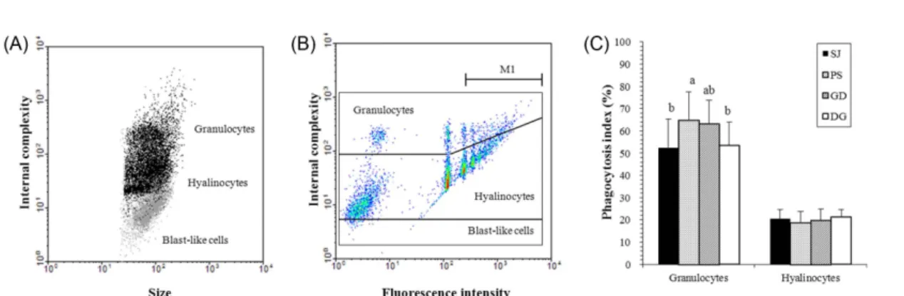
혈구 집단 별 구조적 특징
유세포 분석기를 이용한 참굴 혈구내 세포 형태, 크기 및 내부 밀도에 따라 granulocytes, hyalinocytes, blast-like cells 의 3가지 종류로 구분되었다(Fig. 2A). 이렇게 분류된 혈구 집단내 세포 종류의 구성비는 hyalinocytes의 비율이 평균 67% 이상으로 가장 많았으며, granulocytes가 평균 16% 이며 blast-like cells은 평균 6.4%로 가장 적었다. 지 역 별 혈구의 수를 비교해본 결과 모든 혈구 집단에서 유 의적인 차이가 없었다(Fig. 2B). Granulocytes는 섬진대 교, 평산리, 광도면, 덕곡리에서 측정된 세포수(평균값 ±
Table 1. Shell length (SL) and Condition index (CI) of the wild Crassostrea gigas collected from Seomjin bridge (SJ), Pyeongsan-ri (PS), Gwangdo-myeon (GD), and Deokgok-ri (DG). N, number of sample. SD, standard deviation. Different letters (a-d) in the mean values columns represent significant differences among sampling sites (ANOVA, P < 0.05)
Location N SL (mm) ±SD CI ±SD
SJ 30 46.7
d±5.4 0.289
d±0.114
PS 30 55.8
c±7.4 0.539
b±0.106
GD 30 66.5
b±8.5 0.650
a±0.208
DG 30 80.9
a±9.8 0.381
c±0.085
표준편차)는 각각 306,547 ± 436,639 cells/ml, 154,772 ± 104,760 cells/ml, 166,012 ±88,367 cells/ml, 137,833±21,947 cells/ml였다. Hyalinocytes의 세포수는 섬진대교, 평산리, 광도면, 덕곡리에서 각각 1,141,795 ± 1,610,196 cells/ml, 748,424 ± 581,030 cells/ml, 1,163,151 ± 243,497 cells/ml, 445,166 ±178,929 cells/ml였다. 세포의 크기가 가장 작은 blast-like cells의 세포수는 섬진대교, 평산리, 광도면, 덕곡 리에서 각각 81,816±47,927 cells/ml, 59,397±44,596 cells/ml, 83,915 ±62,146 cells/ml, 49,553±25,925 cells/
ml 였다.
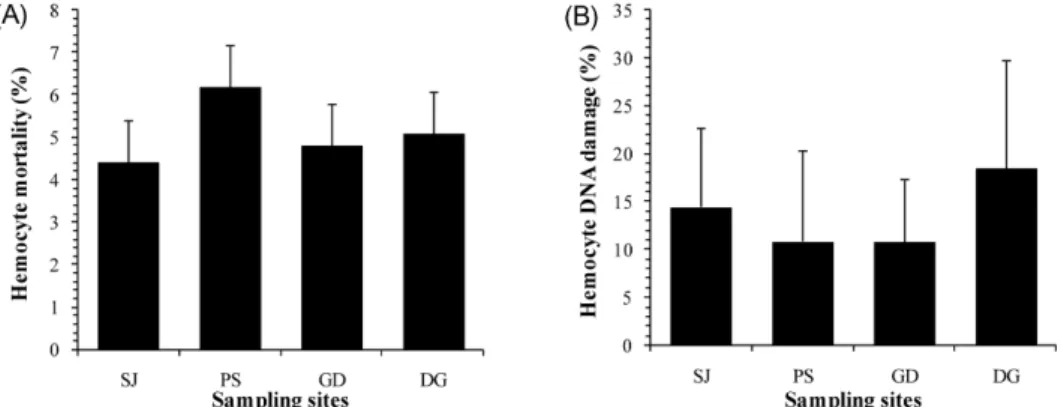
혈구 사멸률과 DNA 손상도
혈구 사멸률과 DNA 손상도는 지역 간의 유의적 차이가 없었다(Fig. 3). 혈구 사멸률(평균값±표준편차)은 섬진대 교, 평산리, 광도면, 덕곡리에서 각각 4.4±1.9%, 6.2±
3.0%, 4.8 ±3.2%, 5.1±1.9%였다. 혈구 DNA 손상도(평균
값±표준편차)는 섬진대교, 평산리, 광도면, 덕곡리에서 각 각 14.5±8.2%, 10.9±9.5%, 10.8±6.6%, 18.5±11.3%였다.
혈구 식세포율
혈구 집단 별 형광 bead를 식작용 할 수 있는지 조사한 결과, granulocytes와 hyalinocytes는 식작용을 하는 반면 blast-like cells은 식작용을 하지 못했다(Fig. 4A, B).
Granulocytes 는 hyalinocytes 보다 평균적으로 2-3배 높은 활발한 식작용 활성을 보였다(Fig. 4C). 지역 간 혈구 식 작용 활성의 차이를 조사한 결과, granulocytes의 식작용 은 평산리의 참굴 혈구가 64.8±12.6%(평균값±표준편차) 로 유의적으로 가장 높았으며(P<0.05), 광도면이 63.2±
10.5%, 덕곡리가 53.4±10.6%, 섬진대교가 52.5±12.7%
로 조사되었다. Hyalinocytes의 식작용 능력은 섬진대교, 평산리, 광도면, 덕곡리에서 20.5±4.1%, 19.0±4.9%, 20.1±
4.7%, 21.2 ±3.4%로 지역 간의 유의적 차이가 없었다.
Fig. 2. Hemocyte type (A) and counts (B) of the wild Crassostrea gigas. Three populations of hemocytes were distinguished: granulocytes, hyalinocytes and blast-like cells. Number of hemocytes populations were not significantly different among sampling sites. SJ, Seomjin bridge; PS, Pyeongsan-ri; GD, Gwangdo-myeon; DG, Deokgok-ri
Fig. 3. Hemocyte mortality (A) and DNA damage (B) of the wild Crassostrea gigas. Mortality and DNA damage of
hemocytes were not significantly different among sampling sites. SJ, Seomjin bridge; PS, Pyeongsan-ri; GD,
Gwangdo-myeon; DG, Deokgok-ri
혈구의 lysosomal membrane stability
혈구의 lysosome membrane이 불안정화 되어 neutral red 가 lysosome membrane으로부터 빠져 나오는 시간을 조사한 결과는 Fig. 5와 같으며, retention time이 짧을수록 혈구의 lysosomal membrane이 상대적으로 불안정 함을 의미한다. 조사결과, 섬진대교의 참굴 혈구의 유지 시간이 53.3±17.2분(평균값±표준편차)으로 가장 짧았으며 광도 면에서 76.0±15.8분으로 가장 길었다(ANOVA, P<0.05).
평산리와 광도면에서는 각각 70.0±15.1분과 76.0±15.8 분으로 조사되었다.
혈구 parameter들 간의 상관관계
이 연구에서 조사한 참굴의 각장, 총 습중량 및 혈구 parameter 들 간의 상관관계를 분석하여 Table 2에 나타냈 다. 참굴의 각장과 총 습중량은 서로 양(+)의 상관관계가 있었으나, 혈구 parameter들과의 상관관계는 보이지 않았 다. 세 종류의 혈구 집단들의 총 수는 각자 서로 양(+) 의 상관관계를 보였으며, 다른 항목들과는 상관관계가 Fig. 4. Flow cytometry determination of the wild Crassostrea gigas hemocytes phagocytosis capacities. (A) Size against
internal complexity dot plot of hemocytes associated (black smear) or not (grey smear) with fluorescent microbeads. (B) Florescence intensity against internal complexity density plot allowed determination of phagocytosis index (M1) for each hemocyte population. (C) Phagocytosis index of C. gigas hemocytes populations. Different letters (a,b) in the mean values columns represent significant differences among sampling sites (ANOVA, P<0.05). SJ, Seomjin bridge; PS, Pyeongsan-ri; GD, Gwangdo-myeon; DG, Deokgok-ri
Fig. 5. Neutral red retention time of the wild Crassostrea gigas. Different letters (a,b) in the mean values columns represent significant differences among sampling sites (ANOVA, P<0.05). SJ, Seomjin bridge; PS, Pyeongsan-ri; GD, Gwangdomyeon;
DG, Deokgok-ri
Table 2. Correlation coefficients among the hemocyte parameters of the Crassostrea gigas collected in this study.
P < 0.05 was regarded as significant correlation. Gr_Count, granulocytes count; Hy_Count, hyalinocytes count;
BL_Count, blast-like cells count; MOT, mortality; DDM, DNA damage; Gr_PHG, granulocytes phagocytosis;
Hy_PHG, hyalinocytes phagocytosis; NRR, neutral red retention time Para-
meters Gr_Count Hy_Count BL_Count MOT DDM Gr_PHG Hy_PHG
Coefficient P Coefficient P Coefficient P Coefficient P Coefficient P Coefficient P Coefficient P Hy_Count 0.895 <.0001 - -
BL_Count 0.638 <.0001 0.701 <.0001 - -
MOT −0.161 0.348 −0.120 0.487 0.251 0.139 - -
DDM −0.213 0.234 −0.267 0.133 -0.018 0.920 0.376 0.031 - -
Gr_PHG −0.171 0.312 −0.005 0.978 0.086 0.611 -0.055 0.748 -0.457 0.007 - -
Hy_PHG −0.010 0.951 0.028 0.870 -0.058 0.732 -0.387 0.020 -0.226 0.198 0.379 0.016 - -
NRR −0.280 0.134 −0.144 0.447 -0.236 0.210 -0.211 0.271 -0.093 0.633 0.456 0.009 -0.103 0.576
없었다. 혈구 사멸률은 DNA 손상도와 양(+)의 관계, hyalinocytes 의 식세포율과는 음(−)의 관계가 관찰되었다.
DNA 손상도는 granulocytes의 식세포율과 음(−)의 상관 관계가 있었다. Granulocytes의 식세포율은 hyalinocytes 의 식세포율과 lysosomal membrane stability에 양(+)의 상관관계가 있었다.
생식소 지수
참굴의 생식소 지수(GI)는 Fig. 6과 같으며 네 지역 간 의 생식소 지수는 유의적인 차이는 없었다. 덕곡리에서는 모든 개체에서 생식세포가 완숙한 시기로 GI는 2.0이였 다. 섬진대교는 완숙기의 25개체와 발달기의 5개체가 관 찰되어 GI는 1.8±0.4(평균값±표준편차)였다. 평산리는 완숙기의 29개체와 발달기의 1개체로 GI는 2.0±0.2였다.
덕곡리는 완숙기의 26개체와 발달기의 4개체로 GI는 1.9 ±0.4였다.
4. 고 찰
유세포 분석기를 이용한 해산 이매패류 혈구 집단의 세포 학적 특성 관찰
유세포 분석기는 유액 상태의 입자나 세포가 일정 감지 지역(sensing point)을 통과할 때 각각의 입자나 세포를 신 속하게 측정하여 한 세포가 갖는 여러 특징(세포의 크기, 세포 내부 밀도, 세포 기능 등)을 동시에 측정할 수 있는 장비이다. 레이저 광원의 직진하는 빛을 통해 세포의 크기 를 산란하는 빛의 정도에 따라 세포의 내부 밀도를 파악
집단을 분류하고, 이들의 면역 관련 기능들을 정량적으로 측정하여 참굴을 비롯한 해산이매패류 혈구의 세포성 면 역기작 측정에 매우 유용한 tool임을 확인하였다. 특히, 이 매패류는 종에 따라 혈구세포내 분포하는 혈구 집단이 다 를 수 있어, 각 종의 고유한 면역학적 특성으로 설명될 수 있다. 해산 이매패류의 혈구 집단의 분류는 연구자에 따라 다소 차이가 있으나, 기본적으로 세포질 내 수많은 과립 을 함유한 과립구(granulocytes)와 세포질 내 과립구가 없는 무과립구(agranulocytes 또는 hyalinocytes)로 나뉜 다(Cheng 1984; Hine 1999). 과립구의 세부 분류는 균질 한(homogeneous) 반면, 무과립구는 종에 따라 다양하게 나뉘어진다. 예를 들면, 대서양굴(C. virginica)과 유럽굴 (Ostrea edulis)에서는 hyalinocytes의 크기에 따라 small hyalinocyte 와 large hyalinocyte로 나뉘었으며(Ashton- Alcox and Ford 1998; Xue et al. 2001; Hégaret et al.
2003), 대서양굴(C. virginica)에서 intermediate cells (Goedken and DeGuise 2004), 참굴(C. gigas)에서 small hyalinocytes (Lambert et al. 2007; Labreuche et al.
2006), 바지락(Tapes philippinarum), Sydney rock oyster (Saccostrea glomerata) 와 강굴(C. ariakensis)에서 blast- like cells 이(Cima et al. 2000; Aladaileh et al. 2007;
Donaghy et al. 2009a) 분류되었다. 이번 연구에서 유세포 분석기를 이용하여 분석된 자연산 참굴 혈구 내 혈구 세포 의 구성은 세포의 크기와 내부 밀도에 따라 granulocytes, hyalinocytes와 blast-like cells의 세 가지 형태로 분류하였 다(Fig. 2A). 분류된 세 종류의 혈구 집단내 구성 비율은 조사 지역 간에 차이는 있었지만 hyalinocytes가 평균 63.1- 75.9% 이상으로 가장 높은 비율을 차지하였고, granulocytes 가 평균 10.8-19.2%, blast-like cells이 5.5-8.2%로 혈구 내 가장 낮은 구성 비율을 나타냈다. 이 결과를 Delaport et al. (2001) 이 조사한 참굴 혈구 집단의 구성 비율 (granulocytes: 14-23%, hyalinocytes: 63-74%)과 비교시 자연산 참굴 혈구의 hyalinocytes의 비율은 비슷한 범위를 나타냈지만, granulocytes의 구성 비율은 다소 낮았다. 이 와 달리, Donaghy et al. (2010)은 2007년 한국의 서해안 Fig. 6. Gonadal Index (GI) of the wild Crassostrea gigas.
0, indifferent stage; 1, developing stage; 2, ripe stage; 3, spawning stage; 4, spent stage. GI were not significantly different among sampling sites.
SJ, Seomjin bridge; PS, Pyeongsan-ri; GD,
Gwangdo-myeon; DG, Deokgok-ri
에 Hebei spirit호 유류 유출 사고 2년이 지난 후, 유출 지 역인 태안 신두리와 대조구 지역인 인천 종현의 양식산 참 굴의 혈구 집단별 구성 비율을 조사하여 hyalinocytes >
blast-like cells > granulocytes의 분포도를 갖고 있다고 설 명하였다. 이러한 혈구내 집단 구성 비율의 차이는 조간대 암반에 부착하여 서식하는 자연산 참굴과 수하식으로 양 식(suspended culture)되는 참굴간의 양식에 따른 성장과 크기의 차이, 자연산 참굴 채집 시기가 여름철인 반면, 양 식산 참굴의 채집 시기는 겨울철로 계절별 영향에 의한 차이일 수도 있으므로 정확한 원인을 파악하기 위해서는 자연산과 양식산 참굴의 계절별 혈구 집단 구성의 변동에 관한 추가적인 연구가 필요하다.
혈림프액 안에 존재하는 혈구의 수를 파악하기 위해서 는 일반적으로 광학현미경 하에서 혈구 계수기(hemo- cytometer) 를 이용하여 세포수를 계수한다. 그러나 세포의 DNA 이중 가닥에 결합하는 형광염색 시약인 SYBR green I 를 처리하면, 유세포 분석기로 시약에 염색된 혈구 세포만을 선택하여 혈림프액 내 혈구의 수를 측정할 수 있다(Choquet et al. 2003). 이 연구에서는 유세포 분석기 를 이용하여 총 혈구 수를 측정하고 혈구 집단의 구성 비 율에 따라 계산한 결과, 참굴의 혈림프액내 실제로 존재하 는 혈구 집단 별 수는 지역 간에 유의적 차이가 없었다 (Fig. 2B). 세 혈구 집단의 수를 합한 총 혈구 수는 Donaghy et al. (2010) 이 2009년 1월에 태안과 인천에서 채집한 양 식산 참굴의 총 혈구 수(태안: 평균 345,233 cells/ml, 인천:
평균 401,201 cells/ml)보다 적게는 2배에서 많게는 4배 가량 많았다. 바지락(R. philippinarum)의 경우에는 총 혈 구 수가 봄과 여름철에는 증가하다가 가을과 겨울철이 되 면 감소하는 경향을 보였다(Soudant et al. 2004; Flye- Saint-Marie et al. 2009). Donaghy et al. (2009b) 는 가을 과 겨울철에 바지락의 총 혈구 수가 줄어드는 이유를 두 가지의 가설을 이용하여 설명하였다. 첫 번째는, 가을과 겨울철에 먹이 부족으로 인하여 세포 분열에 필요한 충분 한 에너지가 제공되지 못하게 됨으로써, 새로운 혈구의 생 산이 줄어들게 되어 혈구의 수가 감소한다는 것이다. 두 번째는, 저수온과 먹이부족으로 인하여 혈구가 영양분의 운반과 소화와 같은 영양학적 활동(nutritional activities) 이 감소하게 됨으로써 혈구의 수가 감소한다는 것이다. 그 러므로 겨울철인 1월에 채집을 한 Donaghy et al. (2010) 연구에서의 참굴 혈구 수는 여름철인 6월에 채집을 한 이 번 연구의 참굴 혈구 수보다 적은 것으로 사료된다. 또한 해산 이매패류의 혈구의 수는 오염 물질의 유입에 따라 증가하거나 감소한다. 구리(Cu)에 노출된 M. edulis(Pipe and Coles 1995; Pipe et al. 1999) 와 C. virginica(Fisher et al. 2000), 카드뮴(Cd)에 노출된 M. edulis(Coles et al.
1995) 에서 총 혈구 수가 증가한 반면, PCBs에 노출된
Chlamys farreri(Liu et al. 2009), 망간(Mn)에 노출된 M.
edulis(Oweson and Hernroth 2009)에서는 총 혈구 수가 감소하였다. 또한, 비오염 지역에 서식하던 M. edulis를 오 염지역으로 이식하였을 때에도 총 혈구 수가 감소한다고 보고하였다(Mayrand et al. 2005). 이러한 연구 결과들과 같이 환경오염에 대한 혈구의 수가 서로 다르게 변동하는 것을 이해하기 위해서는 혈구의 증식과 이동에 대한 구체 적인 연구가 필요하다.
환경스트레스에 의한 참굴 혈구의 면역학적 특성 변화 다양한 genotoxic chemical 등에 의하여 핵내 DNA 사 슬의 염기가 탈락하는 손상이 발생한다(Lee and Steinert 2003). 이러한 DNA가 손상되는 정도를 측정하는 방법으 로는 comet assay와 유세포 분석기가 일반적으로 사용되 고 있다. Comet assay인 경우는 민감도가 높긴 하지만 유 세포 분석기를 이용한 방법에 비해 시간과 비용이 많이 든다. 반면, 단일 세포의 핵내 DNA 이중 가닥에 부착할 수 있는 형광염색소 propidium iodide로 염색하여, 염색된 세포를 유세포 분석기로 측정하면 단시간에 세포내 핵산 의 양과 DNA 손상도를 측정할 수 있다(Darzynkiewicz et al. 2004; da Silva et al. 2005; Grigoryan et al. 2007).
Park et al. (2009) 은 카드뮴에 노출시킨 바지락의 DNA 손상도를 유세포 분석기를 이용하여 측정한 결과, 대조구 에 비해 노출된 바지락 혈구의 DNA 손상도가 증가함을 확인하였다. 이 연구에서는 참굴 혈구의 DNA 손상도가 지역 간의 유의적인 차이는 없었지만, 광양만의 내만인 섬 진대교, 진해만의 외만인 덕곡리가 평균적으로 약 1.5배 높은 것으로 측정되었다. 그러므로 섬진대교와 덕곡리 지 역의 genotoxicity가 평산리와 광도면보다 높은 것으로 판 단된다. 혈구 DNA의 손상이 유발되면 apoptosis나 necrosis 로 발전하게 되고 결국 혈구가 사망하게 된다(Lee and Steinert 2003). 이 연구에서도 참굴 혈구내 면역 인자들의 상관관계 비교시, 혈구의 사멸률과 DNA 손상도가 양(+) 의 상관관계를 보여(Table 2), 오염 물질을 포함한 해양 환경 변화에 따른 참굴 혈구내 두 면역 인자들의 관계가 면역 기능에 중요한 요소임을 알려주고 있다.
이 연구에서는 세 종류의 혈구 집단 중 granulocytes와 hyalinocytes만이 식세포 작용을 하는 것으로 확인되었다.
그 중에서도 granulocytes가 hyalinocytes에 비해 약 3배
정도 활발한 식세포 작용을 하는 것으로 조사되었다. 이러
한 결과는 Mercenaria mercenaria(Tripp 1992), R. decussatus
(López et al. 1997), C. virginica(Goedken and De Guise
2004), Bathymodiolus azoricus(Bettencourt et al. 2009),
C. ariakensis(Donaghy et al. 2009a) 에서 조사된 이매패류
들의 혈구세포 식작용 결과와 일치한다. 네 지역 간의 혈
구의 식세포율은 hyalinocytes의 경우에는 유의적 차이가
리소좀(lysosome)은 세포질 내에 존재하며 다양한 가수 분해 효소를 함유하고 있어 불필요한 물질들을 소화하고 분해하는 기관이다(Luzio et al. 2007). 해양 생물에서 lysosome 은 해독(detoxification)과 방어(defense) 작용에 중요한 역할을 한다(Moore 1980; Lowe et al. 1995a, 1995b). 이러한 기능들은 lysosome membrane stability에 따라 달라지므로, 세포막의 안정도를 측정하여 lysosome 의 기능을 평가할 수 있다(Zhao et al. 2011). Neutral red 는 친유기성(lipophilic) 화학물질로 세포의 세포막에 쉽게 침투할 수 있다(Lowe et al. 1992). Neutral red가 lysosome 내에 보존(retention)될 수 있는 효율은 lysosome 내의 pH 에 의해 좌우되며, lysosome을 산성 상태로 유지시켜 주 는 양성자 펌프(proton pump)에 의해 보존이 유지된다 (Zhao et al. 2011). 그러므로 스트레스를 받지 않은 세포 들은 오랜 시간 동안 neutral red를 가지고 있게 되지만, lysosome의 세포막이나 H
+이온 펌프가 불안정화되면 neutral red가 유출되게 된다. 이러한 원리를 이용하여 lysosome 이 neutral red를 보유하고 있는 시간을 측정하는 neutral red retention(NRR) assay는 굴과 담치류에 다양한 스트레스가 미치는 영향을 조사하는 바이오 마커로 사용 되고 있다(Lowe et al. 1995a, 1995b; Hauton et al. 1998, 2001; Cho and Jeoung 2005; Zhang and Li 2006; Guidi et al. 2010). 이 연구에서는 네 지역간의 NRR 시간이 유 의적인 차이가 있었으며, lysosomal membrane stability는 광도면의 참굴이 가장 높고, 섬진대교의 참굴이 가장 낮은 것으로 조사되었다. 이러한 결과는 혈구 식세포율 조사 결 과와 유사한 경향을 보였다.
참굴의 건강도, 생식소 발달 단계에 따른 혈구 면역 parameter 들과의 상관관계
Flye-Saint-Marie et al. (2009) 는 바지락(R. philiinarum) 의 크기는 식세포율과 혈구 사멸률에 음(−)의 상관 관계가 있다고 설명하였다. 이 연구에서는 채집 지역에 서식하는 참굴들 중 크기가 큰 개체들만 채집을 하였지만, 네 지역 간 참굴의 각장은 유의적인 차이가 있었다(Table 1). 그러
도와 혈구 면역 기능들은 참굴의 생리상태를 평가하는데 중요하게 이용될 수 있을 것이다.
참굴을 비롯한 해산 이매패류는 생식세포가 발달한 후 산란을 하는 번식주기를 가진다. 특히, 산란활동은 상당한 에너지가 소요되는 활동으로(Gosling 2003), 산란 이후의 참굴은 혈구 식세포율, 항균력(antibacterial activity), lysosomal membrane stability 가 감소한다(Cho and Jeong 2005; Li et al. 2010). 그러나, 이 연구에서 관찰된 참굴의 생식소 발달 단계는 대부분의 개체가 산란 전의 완숙기 상태로 일부 개체에서 생식세포 발달이 관찰되었지만, 생 식소 지수는 지역간에 유의적 차이가 없었다. Gagnaire et al. (2006)의 연구에서는 생식세포를 형성하는 이배체 (diploid) 참굴의 식세포능이 생식능력이 없는 삼배체 (triploid) 참굴보다 낮았으며, 식세포능의 차이는 완숙기때 가장 컸음을 확인하였다. 또한, 이배체 굴은 생식세포를 형성하고 발달시키는 활동에 에너지를 소모하기 때문에 식세포 작용과 같은 혈구 기능에 필요한 에너지가 삼배체 굴에 비해 상대적으로 낮기 때문이라고 설명하였다. 따라 서 참굴의 생식소 발달과 산란 활동이 참굴 혈구의 기능 에 영향을 미칠 수 있으므로, 참굴의 혈구 기능 측정 시에 는 개체별 번식상태를 고려해야 할 것이다.
5. 요 약
남해의 광양만과 진해만에 서식하는 자연산 참굴의 건
강성 평가를 위해 세포성 면역을 담당하는 혈구의 기능들
을 유세포 분석기와 Neutral Red Retention(NRR) assay를
이용하여 신속, 정확하게 측정하였다. 광양만과 진해만의
안쪽과 바깥쪽에 서식하는 자연산 참굴의 혈구를 유세포
분석기를 이용하여 형태학적 특성에 따라 혈구의 종류를
분류하고, 혈구 종류별 수, 사멸률, DNA 손상도, 식세포
능을 측정하였다. 또한 NRR assay를 이용하여 혈구의
lysosomal membrane stability 를 측정하였다. 참굴의 혈구
는 granulocytes, hyalinocytes, blast-like cells의 세 가지
종류로 분류되었다. 조사 지역 간의 혈구의 수, 사멸률,
DNA 손상도는 유의적 차이가 없었지만, 식세포율과 lysosomal membrane stability 와 같은 면역 관련 기능들은 유의적 차이가 있었다. 진해만은 내만과 외만 지역에 서식 하는 참굴의 혈구 면역인자들 간에는 유의적 차이가 관찰 되지 않았다. 이에 반해, 광양만에 서식하는 참굴은 내만 지역의 섬진대교가 외만 지역의 평산리 지역에 서식하는 참굴보다 낮은 식세포율과 낮은 lysosomal membrane stability 를 보여 면역력이 저하되어 있는 것으로 추정된다.
하지만, 해양환경 변화와 시료의 면역력과의 상관관계를 이해하기 위해서는 조사지역의 환경적 특성이나 오염정 도, 그리고 시료 내의 오염물질 축적량 등의 객관적인 분 석 결과와의 종합적인 고찰이 필요할 것이다. 유세포 분석 기와 NRR assay를 이용한 참굴 혈구 집단의 형태 변화 및 면역능 측정 기술은 시료의 전처리 없이 빠른 시간 내 에 세포의 특성을 분석할 수 있는 유용한 분석 tool로 활 용될 수 있음을 확인하였다.
사 사
이 연구는 한국해양연구원의 남해 특별관리해역 생태계 건강지수 개발(과제번호 PE98582: Development of Marine Ecosystem Health Index in the special management areas of the South Sea) 의 지원에 의하여 수행되었으며, 지원에 깊은 감사를 드립니다.
참고문헌