ISSN 0378-6471 (Print)⋅ISSN 2092-9374 (Online)
http://dx.doi.org/10.3341/jkos.2016.57.4.614
Original Article
반시야 결손 녹내장안의 중증도에 따른 정상 반시야 신경섬유층 및 신경절세포층 두께 비교
RNFL and Ganglion Cell Complex Thickness in Normal Hemifield According to the Severity of Glaucoma
홍은희⋅신용운⋅임한웅⋅강민호⋅조희윤⋅성민철
Eun Hee Hong, MD, Yong Un Shin, MD, PhD, Han Woong Lim, MD, PhD, Min Ho Kang, MD, PhD, Hee Yoon Cho, MD, PhD, Mincheol Seong, MD, PhD
한양대학교 의과대학 한양대학교구리병원 안과학교실
Department of Ophthalmology, Hanyang University Guri Hospital, Hanyang University College of Medicine, Guri, Korea
Purpose: To analyze the thickness of the circumpapillary retinal nerve fiber layer (cRNFL) and macular ganglion cell complex (mGCC) in apparently normal hemifield areas of glaucomatous eyes with superior or inferior visual hemifield defects according to their severity compared with the same hemifield of normal eyes using Topcon 3D spectral-domain optical coherence tomog- raphy (SD-OCT).
Methods: The present study included 90 normal eyes and 90 glaucomatous eyes with superior or inferior visual hemifield defects that underwent cRNFL and mGCC imaging using 3D SD-OCT. The cRNFL and mGCC parameters were compared between normal hemifield in glaucomatous eyes and the same hemifield in normal eyes. The mean deviation (MD) parameters (Mild: MD
> -6 dB, 54 eyes; Moderate: -6 dB ≥ MD ≥ -12 dB, 60 eyes; Severe: MD < -12 dB, 30 eyes) in glaucomatous eyes were also com- pared between the 3 severity groups.
Results: The average hemifield cRNFL thickness was 93.6 ± 24.2 μm and 118.1 ± 14.1 μm in superior normal hemifield of glau- comatous eyes and controls, respectively, and 107.8 ± 19.1 μm and 124.9 ± 17.1 μm in inferior normal hemifield of glaucomatous eyes and controls, respectively. mGCC thickness was 95.8 ± 5.9 μm and 103.5 ± 7.7 μm in superior normal hemifield of glaucom- atous eyes and controls, respectively, and 93.4 ± 8.2 μm and 104.5 ± 8.2 μm in inferior normal hemifield of glaucomatous eyes and controls, respectively (all p < 0.05). The thickness parameters were decreased in normal hemifield of glaucomatous eyes, which significantly decreased according to the severity (MD) of visual field defect (all p < 0.01).
Conclusions: The measurement of cRNFL and mGCC thickness in normal hemifield of glaucomatous eyes using SD-OCT is useful in detecting structural glaucomatous changes before visual field defects appear.
J Korean Ophthalmol Soc 2016;57(4):614-622
Keywords: Circumpapillary retinal nerve fiber layer (cRNFL), Glaucoma, Hemifield defect, Macular ganglion cell complex (mGCC), Spectral-domain optical coherence tomography (SD-OCT)
■Received: 2015. 12. 10. ■ Revised: 2016. 2. 24.
■Accepted: 2016. 3. 21.
■Address reprint requests to Mincheol Seong, MD, PhD Department of Ophthalmology, Hanyang University Guri Hospital, #153 Gyeongchun-ro, Guri 11923, Korea Tel: 82-31-560-2354, Fax: 82-31-564-9479 E-mail: [email protected]
* This study was presented as a poster at the 114th Annual Meeting of the Korean Ophthalmological Society 2015.
ⓒ2016 The Korean Ophthalmological Society
This is an Open Access article distributed under the terms of the Creative Commons Attribution Non-Commercial License (http://creativecommons.org/licenses/by-nc/3.0/) which permits unrestricted non-commercial use, distribution, and reproduction in any medium, provided the original work is properly cited.
녹내장은 망막신경섬유층두께의 감소와 망막신경절세포 의 감소를 특징으로 한다.1,2 이에 따른 시야결손의 양상은 망막 중심부에서의 망막시신경섬유 축삭의 분포 차이 때문 에 상측과 하측 반시야 사이에 비대칭적으로 나타날 수 있 는데,3 시야 결손은 주로 일측 반시야에서 먼저 나타나는 것으로 알려져 있다.4,5 상측 혹은 하측 반시야 결손이 있는 녹내장 환자에서 정상 반시야에 상응하는 사분면의 망막신
경섬유층 두께를 정상안과 비교한 Badlani et al6의 연구에 서는 정상 반시야에서의 두께가 정상안에 비해 감소되어 있으며 그 감소의 정도가 시야 결손의 중등도와 유의한 관 계가 있음을 밝힘으로써 녹내장성 시야결손이 나타나기 전 에 망막신경섬유층이 얇아지는 구조적 변화가 나타남을 보 인 바 있다. 또한 Inuzuka et al7의 연구 등에서 반시야 결손 을 나타내는 녹내장 환자군에서 결손측 반시야와 정상측 반시야 모두 그에 상응하는 황반부 두께가 감소되어 있으 며, 따라서 시야결손이 나타나기 전인 정상 반시야 구역에 서 구조적 변화가 선행되고, 이러한 구조적 변화들이 시야 결손이 나타나기 전 녹내장 진단의 지표로서 유용하게 사 용될 수 있을 것이라 제안되었다. 최근 optical coherence tomography (OCT) 기술의 발달과 함께 더 정밀한 검사가 가능해지고 있는데, Spectral-domain OCT (SD-OCT)에서 는 스캔속도가 더 빨라(초당 50,000 A-scan) 눈의 움직임에 따른 오류가 더 적고 해상도가 더 높다. SD-OCT를 사용하 게 되면서 황반부 전체의 두께가 아닌 황반부신경절세포복 합층(macular ganglion cell complex, mGCC)의 두께를 측 정하는 것이 가능해졌고, 이를 이용해 시야결손이 반시야 에 국한된 녹내장안에서 정상 반시야 구역의 mGCC 두께 의 감소가 있다는 것을 밝힌 연구들이 있었다.8-11
이 연구들8-11에서는 또한 정상 반시야 구역의 시신경유 두주위망막신경섬유층(circumpapillary retinal nerve fiber layer, cRNFL)이나 mGCC의 두께 감소가 시야 결손의 중 등도와 상관 관계를 보이는지에 대해서도 살펴보았는데, 대상이 되는 녹내장 환자군의 시야 결손 중등도에 따른 분 포를 균등하게 맞추어 시행한 연구는 아직까지 없었다.
한편, mGCC를 구성하는 세 층인 황반부신경섬유층(nerve fiber layer, NFL), 황반부신경절세포층(ganglion cell layer, GCL), 그리고 황반부내망상층(inner plexiform layer, IPL) 은 각각 망막신경절세포의 축삭(axon), 세포체(cell body), 수상돌기(dendrite)에 해당된다. SD-OCT는 내재된 소프트 웨어로 mGCC의 두께를 이 세 층으로 나누어 측정하여, 이 를 이용해 황반부 내측망막의 두께를 더 세분화하여 측정 하는 것이 가능하게 되었다. 그러나 녹내장성 반시야 손상 에서 정상 반시야 망막의 mGCC 두께 감소에 대해 이와 같 이 층을 세분화하여 비교한 연구는 아직까지 발표된 바 없 었다.
본 연구에서는 이 3D SD-OCT의 알고리즘에 따라 측정 한 cRNFL과 mGCC (NFL, GCL+IPL, NFL+GCL+IPL의 각 층) 두께 측정치를 각각 정상군, 녹내장안의 정상 반시 야 및 결손 반시야 간에 비교하고, 녹내장안의 정상 반시야 구역에서 시야결손의 중증도에 따라 그 감소 정도가 유의 한 차이를 보이는지를 비교 분석해 보고자 하였다.
대상과 방법
대상 환자군
2011년 1월 1일부터 2015년 4월 30일까지 내원한 환자 들 중 정상 90안과 반시야에 국한된 국소적 시야결손을 보 이는 정상안압 녹내장 90안을 대상으로 하여 후향적으로 의무기록을 분석하였다. 대상 환자군은 (1) 국소적이거나 전반적인 시신경 테의 얇음 또는 패임이 있거나 양안의 수 직유두함몰비 차이가 0.2 이상, 망막신경섬유층결손, 시신 경유두출혈이 있는 경우 등 녹내장성 시신경손상을 보이고, (2) 최소 1개월 간격으로 시행한 2차례 이상의 연속한 시야 검사상 녹내장성 반시야 손상을 나타낸 군으로 정의하였다.
정상 대조군은 (1) 정상 시신경유두 소견을 보이면서 (2) 시야검사상 위에 기술한 녹내장성 시야결손이 없고, (3) 안 압은 21 mmHg 이하이며 (4) 안구 내 pathology가 없고 (5) 안압 증가의 과거력이나 외상력이 없는 경우로 정의하였다.
당뇨망막병증 등 망막질환, 포도막염, 시야나 시신경에 영 향을 줄 만한 내과 또는 신경과 질환이 있는 환자 및 안구 내 수술력, 레이저 치료력 등의 과거력이나 두개강 내 이상 소견이 있는 경우, 그리고 구면렌즈 대응치가 ±6.0디옵터 이상인 경우는 제외하였다.
녹내장성 반시야 결손은 험프리자동시야계(Humphrey field analyzer, HFA)의 Swedish interactive threshold algo- rithm (SITA)의 central 30-2 검사에서 상측 혹은 하측 반시 야에서 다음 세 가지 기준 중 두 가지 이상을 만족하는 경 우 [(1) Pattern deviation plot의 가장자리를 제외한 부위에 서 인접한 3개 이상 점의 역치가 정상의 5% 미만으로 나타 나고 그중 한 개 이상은 1% 미만이거나, (2) Pattern stand- ard deviation (PSD)이 5% 미만이거나, (3) Glaucoma hemi- field test (GHT) 상 outside normal limits], 그리고 반대측 반시야에서 pattern deviation plot 상 세 개 이상의 연속된 지점에서 역치가 5% 미만인 점이 존재하지 않으면서 두 개 이상의 연속된 지점에서 역치가 1% 미만인 점이 존재하지 않는 경우로 정의하였다. 주시상실 20% 이상 또는 위양성 률과 위음성률 15% 이상의 신뢰도가 떨어지는 경우는 대 상에서 제외하였다. 녹내장안에 대해서는 시야검사의 Mean deviation (MD) 값을 이용하여 시야결손의 중등도에 따라 초기(MD > -6 dB), 중기(-6 dB ≥ MD ≥ -12 dB), 말기(MD
< -12 dB)의 세 군으로 구분하였다.
검사
모든 대상자에서 병력조사, 시력측정, 골드만안압측정, 세극등현미경을 이용한 전안부검사, 앞방각경검사, 안저검 사, 검안경을 이용한 시신경유두검사, 초음파를 이용한 중
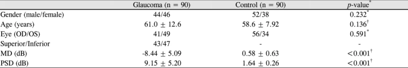
Table 1. Descriptive statistics for demographics and clinical characteristics of the study participants
Glaucoma (n = 90) Control (n = 90) p-value*
Gender (male/female) 44/46 52/38 0.232*
Age (years) 61.0 ± 12.6 58.6 ± 7.92 0.136†
Eye (OD/OS) 41/49 56/34 0.591*
Superior/Inferior 43/47 - -
MD (dB) -8.44 ± 5.09 0.58 ± 0.63 <0.001†
PSD (dB) 9.15 ± 5.20 1.64 ± 0.26 <0.001†
Values are presented as mean ± SD unless otherwise indicated.
MD = mean deviation; PSD = pattern standard deviation.
*Chi-square test; †Independent t-test.
심각막두께측정(Sp-300, Tomey Co., Nagoya, Japan), Humprey field analyzer (model 750, Carl Zeiss Meditec, Dublin, CA, USA)의 central 30-2 SITA-Standard strategy를 이용한 시 야검사, 망막신경섬유층 및 시신경유두사진촬영(KOWA VX-10, Kowa Company Ltd., Tokyo, Japan), 빛간섭단층촬영(3D SD-OCT, software version 7.11, Topcon Corporation, Tokyo, Japan)을 시행하였다.
3D SD-OCT & protocol
본 연구에서는 SD-OCT 중 3D SD-OCT (Topcon Corporation) 를 사용하였고, 3D disc protocol과 3D macular vertical scan protocol을 이용하여 측정한 parameter를 분석에 이용하였 다. 3D SD-OCT는 광원이 840 nm 파장이며, 축해상도는 5 μm 이다. 3D disc protocol로 시신경유두를 중심으로 가로 세로 6 mm 사각형 부위를 512A×128B (각각 512개의 A-scan으 로 구성된 128개의 vertical B-scan) 스캔을 하였다. 내장된 소프트웨어(The built-in analysis software; version 7.11)가 시신경유두 중심을 결정하고 이곳을 중심으로 한 직경 3.4 mm 원의 망막신경섬유층(cRNFL) 두께와 시신경유두 측정치들 이 자동으로 측정되었고, 이 중 cRNFL의 상측, 하측, 평균 두께를 분석에 사용하였다. 또한 3D macular vertical scan 으로 황반부 가로 세로 7 mm 사각형 부위를 512A×128B 스캔하여 황반부내망막두께(mGCC)가 측정되었고, 3D SD-OCT에서는 mGCC 두께 측정 시 이를 NFL, GCL+IPL, NFL+GCL+IPL의 세 가지 측정치로 나누어 측정하는 내장 알고리즘이 사용되고 있어 황반부 6×6 mm2에서 구한 NFL, GCL+IPL, NFL+GCL+IPL (=mGCC) 각각의 상측, 하측, 평균 두께를 분석에 사용하였다. Quality factor 값이 60 미만이거나 중심이 이탈되어 있는 경우는 대상에서 제 외하였다.
통계
통계학적 분석은 SPSS (version 18.0, SPSS Inc., Chicago, IL, USA)를 사용하였다. 녹내장안에서 정상 반시야의 cRNFL
및 mGCC의 두께와 결손측 반시야의 cRNFL 및 mGCC의 두께를 각각 상, 하측 반시야로 나누어 비교하였고(paired t-test), 녹내장안 정상 반시야의 cRNFL 및 mGCC의 두께를 역시 각각 상, 하측 반시야로 나누어 정상안 동측 반시야의 cRNFL 및 mGCC의 두께와 비교하였다(independent t-test).
녹내장안에서 시야결손의 중등도와 cRNFL 및 mGCC의 두께 간의 관계를 분석하기 위해 녹내장안의 정상 반시야 및 결손측 반시야 각각에서 cRNFL 및 mGCC의 두께와 MD, PSD와의 상관 관계를 선형회귀분석을 이용해 분석하 였고, 녹내장안을 MD 값에 따라 분류한 세 군 간 cRNFL 및 mGCC의 두께를 비교하였다(One-way analysis of var- iance [ANOVA]). 이 녹내장안 세 군 각각에서 정상군과 cRNFL 및 mGCC의 두께에 유의한 차이가 있는지를 보기 위 해 녹내장안을 상측 결손과 하측 결손군으로 구분하여 각 정 상 반시야를 정상안 동측 반시야와 비교하였다(independent t-test). p-value 값이 0.05 미만인 경우를 통계적으로 유의한 차이가 있는 것으로 판정하였다.
결 과
정상군 100명 100안과 반시야에 국한된 국소적 시야결손 을 보인 녹내장군 100명 100안 중 image quality가 떨어진 20 안이 제외되어, 최종적으로 총 180명 180안(정상안 90명 90 안과 녹내장안 90명 90안)이 본 연구에 포함되었다. 정상군 의 연령은 평균 58.6 ± 7.92세, 녹내장군의 연령은 평균 61.0
± 12.6세로 통계적으로 유의한 차이가 없었고(p=0.136, in- dependent t-test), 성별 분포에 있어서도 통계적으로 유의한 차이는 없었다(p=0.232, chi-square test). 녹내장군의 43명 은 상측 반시야, 47명은 하측 반시야에 결손이 있었다. MD 는 정상군에서 평균 0.58 ± 0.63 dB, 녹내장군에서 -8.44 ± 5.09 dB, PSD는 정상군에서 평균 1.64 ± 0.26 dB, 녹내장 군에서 평균 9.15 ± 5.20dB로 유의한 차이가 있었다(p<0.001, independent t-test; Table 1). 정상안과 녹내장안에서 cRNFL 및 세분화한 각각의 mGCC 지표를 비교하였고, 모두 녹내
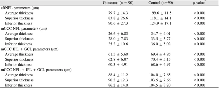
Table 2. Spectrum-domain optical coherence tomography data of the cRNFL thickness and macular ganglion cell complex (mGCC;
NFL, IPL+GCL, NFL+IPL+GCL) thickness parameters in glaucomatous and control eyes
Glaucoma (n = 90) Control (n=90) p-value* cRNFL parameters (μm)
Average thickness 79.7 ± 14.3 99.6 ± 11.5 <0.001
Superior thickness 83.8 ± 26.6 118.1 ± 14.1 <0.001
Inferior thickness 90.6 ± 27.3 124.9 ± 17.1 <0.001
mGCC NFL parameters (μm)
Average thickness 26.6 ± 6.83 34.7 ± 4.01 <0.001
Superior thickness 28.0 ± 7.83 33.5 ± 3.77 <0.001
Inferior thickness 25.2 ± 10.6 36.0 ± 5.02 <0.001
mGCC IPL + GCL parameters (μm)
Average thickness 61.5 ± 5.60 69.4 ± 4.95 <0.001
Superior thickness 62.8 ± 6.07 70.4 ± 5.15 <0.001
Inferior thickness 60.3 ± 4.91 68.6 ± 4.97 <0.001
mGCC NFL + IPL + GCL parameters (μm)
Average thickness 88.4 ± 11.2 104.0 ± 7.65 <0.001
Superior thickness 90.2 ± 12.3 103.5 ± 7.66 <0.001
Inferior thickness 86.2 ± 14.0 104.5 ± 8.20 <0.001
Values are presented as mean ± SD unless otherwise indicated.
cRNFL = circumpapillary retinal nerve fiber layer; mGCC = macular ganglion cell complex; NFL = nerve fiber layer; IPL = inner plexi- form layer; GCL = ganglion cell layer.
*Independent t-test.
Table 3. Comparison of cRNFL thickness and mGCC (NFL, IPL + GCL, NFL + IPL + GCL) thickness parameters in superior
and inferior hemiretina with hemifield defect and normal field and control eyesHemiretina with a hemifield defect
Hemiretina with normal field
Hemiretina of
control eye p-value cRNFL parameters (μm)
Superior 74.1 ± 27.3 93.6 ± 24.2 (n = 43) 118.1 ± 14.1 <0.001*, <0.001† Inferior 74.7 ± 25.7 107.8 ± 19.1 (n = 47) 124.9 ± 17.1 <0.001*, <0.001† mGCC NFL parameters (μm)
Superior 19.1 ± 11.5 31.7 ± 5.56 (n = 43) 33.5 ± 3.77 <0.001*, 0.024†
Inferior 24.7 ± 8.17 30.8 ± 5.53 (n = 47) 36.0 ± 5.02 <0.001*, <0.001†
mGCC IPL + GCL parameters (μm)
Superior 58.3 ± 5.19 63.9 ± 3.51 (n = 43) 70.4 ± 5.15 <0.001*, <0.001†
Inferior 61.8 ± 7.61 62.3 ± 3.78 (n = 47) 68.6 ± 4.97 <0.001*, <0.001†
mGCC NFL + IPL + GCL parameters (μm)
Superior 78.4 ± 14.8 95.8 ± 5.90 (n = 43) 103.5 ± 7.66 <0.001*, <0.001† Inferior 85.0 ± 14.4 93.4 ± 8.20 (n = 47) 104.5 ± 8.20 <0.001*, <0.001† Values are presented as mean ± SD unless otherwise indicated.
cRNFL = circumpapillary retinal nerve fiber layer; mGCC = macular ganglion cell complex; NFL = nerve fiber layer; IPL = inner plexi- form layer; GCL = ganglion cell layer.
*Hemiretina with a hemifield defect vs. Hemiretina with normal field; paired t-test; †Hemiretina with normal field vs Control; independent t-test.
장안에서 유의한 감소를 보였다(Table 2).
cRNFL과 각각의 mGCC 측정치들을 녹내장안 반시야 결손의 위치가 상측 반시야일 때와 하측 반시야일 때로 나 누어서 비교하였다(Table 3). 상측 반시야에 결손이 있을 때와 하측 반시야에 결손이 있을 때 모두 cRNFL 및 세분 화한 mGCC 각 층의 두께는 녹내장안 정상 반시야에서 결손측 반시야에 비해서는 덜 감소되어 있으나(all variables p<0.05, paired t-test) 정상안 동측 반시야에 비해서는 더 감소되어 있는
결과를 보였다(all variables p<0.05, independent t-test).
녹내장안의 정상 반시야 및 결손측 반시야에서 모두 cRNFL 및 mGCC 각 층의 두께 측정치들과 MD 값과는 통 계적으로 유의한 상관관계가 있었고(all variables p<0.05, linear regression), 이는 결손측 반시야에서 더 높은 연관성 을 보였다. 반면 PSD 값은 녹내장안 정상 반시야에서는 cRNFL 두께와만, 결손측 반시야에서는 cRNFL 두께와 mGCC NFL 및 mGCC NFL+GCL+IPL 두께와 유의한 상
Table 4. Correlation between cRNFL and mGCC (NFL, IPL + GCL, NFL + IPL + GCL) thickness parameters and glaucoma se-
verity values in hemiretina with hemiefield defect and normal field, respectivelyMD PSD
R2 p-value* R2 p-value*
cRNFL parameters (μm)
Defected hemifield 0.451 <0.001 0.354 <0.001
Normal hemifield 0.227 <0.001 0.183 <0.001
mGCC NFL parameters (μm)
Defected hemifield 0.430 <0.001 0.421 <0.001
Normal hemifield 0.085 0.005 0.039 0.064
mGCC IPL + GCL parameters (μm)
Defected hemifield 0.100 0.002 0.041 0.055
Normal hemifield 0.119 0.001 0.031 0.096
mGCC NFL + IPL + GCL parameters (μm)
Defected hemifield 0.357 <0.001 0.277 <0.001
Normal hemifield 0.091 0.004 0.030 0.103
cRNFL = circumpapillary retinal nerve fiber layer; mGCC = macular ganglion cell complex; NFL = nerve fiber layer; IPL = inner plexi- form layer; GCL = ganglion cell layer; MD = mean deviation; PSD = pattern standard deviation.
*Linear regression.
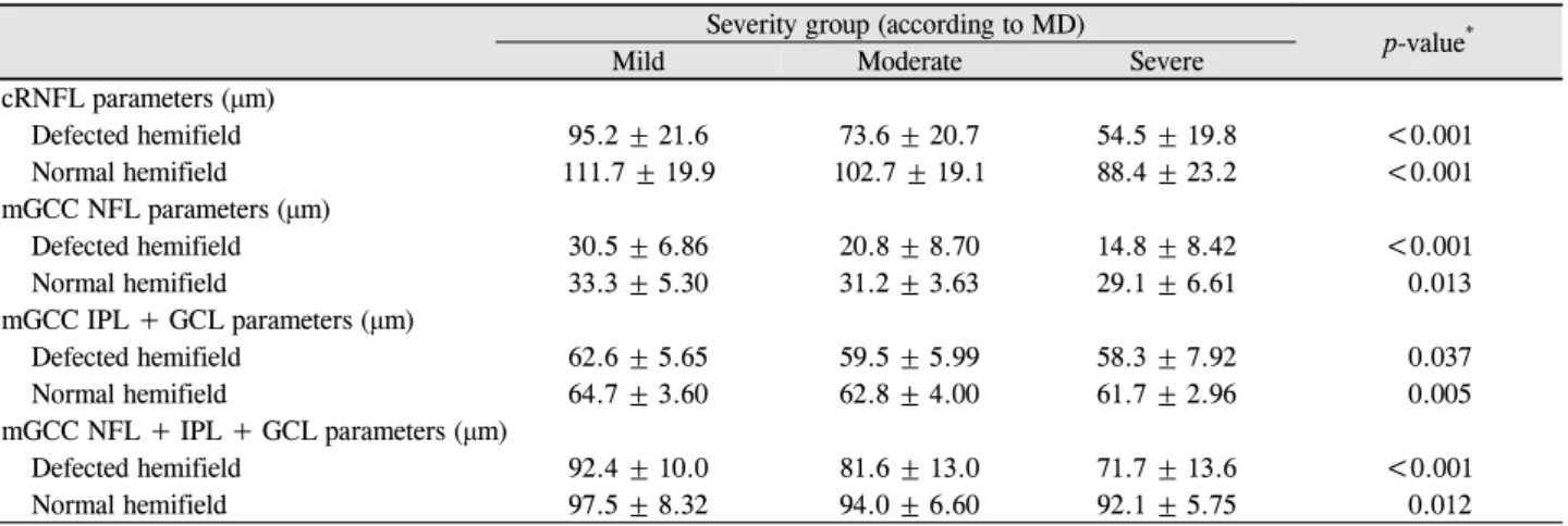
Table 5. Comparison of cRNFL and mGCC (NFL, IPL + GCL, NFL + IPL + GCL) thickness parameters between glaucoma se-
verity groups (mild, moderate and severe groups) according to MD value in hemiretina with hemifield defect and normal field, re- spectivelySeverity group (according to MD)
p-value*
Mild Moderate Severe
cRNFL parameters (μm)
Defected hemifield 95.2 ± 21.6 73.6 ± 20.7 54.5 ± 19.8 <0.001
Normal hemifield 111.7 ± 19.9 102.7 ± 19.1 88.4 ± 23.2 <0.001
mGCC NFL parameters (μm)
Defected hemifield 30.5 ± 6.86 20.8 ± 8.70 14.8 ± 8.42 <0.001
Normal hemifield 33.3 ± 5.30 31.2 ± 3.63 29.1 ± 6.61 0.013
mGCC IPL + GCL parameters (μm)
Defected hemifield 62.6 ± 5.65 59.5 ± 5.99 58.3 ± 7.92 0.037
Normal hemifield 64.7 ± 3.60 62.8 ± 4.00 61.7 ± 2.96 0.005
mGCC NFL + IPL + GCL parameters (μm)
Defected hemifield 92.4 ± 10.0 81.6 ± 13.0 71.7 ± 13.6 <0.001
Normal hemifield 97.5 ± 8.32 94.0 ± 6.60 92.1 ± 5.75 0.012
Values are presented as mean ± SD unless otherwise indicated.
cRNFL = circumpapillary retinal nerve fiber layer; mGCC = macular ganglion cell complex; NFL = nerve fiber layer; IPL = inner plexi- form layer; GCL = ganglion cell layer; MD = mean deviation.
*One-way analysis of variance (ANOVA).
관관계를 보여, MD에 비해서는 통계적 연관성이 다소 떨 어지는 결과를 보였다(linear regression, Table 4). 녹내장안 을 시야결손의 중등도를 반영한 MD 값에 따라 분류한 세 군에서 결손측 반시야와 정상 반시야 모두 중증도가 심한 군일수록 cRNFL 및 mGCC 각 층의 두께가 더 유의하게 감소하는 결과를 보였다(all p<0.05, ANOVA; Table 5).
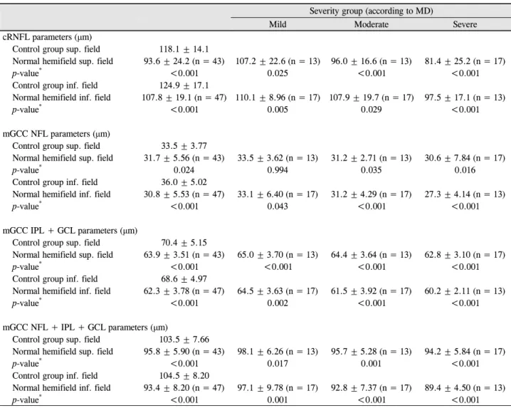
녹내장안 시야결손의 중등도에 따라 분류한 세 군 각각 에서 녹내장안 반시야 결손의 위치가 상측 반시야일 때와 하측 반시야일 때로 나누어서, 녹내장안 정상 반시야와 정 상안 동측 반시야 간의 cRNFL 및 mGCC 각 층의 두께 차 이를 비교하였다(Table 6). 상측 반시야 결손 초기일 때의
mGCC NFL 두께를 제외한 모든 측정치에 있어서 정상안 에 비해 유의한 감소를 보였다(mGCC NFL 초기 상측 p=0.994, 그 외 all p<0.05, independent t-test).
고 찰
녹내장에서 기능적 손상인 시야 결손이 나타나기에 앞서 구조적 손상, 즉 시신경유두와 망막신경섬유층의 변화가 선행된다는 것은 알려져 있다.12-14 또한 최근 OCT의 발달 과 더불어 mGCC의 두께 측정이 가능해지면서 cRNFL 두 께와 더불어 녹내장 초기 진단의 지표로서의 유용성이 연
Table 6. Comparison of cRNFL and mGCC (NFL, IPL + GCL, NFL + IPL + GCL) thickness parameters between glaucoma se-
verity groups (mild, moderate and severe groups) according to MD value in hemiretina with normal field and corresponding hemi- field in normal groupsSeverity group (according to MD)
Mild Moderate Severe
cRNFL parameters (μm)
Control group sup. field 118.1 ± 14.1
Normal hemifield sup. field 93.6 ± 24.2 (n = 43) 107.2 ± 22.6 (n = 13) 96.0 ± 16.6 (n = 13) 81.4 ± 25.2 (n = 17)
p-value* <0.001 0.025 <0.001 <0.001
Control group inf. field 124.9 ± 17.1
Normal hemifield inf. field 107.8 ± 19.1 (n = 47) 110.1 ± 8.96 (n = 17) 107.9 ± 19.7 (n = 17) 97.5 ± 17.1 (n = 13)
p-value* <0.001 0.005 0.029 <0.001
mGCC NFL parameters (μm)
Control group sup. field 33.5 ± 3.77
Normal hemifield sup. field 31.7 ± 5.56 (n = 43) 33.5 ± 3.62 (n = 13) 31.2 ± 2.71 (n = 13) 30.6 ± 7.84 (n = 17)
p-value* 0.024 0.994 0.035 0.016
Control group inf. field 36.0 ± 5.02
Normal hemifield inf. field 30.8 ± 5.53 (n = 47) 33.1 ± 6.40 (n = 17) 31.2 ± 4.29 (n = 17) 27.3 ± 4.14 (n = 13)
p-value* <0.001 0.043 <0.001 <0.001
mGCC IPL + GCL parameters (μm)
Control group sup. field 70.4 ± 5.15
Normal hemifield sup. field 63.9 ± 3.51 (n = 43) 65.0 ± 3.70 (n = 13) 64.4 ± 3.64 (n = 13) 62.8 ± 3.10 (n = 17)
p-value* <0.001 <0.001 <0.001 <0.001
Control group inf. field 68.6 ± 4.97
Normal hemifield inf. field 62.3 ± 3.78 (n = 47) 64.5 ± 3.63 (n = 17) 61.5 ± 3.92 (n = 17) 60.2 ± 2.11 (n = 13)
p-value* <0.001 0.002 <0.001 <0.001
mGCC NFL + IPL + GCL parameters (μm)
Control group sup. field 103.5 ± 7.66
Normal hemifield sup. field 95.8 ± 5.90 (n = 43) 98.1 ± 6.26 (n = 13) 95.7 ± 5.28 (n = 13) 94.2 ± 5.84 (n = 17)
p-value* <0.001 0.017 0.001 <0.001
Control group inf. field 104.5 ± 8.20
Normal hemifield inf. field 93.4 ± 8.20 (n = 47) 97.1 ± 9.78 (n = 17) 92.8 ± 7.37 (n = 17) 89.4 ± 4.50 (n = 13)
p-value* <0.001 0.001 <0.001 <0.001
Values are presented as mean ± SD unless otherwise indicated.
cRNFL = circumpapillary retinal nerve fiber layer; mGCC = macular ganglion cell complex; NFL = nerve fiber layer; IPL = inner plexi- form layer; GCL = ganglion cell layer; MD = mean deviation; sup. = superior; inf. = inferior.
*Independent t-test.
구되고 있다.15,16 정상안에서도 GCL+IPL 층 두께가 시신경 유두 및 cRNFL 두께 측정치들과 유의한 상관관계를 갖는 다는 것이 밝혀진 바 있는데,17 이는 초기 녹내장성 손상과 그 진행을 발견하는 데에 있어 mGCC 및 그중 GCL+IPL 층의 두께가 유용한 지표로 이용될 수 있는 근거로 볼 수 있다. 또한 Nakamura et al18은 반시야에 국한된 시야 결손 을 보이는 녹내장 환자에서 mGCC 두께 및 기능을 반영하는 것으로 볼 수 있는 국소 황반광순응 반응(focal macular pho- topic negative response)이 시야결손이 있는 반시야와 정상 반시야 모두에서 감소하는 것을 밝힌 바 있어, 반시야에 국 한된 결손만 있더라도 정상 반시야 구역에 망막신경절세포 의 소실이 있음을 보였다.
OCT 기술의 발달에 따라 황반부의 전체 망막 두께가 아 닌 내측망막에 국한된 두께를 측정할 수 있게 됨으로써 녹 내장성 변화를 발견할 수 있는 민감도가 증가하였다. Cho et al8은 RTVue-100 (Optovue Inc., Fremont, CA, USA) SD-OCT를 이용해 측정한 mGCC 두께와 시야 감도 간에 유의한 상관관계가 있다고 발표하였다. Inuzuka et al7은 Cirrus HD-OCT (Cirrus; Carl Zeiss Meditec, Dublin, CA, USA)를 이용한 연구에서 망막 두께를 내경계막(internal limiting membrane, ILM)부터 망막색소상피층(retinal pig- ment epithelium, RPE)까지의 두께로 측정하여 반시야 결 손이 있는 녹내장 환자의 정상 반시야 구역에서 망막 두께 의 유의한 감소가 있음을 보였다. 이후의 연구에서는 망막
두께를 mGCC의 두께로 국한하여 정상 반시야 구역에서 mGCC 두께 감소가 시야 감도 변화와 유의한 관계를 보이 며 MD 값에 따른 시야결손 중증도로 분류한 녹내장 단계 에 따라서도 유의한 감소를 보인다고 하였다.9 그러나 이 연구에서 중증도에 따른 분포에 대한 언급은 없었다.
Takagi et al10은 RTVue SD-OCT를 이용해 측정한 연구에 서 반시야 결손이 있는 녹내장 환자의 정상 반시야 구역에 서 정상군에 비해 mGCC 두께가 유의한 감소를 보인다고 한 바 있으며, Na et al11도 마찬가지로 RTVue SD-OCT를 이용한 연구에서 반시야 결손이 있는 녹내장 환자의 정상 반시야 구역에서 정상군에 비해 mGCC와 cRNFL 두께가 모두 감소되어 있고 mGCC 두께와 cRNFL 두께 간에 유의 한 상관관계가 있으며 이 측정치들이 MD, PSD 값과도 유 의한 상관관계를 가진다고 하였다. 그러나 이 연구에서의 녹내장안 평균 MD 값은 -2.44 ± 1.49로, 대상자가 주로 초 기의 녹내장안에 국한되었다는 한계점이 있었다. 이와 같 이 아직까지 결손측 반시야의 시야결손 중등도에 따른 분 포를 균등하게 맞추어 정상 반시야의 cRNFL과 mGCC 두 께 감소 차이를 비교한 연구는 발표된 바 없었다. 또한 앞 서 언급하였듯 NFL, GCL+IPL, NFL+GCL+IPL 세 가지 측 정치로 나눈 알고리즘에 따라 층을 세분화하여 mGCC 두 께 감소를 비교한 연구 역시 아직 발표된 바 없었다.
본 연구에서, 반시야 결손을 보이는 녹내장안의 정상 반 시야 구역 cRNFL과 mGCC 두께는 결손측만큼은 아니지 만 정상안에 비해 감소되어있는 결과를 보였다(Table 3).
이는 Na et al11의 RTVue SD-OCT를 이용한 연구 결과와도 일치하는데, 이것은 녹내장안의 정상 반시야 구역에도 아 직 기능적 손상인 시야 결손이 나타나기 전이지만 선행되 고 있는 구조적 손상이 있다는 것을 의미한다고 볼 수 있 다. 또한 정상 반시야 구역의 cRNFL과 mGCC 두께의 각 측정치가 모두 MD 값과 유의한 상관관계를 가지며(Table 4) MD 값에 따른 시야결손 중등도로 분류한 세 군 간의 비 교에서도 각 측정치들이 모두 중등도가 심한 군일수록 더 유의하게 감소한다는 결과(Table 5)를 보였다. 이는 Cho et al8이 mGCC 두께와 시야 감도 간에 유의한 상관관계가 있 다고 하였고 Inuzuka et al9이 MD 값에 따른 시야결손 중등 도로 분류한 군 간의 비교에서도 시야결손이 심할수록 mGCC와 cRNFL의 두께 감소가 유의하게 증가한다고 발 표한 것과 일치하는 점이다. Na et al11 역시 정상 반시야의 cRNFL과 mGCC 두께가 HFA의 시야 지표인 MD, PSD와 유의한 상관관계를 갖는다는 것을 보였는데, 본 연구에서 는 PSD의 경우 유의한 관계를 갖지 않는 것으로 나타났다.
본 연구에서는 cRNFL과 mGCC의 각 두께 측정치들을 상측 반시야 결손인 경우와 하측 반시야 결손인 경우로 나
누어서 각각의 데이터를 제시하고 비교하였는데, 이는 상 측 반시야와 하측 반시야 사이에는 망막시신경섬유 축삭 분포의 차이가 있고 구조적 변화가 비대칭적으로 일어난다
는3-6,19 점을 고려하여 좀 더 정확한 결과를 얻기 위한 것이
었다. 실제로 정상군에서도 상, 하측 두께 측정치들 간에 차이가 있었다(Table 2; all p<0.05, not shown on Table).
또한 주로 초기의 환자들을 다뤘던 이전 연구11와는 달리 본 연구에서는 MD 값에 따른 초기, 중기, 말기 각 군의 분 포가 각각 30명으로 균일하여, 시야결손 중증도에 따른 각 군 간의 비교가 가능했다. 이전의 연구들과의 또 다른 차이 점으로 본 연구에서는 Topcon의 3D SD-OCT를 사용하였 다는 점을 들 수 있는데, 3D macular vertical scan protocol 에 따라 mGCC를 NFL과 GCL+IPL로 나누어 분석이 가능 했다. 이에 따라 정상 반시야 구역의 cRNFL과 mGCC 각 층의 두께를 MD 값에 따른 각 군별로 정상군과 비교해 본 결과, 시야결손 초기군에서는 상측 결손군의 mGCC NFL 두께를 제외하고는 정상군에 비해 유의한 감소를 보였고, 중기, 말기군에서는 cRNFL과 mGCC의 모든 측정치에 있 어 정상군에 비해 유의한 감소를 보였다(Table 6). 또한 세 군 간에도 시야 결손의 중등도가 증가함에 따라 그 감소 정 도에 있어 유의한 차이가 있었다(Table 5). 상측 결손의 초 기 단계에서 mGCC NFL 층의 두께가 유의한 감소를 보이 지 않은 것은 망막신경절세포복합체의 손상에 있어 시기적 으로 세포체와 수상돌기의 손상이 먼저 일어나고 축삭의 손상이 조금 더 나중에 일어나기 때문일 수 있다고 유추해 볼 수 있겠으나, 이에 대해서는 추가적인 연구가 필요할 것 으로 보인다. 하측 결손의 경우 상측과 달리 초기에도 mGCC NFL 층의 두께가 정상군에 비해 유의한 감소를 보 였는데, 이와 같은 상하측 간의 차이는 망막시신경섬유 축 삭 분포 및 구조적 변화 진행에 있어서의 비대칭성3 때문인 것으로 보이며, 마찬가지로 이에 대해서는 추가적인 연구 가 필요할 것으로 생각된다.
이번 연구의 제한점으로는 우선 연구 대상자의 수를 들 수 있다. 시야결손의 중증도에 따른 각 군별 30명으로, 더 많은 수의 대상자에 대한 연구를 시행하지 못했다는 한계 가 있지만, 기존의 연구들과 견주어 볼 때 전체 환자 수가 적은 편은 아니었으며 본 연구에서처럼 시야 결손의 중등 도에 따른 세 군 간 균등한 분포하에 시행된 연구가 없었던 점은 본 연구의 장점이라 할 수 있겠다. 또한 MD 값은 백 내장 등 매체 혼탁의 영향을 받는 것으로 알려져 있어 중증 도 반영에 한계가 있을 수 있다는 점 역시 한계로 들 수 있 다. 이러한 영향을 덜 받는 것으로 알려져 있는 시야 지표 인 visual field index (VFI)는 비교적 최근 HFA 검사 결과 에 나오기 시작하여 VFI 값을 이용 가능한 환자 수가 상대
적으로 적어 본 연구에서는 사용하지 못하였으나, 추후 연 구에서 시야결손의 중증도를 반영하는 지표로 VFI의 사용 을 고려해 볼 수 있을 것으로 보인다.
결론적으로, 본 연구에서는 시야검사상 반시야 결손을 나타내는 녹내장 환자의 정상 반시야 구역 cRNFL 및 mGCC 각 층의 두께가 정상군에 비해 감소되어 있었으며, 그 감소 정도는 시야결손의 중증도와 유의한 관계를 가진 다는 것을 보였다. 이는 반시야 결손 녹내장 환자에서 반대 측의 정상 시야에 상응하는 구역에도 결손측의 시야결손 정도에 따라 구조적 손상이 진행되고 있다는 것을 나타내 며, 시야결손이 나타나기 전의 녹내장안에서 cRNFL 및 mGCC 두께 측정의 진단적 유용성을 다시 한 번 확인할 수 있는 결과로 볼 수 있을 것이다.
REFERENCES
1) Quigley HA, Addicks EM, Green WR. Optic nerve damage in hu- man glaucoma. III. Quantitative correlation of nerve fiber loss and visual field defect in glaucoma, ischemic neuropathy, papilledema, and toxic neuropathy. Arch Ophthalmol 1982;100:135-46.
2) Quigley HA, Dunkelberger GR, Green WR. Retinal ganglion cell atrophy correlated with automated perimetry in human eyes with glaucoma. Am J Ophthalmol 1989;107:453-64.
3) Choi JA, Park HYL, Jung KI, et al. Difference in the properties of retinal nerve fiber layer defect between superior and inferior visual field loss in glaucoma. Invest Ophthalmol Vis Sci 2013;54:6982-90.
4) Duggan C, Sommer A, Auer C, Burkhard K. Automated differ- ential threshold perimetry for detecting glaucomatous visual field loss. Am J Ophthalmol 1985;100:420-3.
5) Susanna R Jr, Nicolela MT, Soriano DS, Carvalho C. Automated perimetry: a study of the glaucoma hemifield test for the detection of early glaucomatous visual field loss. J Glaucoma 1994;3:12-6.
6) Badlani V, Shahidi M, Shakoor A, et al. Nerve fiber layer thickness in glaucoma patients with asymmetric hemifield visual field loss. J Glaucoma 2006;15:275-80.
7) Inuzuka H, Kawase K, Sawada A, et al. Macular retinal thickness in glaucoma with superior or inferior visual hemifield defects. J
Glaucoma 2013;22:60-4.
8) Cho JW, Sung KR, Lee S, et al. Relationship between visual field sensitivity and macular ganglion cell complex thickness as meas- ured by spectral-domain optical coherence tomography. Invest Ophthalmol Vis Sci 2010;51:6401-7.
9) Inuzuka H, Kawase K, Yamada H, et al. Macular ganglion cell complex thickness in glaucoma with superior or inferior visual hemifield defects. J Glaucoma 2014;23:145-9.
10) Takagi ST, Kita Y, Yagi F, Tomita G. Macular retinal ganglion cell complex damage in the apparently normal visual field of glaucom- atous eyes with hemifield defects. J Glaucoma 2012;21:318-25.
11) Na JH, Kook MS, Lee Y, et al. Detection of macular and circum- papillary structural loss in normal hemifield areas of glaucomatous eyes with localized visual field defects using spectral-domain opti- cal coherence tomography. Graefes Arch Clin Exp Ophthalmol 2012;250:595-602.
12) Sommer A, Katz J, Quigley HA, et al. Clinically detectable nerve fiber atrophy precedes the onset of glaucomatous field loss. Arch Ophthalmol 1991;109:77-83.
13) Quigley HA, Katz J, Derick RJ, et al. An evaluation of optic disc and nerve fiber layer examinations in monitoring progression of early glaucoma damage. Ophthalmology 1992;99:19-28.
14) Zeyen TG, Caprioli J. Progression of disc and field damage in early glaucoma. Arch Ophthalmol 1993;111:62-5.
15) Leung CK, Chan WM, Yung WH, et al. Comparison of macular and peripapillary measurements for the detection of glaucoma: an optical coherence tomography study. Ophthalmology 2005;112:
391-400.
16) Lalezary M, Medeiros FA, Weinreb RN, et al. Baseline optical co- herence tomography predicts the development of glaucomatous change in glaucoma suspects. Am J Ophthalmol 2006;142:576-82.
17) Tham YC, Cheung CY, Koh VT, et al. Relationship between gan- glion cell-inner plexiform layer and optic disc/retinal nerve fibre layer parameters in non-glaucomatous eyes. Br J Ophthalmol 2013;97:1592-7.
18) Nakamura H, Hangai M, Mori S, et al. Hemispherical focal mac- ular photopic negative response and macular inner retinal thickness in open-angle glaucoma. Am J Ophthalmol 2011;151:494-506.e1.
19) Kee C, Cho C. Evaluation of retinal nerve fiber layer thickness in the area of apparently normal hemifield in glaucomatous eyes with optical coherence tomography. J Glaucoma 2003;12:250-4.
= 국문초록 =
반시야 결손 녹내장안의 중증도에 따른 정상 반시야 신경섬유층 및 신경절세포층 두께 비교
목적: 시야검사상 반시야결손을 나타내는 녹내장 환자에서 스펙트럼영역 빛간섭단층촬영(spectral-domain optical coherence tomography, SD-OCT)으로 측정한 정상 반시야의 시신경유두주위 망막신경섬유층(circumpapillary retinal nerve fiber layer, cRNFL) 및 황반부신경절세포층(macular ganglion cell complex, mGCC) 두께를 정상군과 비교하고, 시야결손 중증도에 따른 차이를 알아보고자 하였다.
대상과 방법: 정상안 90안과 시야검사상 반시야결손을 나타내는 녹내장안 90안에서 SD-OCT를 이용하여 녹내장안 정상 반시야와 정상안 동측 반시야의 cRNFL과 mGCC 두께를 비교하였고, 시야결손의 중증도에 따라 초, 중, 말기 세 군으로 나누어 비교하였다.
결과: 녹내장안 정상 반시야와 정상안 동측 반시야에서 각각 평균 cRNFL 두께는 상측 93.6 ± 24.2 μm, 118.1 ± 14.1 μm, 하측 107.8
± 19.1 μm, 124.9 ± 17.1 μm, mGCC 두께는 상측 95.8 ± 5.9 μm, 103.5 ± 7.7 μm, 하측 93.4 ± 8.2 μm, 104.5 ± 8.20 μm (all p<0.05)로 녹내장안 정상 반시야에서 유의한 감소를 보였으며, 시야결손의 중증도 증가에 따라 더 감소하는 양상을 보였다(all p
<0.01). 녹내장안의 시야결손 중증도에 따라 분류한 세 군에서도 nerve fiber layer 두께 외의 모든 측정치에서 정상안에 비해 유의한 감소를 보였다.
결론: SD-OCT를 이용한 cRNFL 및 mGCC 두께 측정은 시야 변화가 나타나기 전의 정상 반시야에서 구조적 변화를 반영하는 초기 검사로써 유용하게 사용될 수 있을 것이다.
<대한안과학회지 2016;57(4):614-622>