Radiological Evaluation of the Initial Fixation between Cortical Bone Trajectory and
Conventional Pedicle Screw Technique for Lumbar Degenerative Spondylolisthesis
Koshi Ninomiya, Koichi Iwatsuki, Yu-Ichiro Ohnishi, Toshiki Yoshimine
Department of Neurosurgery, Osaka University Graduate School of Medicine, Osaka, Japan
Study Design: Retrospective study.
Purpose: To compare initial fixation using the cortical bone trajectory (CBT) technique versus conventional pedicle screws (PS) in ra- diographs of postsurgical lumbar degenerative spondylolisthesis.
Overview of Literature: Few reports have documented the holding strength of CBT technique for spondylolisthesis cases.
Methods: From October 2009 to June 2014, 21 cases of degenerative spondylolisthesis were surgically treated in our institution. Ten were treated with conventional PS technique and 11 of with CBT technique. Mean lumbar lordosis and percent slippage were evalu- ated preoperatively, immediately after surgery, and 6 months and 1 year postoperatively using radiographs. We also investigated percent loss of slip reduction.
Results: There were statistically significant differences between preoperative percent slippage and postoperative slippage in both PS and CBT procedures over 1 year, and both techniques showed good slip reduction. On the other hand, lumbar lordosis did not change significantly in either the PS or CBT groups over 1 year.
Conclusions: CBT technique showed similarly good initial fixation compared with the PS procedure in the treatment of lumbar de- generative spondylolisthesis.
Keywords: Cortical bone trajectory; Conventional pedicle screw; Lumbar degenerative spondylolisthesis
Copyright Ⓒ 2016 by Korean Society of Spine Surgery
This is an Open Access article distributed under the terms of the Creative Commons Attribution Non-Commercial License (http://creativecommons.org/licenses/by-nc/3.0/) which permits unrestricted non-commercial use, distribution, and reproduction in any medium, provided the original work is properly cited.
Asian Spine Journal • pISSN 1976-1902 eISSN 1976-7846 • www.asianspinejournal.org
Received Jul 12, 2015; Revised Aug 25, 2015; Accepted Aug 27, 2015 Corresponding author: Koshi Ninomiya
Department of Neurosurgery, Osaka University Graduate School of Medicine, 2-2 Yamadaoka, Suita 565-0871, Japan
Tel: +81-6-6879-3652, Fax: +81-6-6879-3659, E-mail: [email protected]
ASJ A SJ
Introduction
Cortical bone trajectory (CBT) was first described by Santoni et al. [1] in 2009. Compared to traditional pedicle screw (PS) insertion, CBT is thought to be more effec- tive for initial fixation by maximizing screw-cortical bone contact. The authors demonstrated a 30% increase in uni-
axial yield pullout load and equivalency in mixed loading for the CBT screw compared with traditional PSs in their human cadaver study.
Matsukawa et al. [2] reported a significant difference between the mean maximum intraoperative insertional torque of CBT screws and traditional screws. We also con- firmed in an in vivo study that the pars interarticularis is a
very important structure for the CBT technique in spon- dylolysis and non-spondylolysis cases. In the study, the mean maximum insertional torque of non-spondylolysis cases was twice as high as that of spondylolysis cases (Asian Spine Journal, in press).
However, higher pullout strength and insertional torque do not always reflect favorable initial fixation. As far as initial fixation is concerned, some postoperative radio- logical evaluation may be needed.
Therefore, we evaluated initial fixation through radio- graphs of patients with lumbar degenerative spondylolis- thesis treated by the CBT technique compared to those treated by the conventional PS procedure.
Materials and Methods
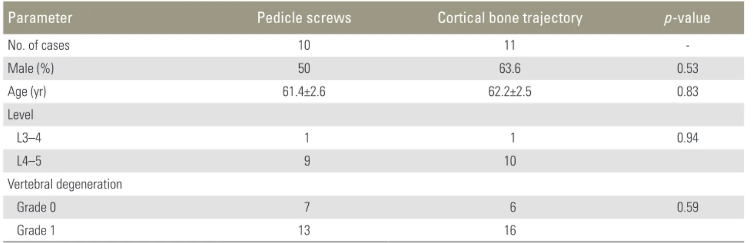
Between October 2009 and June 2014, 21 patients with Meyerding grade I–II degenerative spondylolisthesis un- derwent posterior lumbar interbody fusion (PLIF) with conventional PS (10 patients) or CBT (11 patients) proce- dure in our institution. Cases of spondylolytic spondylo- listhesis were excluded. All patients had lower extremity pain before surgery. We used conventional PS technique from 2009 to 2012, and CBT from 2012 to 2014. All pa- tients had single-level fusion performed. Operative level, age, and sex distribution of the two groups did not differ (Table 1).
For evaluation of vertebral degeneration, all vertebrae on lumbar radiographs were classified using a semi-quan- titative method [3]. This assessment of vertebral fractures can be performed quickly on a routine basis and cor-
relates moderately well with quantitative morphometry.
Vertebrae are graded on visual inspection and without direct vertebral measurement as normal (grade 0), mildly deformed (grade 1, approximately 20%–25% reduction in anterior, middle, and/or posterior height and a reduc- tion of area 10%–20%), moderately deformed (grade 2, approximately 25%–40% reduction in any height and a re- duction in area 20%–40%), and severely deformed (grade 3, approximately more than 40% reduction in any height and area). There were 7 grade 0 vertebrae and 13 grade 1 vertebrae in the PS-treated group, and 6 grade 0 vertebrae and 16 grade 1 vertebrae in the CBT-treated group; no statistical differences were evident (Table 1). There were no grade 2 and grade 3 vertebrae in this study.
1. Surgical procedures
The conventional PS procedure was performed in the standard fashion reported previously by Weinstein et al.
[4]. Screws were 6–6.5 mm in diameter and 40–55 mm long (Easyspine multiaxial screw, Alphatec Spine, Tokyo, Japan; Capstone system screw, medtronic sofamor danek, Osaka, Japan). Two titanium (Telamon, Medtronic so- famor Danek; Novel, alphatec Spine) or polyether ether ketone (peek) (Capstone; Medtronic sofamor Danek) in- terbody cages were used.
CBT was performed under lateral fluoroscopy. We used the isthmus of the lamina as an anatomical landmark for entry [5]. Screws were placed 3 mm inside the isthmus and inserted cephalad to and laterally from the isthmus. Same- size tapping was performed. The screws were 4.5–5 mm
Table 1. Demographic data between PS and CBT
Parameter Pedicle screws Cortical bone trajectory p-value
No. of cases 10 11 -
Male (%) 50 63.6 0.53
Age (yr) 61.4±2.6 62.2±2.5 0.83
Level
L3–4 1 1 0.94
L4–5 9 10
Vertebral degeneration
Grade 0 7 6 0.59
Grade 1 13 16
Values of age are presented as mean±standard error.
PS, pedicle screws; CBT, cortical bone trajectory.
in diameter and 2–35 mm in length, with a 4 mm pitch (Zodiac polyaxial screw, Alphatec Spine). One titanium or PEEK interbody cage (NOVEL, Alphatec Spine) was used.
In both techniques, cages were packed with autologous bone graft, and bones were also used for posterolateral bone fusion.
2. Radiological assessment
Radiological outcome was evaluated by comparing per- cent slippage and lumbar lordosis before surgery with that immediately after surgery, 6 months later, and 1 year later.
Percent loss of slip reduction was also checked. Lumbar lordosis was measured from the superior end plate of L1 to the superior end plate of L5.
3. Statistical analyses
JMP Pro 11 (SAS Institute Inc., Cary, NC, USA) was used for statistical analyses. Statistical significance was defined
as p<0.05. Percent slippage and lumbar lordosis were ana- lyzed using one-way analysis of variance (ANOVA) with Tukey’s test. Also, percent loss of slip was analyzed using ANOVA and the chi-square test was used for demograph- ic data between PS and CBT.
Results
Figs. 1 and 2 depict representative cases of conventional PS and CBT techniques.
One patient treated with CBT had spacer backout 2 weeks after surgery, and he underwent reoperation. He was excluded from the analysis at 6 months and 1 year after the operation. Other patients had no complications and experienced a good clinical course. In conventional PS cases, lower extremity pain disappeared in 70% patients after the operation through 1 year. In CBT cases, 73% of the patients were free from pain after the operation.
Fig. 3 shows the time course of the percent slippage in each case. The percent slippage decreased from 12.7%
Fig. 1. Plain radiographs of a 67-year-old patient taken before (A, B), immediately after surgery (C, D), and 1 year after sur- gery (E, F) using the conventional pedicle screws technique.
A B C D
E F
before surgery to 5.0% after 1 year using the PS technique.
Using the CBT technique, the percent slippage decreased from 11.1% before surgery to 3.2% after 1 year. Both tech- niques demonstrated a significant difference between pre-
and postoperation. In addition, there was no significant difference of loss of percent slip in 6 months and in 1 year between both procedures (Table 2, Suppl. Table 1).
The lumbar lordosis did not change significantly in ei- Fig. 2. Plain radiographs of a 48-year-old patient before (A, B), right after (C, D) and at 1 year after (E, F) surgery with the cortical bone trajectory technique.
A B C D
E F
Pre-OP
12.7±1.9 Post-OP
3.4±1.9 6 mo
3.8±1.9 1 yr
5.0±1.9 16
14 12 10 8 6 4 2 0 (%)
*p<0.05, **p<0.01 PS
** * *
Pre-OP
11.1±1.5 Post-OP
2.0±1.5 6 mo
3.2±1.6 1 yr
3.2±1.6 16
14 12 10 8 6 4 2 0 (%)
**p<0.01, ***p<0.001 CBT
*** ** **
Fig. 3. Bar diagram showing the significant improvement of the percent slip (mean±standard error) with PS technique (A) and CBT technique (B). PS, pedicle screws; CBT, cortical bone trajectory; Pre-OP, preoperative; Post-OP, postoperative.
A B
ther PS or CBT groups over 1 year (Suppl. Table 2, Fig. 4).
Discussion
CBT has a more medial-to-lateral and shorter path than the traditional technique for spinal fusion, and is thought to be effective for severely degenerated vertebrae because the screws are primarily stabilized in the posterior ele- ments.
In vivo insertional torque of CBT screws was first re- ported by Matsukawa et al. [2]. The authors described a significant difference between the mean maximum inser- tional torque of CBT screws and traditional screws, with the former being almost twice as high as the latter.
Several studies [6-8] have reported that the insertional torque of PSs is highly correlated with pullout strength.
Other studies described screw loosening caused primar- ily by cyclic caudocephalad toggling at the bone-screw interface [9] and that insertional torque can predict screw loosening [10].
Therefore, when we consider holding power, or initial fixation generated by PSs, we must evaluate postopera- tive radiographs. In the present study of degenerative spondylolisthesis cases, percent slip, loss of slip reduction, and lumbar lordotic angle were evaluated for this initial
fixation.
Biomechanically, slip reduction is thought to be achieved by anteroposterior directional force, and main- tenance of lordosis achieved by craniocaudal directional force. This realignment might enhance good bone fusion and clinical outcome [11], and prevent adjacent-segment disease.
Anterior column augmentation with PLIF using inter- vertebral spacers in addition to PS fixation can produce a superior fusion rate and improve clinical outcomes in spondylolisthesis [12-14]. Recently, Takata et al. [15]
proposed a hybrid technique of CBT and pedicle screw- ing for minimally invasive spine reconstruction surgery.
According to the authors, the hybrid CBT-PS technique allows sufficient holding strength of the slipped vertebra compared with other conventional PS procedures. As far as slip reduction and a low percentage of slip loss that is maintained of the initial fixation, our pure CBT screwing for degenerative spondylolisthesis also showed similar good holding power compared with the PS technique.
There was no significant difference between preopera- tive and postoperative lumbar lordosis in either CBT or conventional PS cases. However, our observations of a slight lordotic change in PS cases and the occurrence of one spacer backout in a CBT case indicates the need Table 2. Loss of % slip
Time since surgery PS (%) CBT (%) p-value
6 mo 0.59±0.57 0.98±0.57 0.63
6 mo–1 yr 0.1±0.64 0.0±0.64 0.96
Values are presented as mean±standard error.
PS, pedicle screws; CBT, cortical bone trajectory.
Fig. 4. Bar diagram showing that the lumbar lordosis (mean±standard error) did not significantly change with the PS technique (A) or CBT technique (B). PS, pedicle screws; CBT, cortical bone trajectory.
Pre-OP
24.6±3.9 Post-OP
25.4±3.9 6 mo
27.4±3.9 1 yr 29.5±3.9 34
32 30 28 26 24 22 20
(°) PS
Pre-OP
24.1±2.4 Post-OP
23.9±2.4 6 mo
24.4±2.5 1 yr 24.0±2.5 34
32 30 28 26 24 22 20
(°) CBT
A B
for further investigation of which technique is better for the maintenance of lordosis. Nevertheless, our results do demonstrate that CBT is well indicated for elderly or osteoporotic patients compared to the conventional PS technique because of its less invasive nature.
This study has some limitations. Most cases were of grade I spondylolisthesis using several different type cages, so we cannot extrapolate to patients with more se- vere slippage. Second, this study was a small case series.
Further investigations with a larger patient populations are needed.
Despite these limitations, CBT screwing showed suf- ficient holding power in this study. Therefore, pending conformation in future studies, the CBT technique is expected to yield good bone fusion and good long-term clinical outcomes.
Conclusions
The CBT technique shows similarly good initial fixation compared with the PS procedure for lumbar degenerative spondylolisthesis.
Conflict of Interest
No potential conflict of interest relevant to this article was reported.
Supplementary Materials
Suppl. Table 1. % slip change
Supplemental data can be found at: http://www.asian- spinejournal.org/src/sm/asj-10-251-s001.pdf.
Suppl. Table 2. Lumbar lordosis change (°)
Supplementary material can be found at: http://www.
asianspinejournal.org/src/sm/asj-10-251-s002.pdf.
References
1. Santoni BG, Hynes RA, McGilvray KC, et al. Cortical bone trajectory for lumbar pedicle screws. Spine J 2009;9:366-73.
2. Matsukawa K, Yato Y, Kato T, Imabayashi H, Asa- zuma T, Nemoto K. In vivo analysis of insertional torque during pedicle screwing using cortical bone trajectory technique. Spine (Phila Pa 1976) 2014;39:
E240-5.
3. Genant HK, Wu CY, van Kuijk C, Nevitt MC. Ver- tebral fracture assessment using a semiquantitative technique. J Bone Miner Res 1993;8:1137-48.
4. Weinstein JN, Rydevik BL, Rauschning W. Anatomic and technical considerations of pedicle screw fixa- tion. Clin Orthop Relat Res 1992;(284):34-46.
5. Iwatsuki K, Yoshimine T, Ohnishi Y, Ninomiya K, Ohkawa T. Isthmus-guided cortical bone trajectory for pedicle screw insertion. Orthop Surg 2014;6:244- 8.
6. Daftari TK, Horton WC, Hutton WC. Correlations between screw hole preparation, torque of insertion, and pullout strength for spinal screws. J Spinal Disord 1994;7:139-45.
7. Myers BS, Belmont PJ Jr, Richardson WJ, Yu JR, Harper KD, Nightingale RW. The role of imaging and in situ biomechanical testing in assessing pedicle screw pull-out strength. Spine (Phila Pa 1976) 1996;
21:1962-8.
8. Zdeblick TA, Kunz DN, Cooke ME, McCabe R.
Pedicle screw pullout strength. Correlation with in- sertional torque. Spine (Phila Pa 1976) 1993;18:1673- 6.
9. Law M, Tencer AF, Anderson PA. Caudo-cephalad loading of pedicle screws: mechanisms of loosening and methods of augmentation. Spine (Phila Pa 1976) 1993;18:2438-43.
10. Okuyama K, Abe E, Suzuki T, Tamura Y, Chiba M, Sato K. Can insertional torque predict screw loosen- ing and related failures? An in vivo study of pedicle screw fixation augmenting posterior lumbar inter- body fusion. Spine (Phila Pa 1976) 2000;25:858-64.
11. Kawakami M, Tamaki T, Ando M, Yamada H, Hashi- zume H, Yoshida M. Lumbar sagittal balance influ- ences the clinical outcome after decompression and posterolateral spinal fusion for degenerative lumbar spondylolisthesis. Spine (Phila Pa 1976) 2002;27:59- 64.
12. Okuyama K, Kido T, Unoki E, Chiba M. PLIF with a titanium cage and excised facet joint bone for de- generative spondylolisthesis--in augmentation with a pedicle screw. J Spinal Disord Tech 2007;20:53-9.
13. Patil SS, Rawall S, Nagad P, Shial B, Pawar U, Nene AM. Outcome of single level instrumented posterior lumbar interbody fusion using corticocancellous laminectomy bone chips. Indian J Orthop 2011;45:
500-3.
14. Greiner-Perth R, Boehm H, Allam Y, Elsaghir H, Franke J. Reoperation rate after instrumented poste- rior lumbar interbody fusion: a report on 1680 cases.
Spine (Phila Pa 1976) 2004;29:2516-20.
15. Takata Y, Matsuura T, Higashino K, et al. Hybrid
technique of cortical bone trajectory and pedicle screwing for minimally invasive spine reconstruction surgery: a technical note. J Med Invest 2014;61:388- 92.