http://dx.doi.org/10.12671/jkfs.2016.29.2.121
121
Copyright ⓒ 2016 The Korean Fracture Society. All rights reserved.
This is an Open Access article distributed under the terms of the Creative Commons Attribution Non-Commercial License (http://creativecommons.org/licenses/
by-nc/4.0) which permits unrestricted non-commercial use, distribution, and reproduction in any medium, provided the original work is properly cited.
Received January 4, 2016 Revised February 1, 2016 Accepted February 1, 2016
Address reprint requests to: Joon Yub Kim, M.D., Ph.D.
Department of Orthopaedic Surgery, Seonam University College of Medicine Myongji Hospital, 55 Hwasu-ro 14beon-gil, Deogyang- gu, Goyang 10475, Korea
Tel: 82-31-810-6530ㆍFax: 82-31-969-0500 E-mail: [email protected]
Financial support: None. Conflict of interest: None.
쇄골 간부 골절 수술 후 발생한 감각저하:
금속판 위치와 연관성 및 임상상
김성훈⋅김준엽* ⋅고경환
†⋅정명곤*⋅조재호*
국민건강보험 공단 일산병원 정형외과, 서남대학교 의과대학 명지병원 정형외과*, 인제대학교 일산백병원 정형외과†
Hypoesthesia after Open Reduction and Plate Fixation of Clavicular Midshaft Fractures: Correlation with Plate Location and Clinical Features of Hypoesthesia
Seong Hun Kim, M.D., Joon Yub Kim, M.D., Ph.D.* , Kyoung Hwan Koh, M.D.
†, Myung Gon Jung, M.D.*, Jae Ho Cho, M.D.*
Department of Orthopaedic Surgery, National Health Insurance Service Ilsan Hospital, Department of Orthopaedic Surgery, Seonam University College of Medicine Myongji Hospital*, Department of Orthopaedic Surgery, Inje University Ilsan Paik Hospital†, Goyang, Korea
Purpose: The aim of this study is to evaluate the correlation between the location of the plate and the incidence of clavicular hypoesthesia and the clinical features of patients with clavicular hypoesthesia after open reduction and internal fixation of clavicular midshaft fractures.
Materials and Methods: Seventy-eight patients who underwent open reduction and plate fixation for clavicle midshaft frac- tures between March 2013 and October 2014 were assessed for eligibility. The total clavicular length (A), the distance to the medial end of the plate from the sternoclavicular joint (B), and the distance to the lateral end of the plate from the sternoclavicular joint (C) were measured. Correlation between the location of the clavicular plate and the incidence of clav- icular hypoesthesia was evaluated. In addition, the severity, and recovery of hypoesthesia were evaluated. Patient satisfaction, pain visual analogue scale were evaluated regarding hypoesthesia.
Results: The incidence of hypoesthesia was 32.1% (25/78 patients). No correlation was observed with respect to the location of the clavicular plate and the incidence of clavicular hypoesthesia (p=0.666 at the medial end, p=0.369 at the lateral end).
Recovery from hypoesthesia was observed in 23 out of 25 patients (p=0.008). Patient satisfaction and pain showed negative correlation with the incidence of hypoesthesia (p=0.002 and p=0.022).
Conclusion: There was no correlation between clavicular hypoesthesia and the plate location. Although most cases of hypo- esthesia were recovered, we should try to avoid hypoesthesia due to the negative ‘correlation’ with patient satisfaction and pain.
Key Words: Clavicle, Fracture fixation, Hypoesthesia, Correlation study
Introduction
Midshaft clavicle fractures account for 80% of all types of clavicle fractures.1-7) The mainstay of treatment for displaced midshaft clavicle fractures is an open reduction and plate fixation.2,8) It is more beneficial than con-
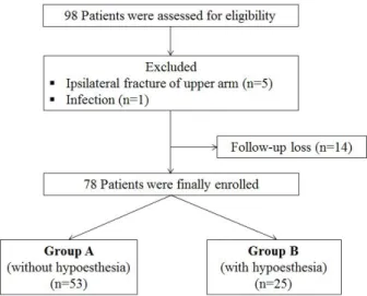
Fig. 1. Flowchart of patient enrollment.
servative treatment in terms of the incidence of mal- union and nonunion and early motion.7-10) However, there might be some complications with regard to open reduction and plate fixation such as infections, hypo- esthesia, irritation of the skin, and hardware failure.11) Clavicular hypoesthesia is a common complication after surgery for clavicle midshaft fractures and its effect on clinical outcome is known to be insignificant with respect to function.2,6,11,12) The permanent hypoesthesia might oc- cur in some patients and they might severely suffer from this complication and could be unsatisfied with those complications.6,11,13) However, we are still less informed about the clinical implications of clavicular hypoesthesia.
Supraclavicular nerve injury during the operation is known to be a pathology of clavicular hypoesthesia.6,14,15) The supraclavicular nerve arises from the roots of C3 and C4 and the intermediate and lateral branches of the supraclavicular nerve are the main branches of innerva- tion around the shoulder.6,14) The anatomical safe zone without any disruption of the supraclavicular nerve dur- ing clavicle surgery is known to be located at the narrow area near the sternoclavicular (SC) joint and the acromio- clavicular joint irrespective of anatomical variations.14) The vertical incision and the minimally invasive approach rather than the transverse incision along the long axis of the clavicle are known to reduce the incidence of clav- icular hypoesthesia.6) However, the minimally invasive exposure and the vertical incision might be insufficient to expose the long spiral type of fractures.
We hypothesized that clavicular hypoesthesia might be clinically correlated with the satisfaction of patients rath- er than the other functional problems and there might be a anatomical correlation between the location of the clavicular plate and the incidence of hypoesthesia. We postulated that the extent of the approach was propor- tionally related to the length of the plate that we used during surgery. Therefore, the analysis regarding the ex- tent of the approach could be performed by the radio- logical analysis of plate location. The aims of this study were to verify the correlation between the location of clavicular plates and the incidence of clavicular hypo- esthesia and to identify clinical features of clavicular hy- poesthesia including the patient’s satisfaction and pain.
Materials and Methods
The institutional review board of the Seonam University College of Medicine Myongji Hospital approved this study (MJH 15-013).
1. Cohort
Ninety-eight patients who received the open reduction and internal fixation for the displaced midshaft clavicle fractures between March 2013 and October 2014 at two hospitals were found to be eligible for this retrospective study. Exclusion criteria were ipsilateral upper extremity fractures such as scapular fracture (n=4) and humeral shaft fracture (n=1), patients with infection (n=1) and frac- tures in disabled patients, and 14 patients were excluded due to loss of follow-up. A total of 78 patients were fi- nally enrolled in this study (Fig. 1). Mean age of the 78 enrolled patients (58 men, 20 women) was 41.0±17.6 years (range, 15-71 years). The mean follow-up period was 12.6±3.0 months (range, 6-18 months). Demo- graphic characteristics and radiological variables of pa- tients in groups A and B are summarized in Table 1.
2. Operation technique
We performed the operation with the patients in the beach chair position. Before the start of the operation, the fracture site was marked after confirmation by the
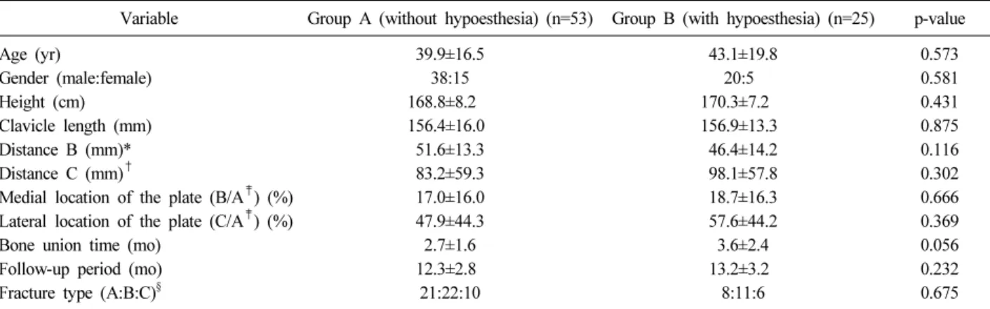
Table 1. Demographics and Radiologic Variables of the Cohort
Variable Group A (without hypoesthesia) (n=53) Group B (with hypoesthesia) (n=25) p-value
Age (yr) 39.9±16.5 43.1±19.8 0.573
Gender (male:female) 38:15 20:5 0.581
Height (cm) 168.8±8.2 170.3±7.2 0.431
Clavicle length (mm) 156.4±16.0 156.9±13.3 0.875
Distance B (mm)* 51.6±13.3 46.4±14.2 0.116
Distance C (mm)† 83.2±59.3 98.1±57.8 0.302
Medial location of the plate (B/A‡) (%) 17.0±16.0 18.7±16.3 0.666
Lateral location of the plate (C/A‡) (%) 47.9±44.3 57.6±44.2 0.369
Bone union time (mo) 2.7±1.6 3.6±2.4 0.056
Follow-up period (mo) 12.3±2.8 13.2±3.2 0.232
Fracture type (A:B:C)§ 21:22:10 8:11:6 0.675
Values are presented as mean±standard deviation or ratio. *Distance from the sternoclavicular joint to the medial end of the plate. †Dis- tance from the sternoclavicular joint to the lateral end of the plate. ‡Total clavicular length. §Fracture types are categorized by AO classification.
Fig. 2. Radiologic evaluation using the clavicle anteropo- sterior X-ray. A: The total length of the clavicle. B: The distance from the sternoclavicular joint to the medial end of the plate. C: The distance from the sternoclavicular joint to the lateral end of the plate.
c-arm. Then, a transverse skin incision along the long axis of the clavicle was performed such that the center of the incision was at the center of the fracture. During the operation, the procedure for identification of every branches of the supraclavicular nerve was not perfor- med. We used the lag screws for comminuted fractures, and locking compression plates (Acumed, Portland;
Synthes, Oberdorf, Netherlands) were applied to stabilize the fractures.
3. Postoperative rehabilitation
Arm sling was applied for 2 weeks and the pendulum and active assisted exercises were started immediately af- ter the operation. However, weight bearing onto the af- fected shoulder and contact sports were prohibited until postoperative 12 weeks.
4. Radiological evaluation
Radiologic evaluation was performed by using the clavicle anteroposterior (AP), cephalic and caudal tilt views. The location of the clavicular plate was assessed by two 10-year experienced orthopedic surgeons (J.Y.K., S.H.K.) in shoulder surgery on a clavicle AP X-ray. The total clavicular length (A), the distance to the medial end of the plate from the SC joint (B), and the distance to the lateral end of the plate from the SC joint (C) were measured and the distance to the medial end of
the plate to the total length of the clavicle (B/A) and the distance to the lateral end of the plate to the total length of the clavicle (C/A) w ere calculated (Fig. 2). The bone union was evaluated at 4, 6, 12, 18, and 24 weeks postoperatively. Union was defined as callus formation or filling of hairline like gap during the follow-up.
5. Clinical outcome evaluation
Hypoesthesia was evaluated before the surgery and at postoperative 4 weeks of follow-up.13) We asked the pa- tients whether they could identify the light touch sensa- tion caused by the finger contact just distal to the clavi-
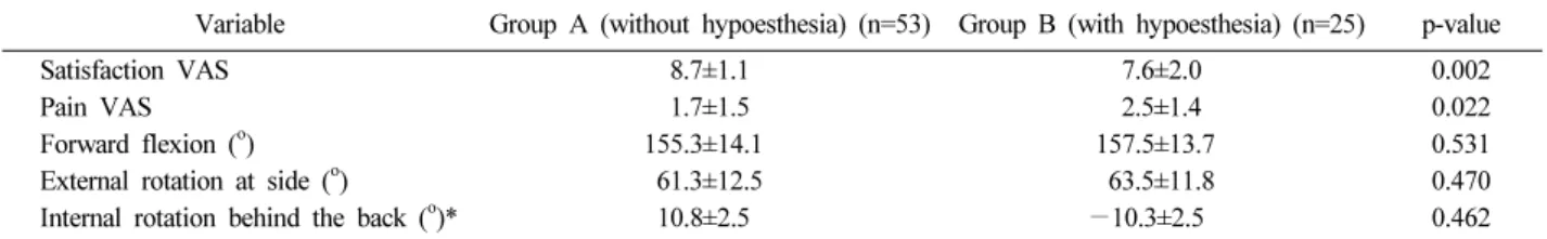
Table 2. Clinical Features of Clavicular Hypoesthesia
Variable Group A (without hypoesthesia) (n=53) Group B (with hypoesthesia) (n=25) p-value
Satisfaction VAS 8.7±1.1 7.6±2.0 0.002
Pain VAS 1.7±1.5 2.5±1.4 0.022
Forward flexion (o) 155.3±14.1 157.5±13.7 0.531
External rotation at side (o) 61.3±12.5 63.5±11.8 0.470
Internal rotation behind the back (o)* 10.8±2.5 −10.3±2.5 0.462
Values are presented as mean±standard deviation. *Measured by the vertebral level that was possible for the patient to reach with the thumb and numbered serially as 1 to 12 for the 1st to 12th thoracic vertebrae, 13 to 17 for the 1st to 15th lumbar vertebrae, and 18 for any level below the sacral vertebrae. VAS: Visual analogue scale.
cle or incision site and anterior chest and compared it to that on the contralateral side. We tried to differentiate the hypoesthesia with tingling sensation or hyperesthesia.
The severity of hypoesthesia was also assessed in three grades or as mild, moderate, and severe, and the loca- tion of hypoesthesia was assessed as medial or lateral.
The recovery of hypoesthesia was assessed at a month interval. Pain visual analogue scale (VAS; 0 to 10, with 10 defined as the worst pain), range of motion (ROM) of the shoulder, and satisfaction VAS (0 to 10, with 10 defined as highest satisfaction) were evaluated at the fi- nal follow-up (minimum 6 months). Patients without hy- poesthesia were designated as group A and patients with hypoesthesia were designated as group B.
6. Sample size calculation
At the start of the study, the sample size was calcu- lated to detect 10% difference of satisfaction VAS of pa- tients with clavicular hypoesthesia. The 10% difference was determined to be clinically significant based on the pilot study in the author’s hospital that included 20 patients. A sample size of 96 patients was required for a power of 80% at a type I error level of 0.05 and for an expected dropout rate of 20%. We also performed the post-hoc analysis with the results of satisfaction VAS in each group and the power was calculated 73%.
7. Statistical analyses
All statistical analyses were performed using the PASW software package ver. 18.0 (IBM Co., Armonk, NY, USA). Student t-test was used to evaluate differences be- tween the groups for continuous variables, and the chi-square or Fisher’s exact test was used for compar-
ison of categorical variables.
A p<0.05 was considered to indicate statistical signifi- cance.
Results
There were no significant demographic differences be- tween the two groups (all p>0.05). We found that ev- ery case of enrolled clavicle fractures revealed no pre- operative hypoesthesia, and the incidence of post- operative hypoesthesia was 32.1% (25 out of 78 pa- tients). Twenty-two patients reported that their symptoms were mild rather than moderate (p=0.917). The locations of hypoesthesia were evenly distributed (medial in 12 cases, lateral in 13 cases). The recovery from hypo- esthesia was observed in 23 out of 25 patients (p=0.008) and mean recovery time was 8.2±4.1 months (range, 3-15 months).
The medial end location of the plate was at a mean 17.5%±16.0% of the total length of the clavicle and the lateral end location of the plate was at a mean 51.0%±44.3% of the total length of the clavicle. No cor- relation was observed with respect to the location of the clavicular plate and the incidence of clavicular hypo- esthesia (p=0.666 at the medial end and p=0.369 at the lateral end).
Hypoesthesia was related to the satisfaction VAS (p=0.002). Interestingly, we observed that the pain VAS was also related to the incidence of hypoesthesia (p=0.022).
There was no significant difference with respect to the shoulder ROM between the two groups (Table 2).
Discussion
According to the results of our study, there was no correlation between the location of the plate and the in-
cidence of clavicular hypoesthesia. The incidence of clavicular hypoesthesia in this study was 32.1% and most of the cases of hypoesthesia recovered finally, which was similar to the previous results.13)
The universal transverse skin incision along the long axis of the clavicle might cause injury to branches of the supraclavicular nerve more often than the other ap- proaches such as vertical incision and minimally invasive incision and it might cause permanent neurologic dam- age or worsen the clinical outcomes.6) Some of the ana- tomical studies found that there might be two (medial and lateral) or three branches (medial, intermediate, and lateral) of the supraclaviclular nerve that vertically pass over the clavicle and the anatomical locations of these branches to the whole length of the clavicle were docu- mented to be predictable.6,14,15) However, the clinical correlation between incidence of supraclavicular nerve injury and anatomical location of the plate or extent of the approach during clavicle operation has not enoughly studied.
We hypothesized that there might be some correlation between plate location and the incidence of clavicular hypoesthesia as well as the clavicular hypoesthesia was related to the satisfaction of patients or other clinical variables such as pain of the shoulder.
In the current study, clavicular plates were located be- tween mean 5.0±1.4 cm and 8.8±5.9 cm or between mean 17.5%±16% and 51%±44.3% from the SC joint.
We observed that both the absolute distance of the lat- eral end of the plate from the SC joint (B) and the rel- ative location of the lateral end of the plate to the whole length of the plate (B/A) were in wide range;
however, the incidence of hypoesthesia was not affected by these variations in the location of the plate or extent of the approach. We considered that this might be somewhat due to the presence or absence of the inter- mediate branch of the supraclavicular nerve.14) According to Nathe et al.,14) the intermediate branch was observed in about half of the normal population and the distance from the SC joint to the intermediate branch w as be- tween 26.9% and 64.3% the length of the clavicle.
These characteristics related to the intermediate branch of the supraclavicular nerve might have resulted in the unpredictable incidence of hypoesthesia.
In this study, the satisfaction VAS score was statisti-
cally decreased in patients with clavicular hypoesthesia.
However, according to the results of a previous study comparing the overall satisfaction between the vertical and horizontal incisions during the clavicle fracture oper- ation, the patients were significantly more satisfied with the horizontal incision, and it might be due to the dif- ference in satisfaction with the scar.6) Previous studies reported that there were several patients who suffered from severe hypoesthesia after the clavicle operation.6,13) However, we observed that most of the patients en- rolled in this study reported mild hypoesthesia. Only two out of twenty-five patients reported moderate hypoesthesia. The high prevalence of mild hypoesthesia rather than moderate and severe hypoesthesia might be due to the late evaluation period at postoperative 4 weeks as well as the possible bias among the physi- cians’ evaluations. The patients might represent dimin- ished symptoms at postoperative 4 weeks compared to the postoperative two week period and physicians might evaluated the degree of hypoesthesia as mild to acquire better surgical results.
Interestingly, we observed that the pain VAS in group B (with hypoesthesia) was significantly worse than that in group A (without hypoesthesia) at the final follow-up.
We thought this might be due to the neuralgic pain during recovery that occurred in some cases of injury to branches of the supraclavicular nerve. Previous studies reported that hypoesthesia was not related to the shoulder function which was composed of pain, should- er motion, and muscle power. In this study, we did not perform functional evaluation by using scores; however, the pain VAS and ROM of the shoulder were evaluated.
No significant differences with ROMs were observed be- tween groups.
There are some limitations to this study. First, we did not identify the every branch of the supraclavicular nerve during surgery; therefore, we could not verify the actual relationship between injury to branches of the su- praclavicular nerve and clavicular hypoesthesia. Second, the degree of hypoesthesia was evaluated in only three categories such as mild, moderate, and severe which might be inappropriate for evaluating the exact degree of hypoesthesia. Finally, the post-hoc analysis revealed the 73% of power which meant that we might not made the strong conclusion with this study regarding
satisfaction of patients.
To the best of the authors’ knowledge, this is the first study to investigate the correlation betw een the lo- cation of the plate or the extent of approach and the incidence of hypoesthesia although radiological evalua- tion was performed by using the clavicle AP X-ray.
Conclusion
We could not predict the incidence of hypoesthesia by the location of clavicular plates. Clinically, the sat- isfaction and pain of patients might be related to the in- cidence of clavicular hypoesthesia and larger size cohort study might be needed in the future to get the strong conclusion regarding clavicular hypoesthesia.
References
1) Beirer M, Postl L, Crönlein M, et al: Does a minimal invasive approach reduce anterior chest wall numbness and postoperative pain in plate fixation of clavicle frac- tures? BMC Musculoskelet Disord, 16: 128, 2015.
2) Canadian Orthopaedic Trauma Society: Nonoperative treatment compared with plate fixation of displaced mid- shaft clavicular fractures. A multicenter, randomized clin- ical trial. J Bone Joint Surg Am, 89: 1-10, 2007.
3) Hill JM, McGuire MH, Crosby LA: Closed treatment of displaced middle-third fractures of the clavicle gives poor results. J Bone Joint Surg Br, 79: 537-539, 1997.
4) Jeray KJ: Acute midshaft clavicular fracture. J Am Acad Orthop Surg, 15: 239-248, 2007.
5) Khan LA, Bradnock TJ, Scott C, Robinson CM: Fractures of the clavicle. J Bone Joint Surg Am, 91: 447-460, 2009.
6) Wang K, Dowrick A, Choi J, Rahim R, Edwards E:
Post-operative numbness and patient satisfaction following plate fixation of clavicular fractures. Injury, 41: 1002-1005, 2010.
7) Zlowodzki M, Zelle BA, Cole PA, Jeray K, McKee MD;
Evidence-Based Orthopaedic Trauma Working Group:
Treatment of acute midshaft clavicle fractures: systematic review of 2144 fractures: on behalf of the Evidence-Based Orthopaedic Trauma Working Group. J Orthop Trauma, 19: 504-507, 2005.
8) McKee MD, Pedersen EM, Jones C, et al: Deficits fol- lowing nonoperative treatment of displaced midshaft clav- icular fractures. J Bone Joint Surg Am, 88: 35-40, 2006.
9) Postacchini R, Gumina S, Farsetti P, Postacchini F:
Long-term results of conservative management of midshaft clavicle fracture. Int Orthop, 34: 731-736, 2010.
10) Smekal V, Oberladstaetter J, Struve P, Krappinger D:
Shaft fractures of the clavicle: current concepts. Arch Orthop Trauma Surg, 129: 807-815, 2009.
11) Christensen TJ, Horwitz DS, Kubiak EN: Natural his- tory of anterior chest wall numbness after plating of clavi- cle fractures: educating patients. J Orthop Trauma, 28:
642-647, 2014.
12) Shen WJ, Liu TJ, Shen YS: Plate fixation of fresh dis- placed midshaft clavicle fractures. Injury, 30: 497-500, 1999.
13) Wang L, Ang M, Lee KT, Naidu G, Kwek E:
Cutaneous hypoesthesia following plate fixation in clavicle fractures. Indian J Orthop, 48: 10-13, 2014.
14) Nathe T, Tseng S, Yoo B: The anatomy of the supra- clavicular nerve during surgical approach to the clavicular shaft. Clin Orthop Relat Res, 469: 890-894, 2011.
15) OʼNeill K, Stutz C, Duvernay M, Schoenecker J: Supra- clavicular nerve entrapment and clavicular fracture. J Orthop Trauma, 26: e63-e65, 2012.
Copyright ⓒ 2016 The Korean Fracture Society. All rights reserved.
This is an Open Access article distributed under the terms of the Creative Commons Attribution Non-Commercial License (http://creativecommons.org/licenses/
by-nc/4.0) which permits unrestricted non-commercial use, distribution, and reproduction in any medium, provided the original work is properly cited.
http://dx.doi.org/10.12671/jkfs.2016.29.2.121
쇄골 간부 골절 수술후 발생한 감각저하:
금속판 위치와 연관성 및 임상상
김성훈⋅김준엽* ⋅고경환
†⋅정명곤*⋅조재호*
국민건강보험 공단 일산병원 정형외과, 서남대학교 의과대학 명지병원 정형외과*, 인제대학교 일산백병원 정형외과†
목 적: 쇄골 간부 골절에서 관혈적 정복술 및 내고정술후 발생하는 감각저하의 발생과 금속판 위치와의 관계 및 임상 양상을 평가하고자 한다.
대상 및 방법: 2013년 3월에서 2014년 10월 사이에 쇄골 간부 골절로 수술받은 78명의 환자를 대상으로 하였다. 전체 쇄골의 길이(A), 흉쇄관절에서 쇄골 금속판 내측단까지의 거리(B), 흉쇄관절에서 쇄골 금속판 외측단까지의 거리(C)를 측정하였으며, 쇄골 금속판의 위치와 감각저하 발생과의 관계를 방사선적으로 평가하였다. 또한 감각저하의 심한 정도, 회복, 환자 만족도, 통증 척도 및 운동범위도 측정하였다.
결 과: 감각저하 발생률은 32.1% (25/78)였다. 금속판의 위치와 감각저하는 연관성이 없었다(내측단, p=0.666; 외측단, p=0.369). 감각저하에서의 회복은 대부분 이루어졌다(23/25, p=0.008). 감각저하 발생은 환자 만족도와 통증 정도에 음성적인 영향이 있었다(환자 만족도, p=0.002; 통증 정도, p=0.022).
결 론: 쇄골 간부 수술 후 감각저하 발생은 금속판의 위치와 연관이 없었다. 감각저하가 발생하더라도 대부분 회복되었지만, 환자 만족도와 통증정도에 음성적인 영향을 미치므로 수술시 감각저하 발생을 줄이기 위해 노력해야 한다.
색인 단어: 쇄골, 골절 고정술, 감각저하, 연관성 연구
접수일 2016. 1. 4 수정일 2016. 2. 1 게재확정 2016. 2. 1 교신저자 김준엽
10475, 고양시 덕양구 화수로14번길 55, 서남대학교 의과대학 명지병원 정형외과 Tel 031-810-6530, Fax 031-969-0500, E-mail [email protected]
127