Journal of the Korea Academia-Industrial cooperation Society
Vol. 18, No. 8 pp. 84-87, 2017
https://doi.org/10.5762/KAIS.2017.18.8.84 ISSN 1975-4701 / eISSN 2288-4688
84
뇌과관류증후군에서 보일 수 있는 정맥울혈 1례
봉정빈, 강현구*
조선대학교의과대학 신경과학 교실
Venous Congestion in Cerebral Hyperperfusion Syndrome : A Case Report
Jeong Bin Bong, Hyun Goo Kang
*Department of Neurology, Chosun University School of Medicine
요 약 뇌과관류증후군은 경동맥 스텐트 삽입술 또는 내막 절제술 시행 시 발생할 수 있는 드문 합병증으로 대사 요구량보 다 관류가 더 많은 상태를 유발하는 다양한 기전에 의해 발생한다. 주 임상 증상은 편측성 두통, 고혈압, 발작 및 국소 신경계 결손이 있으며, 심한 경우 지주막하 출혈 및 뇌실질 출혈로 영구적 장애 또는 사망까지 유발할 수 있다. 일반적으로 뇌과관 류증후군은 두개경유도플러, 관류 뇌자기공명영상 및 단일광자방출컴퓨터단층촬영으로 진단할 수 있다. 저자들의 연구에서 는 내경동맥 스텐트 이후 확인한 혈관조영술에서 의미있는 정맥울혈 증상을 보여 뇌과관류증후군을 진단할 수 있었다. 환자 는 증상성 양쪽 내경동맥 협착을 보이고 있었고, 협착으로 인해 곁순환 동맥들이 잘 발달하게 되었다. 이렇게 곁순환 동맥이 잘 발달된 상태에서 환자에게 내경동맥 스텐트를 삽입한 이후 대뇌 혈류량이 증가되며 혈류의 방향이 바뀌어 정맥 울혈이 생길 수 있으며, 경동맥 스텐트 삽입술 또는 내막 절제술 시행 이후 정맥울혈이 보일 시 뇌과관류증후군을 예측할 수 있다.
이 연구는 내경동맥 스텐트 삽입 후 바로 시행하는 혈관 조영술을 통해서 뇌과관류증후군을 확진할 수 있음을 보여준 1례의 보고이다.
Abstract Cerebral hyperperfusion syndrome (CHS) is a rare complication that can occur when conducting stent insertion or endarterectomy in patients with carotid artery stenosis and is known to be caused by various mechanisms when the blood volume abruptly increases. The main clinical symptoms are unilateral headache, hypertension, seizure, and focal neurologic deficit. Subarachnoid hemorrhage and parenchymal hemorrhage may lead to permanent impairment or death in severe cases. CHS can be predicted by using transcranial Doppler, perfusion magnetic resonance imaging, and single photon emission computed tomography. In our case report, a patient developed CHS subsequent to significant venous congestion caused by carotid artery stent insertion. The patient had preexisting, symptomatic bilateral carotid artery stenosis. Venous congestion occurs when the direction of blood flow changes because of increased blood volume in patients with well-developed collateral vessels. We believe that CHS can be predicted from this finding. This study reports the possibility that CHS could be confirmed by cerebral angiography after insertion of the internal carotid stent.
Keywords : Cerebral hyperperfusion syndrome, Carotid artery stent, Collateral vessels, Digital subtraction angiography, Venous congestion
*Corresponding Author : Hyun Goo Kang (Chosun Univ.) Tel: +82-62-220-3182 email: [email protected] Received May 17, 2017
Accepted August 17, 2017
Revised (1st June 29, 2017, 2nd June 30, 2017) Published August 31, 2017
1. 서론
뇌과관류증후군(cerebral hyperperfusion syndrome,
CHS)은 경동맥 스텐트 삽입술 또는 내막 절제술 시행 시 발생할 수 있는 드문 합병증으로, 대사 요구량보다 관류가 더 많은 상태 또는 시술 이전 관류량보다 100%
뇌과관류증후군에서 보일 수 있는 정맥울혈 1례
85
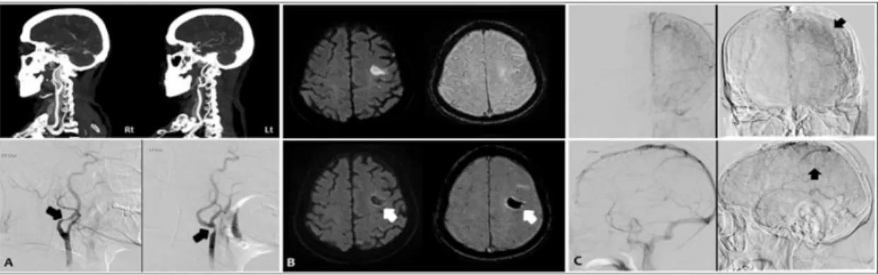
Fig. 1. (A) Bilateral carotid artery stenosis on brain CT angiography and transfemoral cerebral angiography (asymptomatic right proximal intracranial artery stenosis: 70% (arrow), symptomatic left distal common carotid artery stenosis:
65% (arrow) ). (B) Acute cerebral infarction on diffusion-weighted image (DWI) and susceptibility-weighted image (SWI) on admission (upper figure). Hemorrhagic transformation on DWI and SWI (arrow) after stent insertion in the left carotid artery (lower figure). (C) Venous congestion was observed in the left frontoparietal lobes after stent insertion (arrow).
이상 증가한 경우로 정의한다[1]. 뇌과관류증후군은 경 동맥 협착으로 뇌혈관들이 장기간 저혈류에 노출되었을 때 만성적으로 혈관의 자동 조절능이 소실되게 되는데, 이때 혈류량이 과도하게 증가하게 되면 혈관-뇌-장벽의 파괴, 혈관 외 독소 방출, 혈관성 뇌부종 및 뇌출혈 등의 이유로 뇌기능에 저하가 생겨 발생한다[2]. 뇌과관류증 후군을 유발할 수 있는 위험 요인으로는 심한 협착, 곁 순환의 저형성, 높은 혈압 및 혈관-뇌-장벽의 파괴 등이 있을 수 있는데[3-7], 뇌과관류증후군의 드문 유병률 (0.7%-3%)로 인해 정확한 병태생리학적 기전은 아직 밝혀지지 않았다[3-5]. 이 증후군의 주요 임상 증상에는 편측성 두통, 고혈압, 발작 및 국소 신경계 결손이 있으 며, 심한 경우 지주막하 출혈 및 뇌실질 출혈로 영구적 장애 또는 사망까지 유발할 수 있다. 이 증후군이 내막 절제술에서는 술 후 6일째에 가장 많이 나타나지만, 스 텐트 삽입술에서는 12시간 이내에 가장 자주 나타난다 고 알려져 있다[8]. 저자들은 경동맥 스텐트 삽입술 직 후 운동 실어증을 보이다가 24시간 이내 호전된 환자에 서 시술 전후의 뇌혈관 조영술에서 정맥 울혈을 확인하 였고 이를 통해 뇌과관류증후군을 진단한 증례를 경험 하여 보고한다.
2. 증례
76세 남자가 내원 3시간 30분 전 발생한 우반신 마비
를 주소로 내원하였다. 4년 전 관상동맥 성형술 후 cilostazol 100mg, rosuvastatin 5mg 복용 중이었으며, 그 외 고혈압, 당뇨 및 흡연 등의 위험인자는 없었다. 신 경학적 검사에서 의식은 명료하였으나 우측 상하지에 Medical research council (MRC) grade 1/2의 근력약화 와 피질경유운동실어증이 관찰되었고 National Institute of Health (NIH) 뇌졸중척도는 8점이었다. 뇌 전산화단 층촬영에서 이상소견은 없었으나 혈관조영술에서 좌측 총경동맥 원위부 및 우측 내경동맥 근위부에 심한 협착 소견이 관찰되었다(Fig. 1A). 급성기 뇌경색으로 판단하 여 조직플라스미노겐활성제를 정맥 내 투여하였고, 4시 간 후 신경학적 검사에서 우측 상하지 MRC grade 4/4, 경미한 조음장애만 보여 NIH 뇌졸중척도는 3점으로 호 전되었다. 혈전용해제 투여 24시간 뒤 시행한 뇌 자기공 명영상에서 좌측 중대뇌동맥 영역에 주로 피질 부위에 다발성 뇌경색이 관찰되었다. 3일 뒤 양측 경동맥 협착 정도를 확인 위해 카테터혈관조영술을 시행하였으며, 그 결과 NASCET 기준으로 좌측 총경동맥 원위부에 65%
및 우측 내경동맥 기시부에 70% 정도의 협착이 관찰되 었다. 이에 증상성 좌측 경동맥 협착에 대한 스텐트 삽입 을 시행하였으며, 시술 전 신경학적 후유증은 우측 하지 MRC grade 4+, 혈압은 130/80 mmHg였다. 혈관중재술 을 통해 정상적으로 스텐트를 삽입하였고 시술 중 환자 의 혈압은 정상적으로 유지되었다. 하지만 시술 종료 직 후 신경학적 검사에서 우측 상하지 MRC grade 2/3로 근력 악화 및 운동 실어증이 새로 관찰되었고, 혈압은
한국산학기술학회논문지 제18권 제8호, 2017
86 150/100 mmHg로 시술 전보다 약간 높게 측정되었다.
시술 중 새로운 뇌경색의 발생 가능성을 확인하기 위해 즉시 뇌 자기공명영상을 촬영하였고, 확산강조영상 (diffusion-weighted imaging)에서는 이전 영상과 비교 시 새로 발생한 뇌경색 병변 관찰되지 않았으나, 감수성 강조영상(susceptibility-weighted imaging)에서는 이전 경색 부위로 출혈성 변성이 관찰되었다(Fig. 1B). 또한 시술 종료 전 시행한 뇌혈관조영술에서 정맥상(venous phase)에서 울혈 소견이 관찰되었다(Fig. 1C). 이에 뇌과 관류증후군이 의심되어 정맥 내 베타차단제(labetalol)를 사용하여 수축기 혈압을 120mmHg 이하로 엄격하게 조 절하였고, 시술 종료 6시간 뒤 처음 증상 발생시보다 우 측 상하지 위약감과 실어증은 호전 추세를 보였다. 시술 종료 18시간 뒤 악화되었던 신경학적 증상은 모두 호전 되었고, 퇴원 1개월 뒤 추적관찰 시에도 상태는 변화 없 었다.
3. 고찰
본 증례는 양측 경동맥 협착이 있었던 환자에서 좌측 중대뇌동맥 영역에 급성 뇌경색이 발생한 경우로, 증상 성 좌측 경동맥협착에 대해 스텐트 삽입술 직후 신경학 적 증상이 발생하였다가 24시간 이내에 호전되어 뇌과 관류증후군으로 확인한 경우이다. 현재까지 뇌과관류증 후군을 진단하는 방법으로는 두개경유 도플러 초음파 (TCD)와 기능자기공명영상(functional MRI)을 이용하 여 뇌혈관 예비능을 평가하거나, 뇌관류 상태를 알려주 는 검사(SPECT, 관류MR)들을 통해 진단하지만 많은 비 용과 시간이 필요하다[9]. 최근 49명의 환자를 대상으로 가로정맥굴의 협착이 있는 환자와 없는 환자에서의 뇌과 관류증후군의 발생빈도를 비교하여 정맥배출의 저하가 스텐트 이후 뇌과관류증후군의 발생에 주요 역할을 할수 있다는 보고가 있었다[10]. 위 연구에서는 내경동맥으로 들어온 혈류가 내경정맥으로 빠져나가는 시간을 측정하 여 뇌과관류증후군의 발생을 예측하였다. 정맥배출의 저 하가 뇌관류증후군의 발생을 예측한다는 점에서 본 증례 와 유사하지만 본 증례는 스텐트 시술시 뇌혈관 조영술 에서 정맥울혈을 직접 확인할 수 있었다는 것에 차이가 있다.
본 증례에서 스텐트 삽입술 후 발생한 신경학적 증상 의 원인으로 뇌경색의 재발을 고려하여 확산강조영상으
로 확인하였으나 새로운 병변은 관찰되지 않아 뇌경색의 재발은 배제하였다. 대신 시술 후 증가된 혈압과 감수성 강조영상에서 이전 경색 부위로 출혈성 변성이 발생되었 다는 점, 그리고 스텐트 삽입 후 시술 종료 직전 시행한 뇌혈관 조영술에서 좌측 중대뇌동맥 영역에 정맥울혈이 관찰된 점, 그리고 엄격한 혈압 조절 후 짧은 시간안에 모든 증상들이 호전되었던 점 등으로 미루어 뇌과관류증 후군을 진단할 수 있었다[9-10].
스텐트 삽입 후 뇌혈관 조영술에서 관찰되었던 정맥 울혈 소견은 자동 조절능이 소실된 혈관들이 만성적으로 확장되어 있는 상태에서 경동맥 확장으로 인한 갑작스런 혈류량 증가로 인해 정맥으로의 혈류 배액이 늦어짐에 따라 나타나는 소견이거나, 기존에 곁순환 혈류가 뒤채 움혈관(backfilling artery)을 통해 역방향으로 들어오다 가 경동맥에서 올라오는 혈류량이 증가함에 따라 혈류 방향이 정상적으로 바뀌면서 발생하는 소견일 수 있으며 스텐트 삽입술 후 혈관 조영술을 통해 정맥울혈의 유무 를 확인함으로써 뇌과관류증후군을 예측할 수 있겠다 [10].
4. 결론
이는 내막절제술이나 스텐트삽입술 전 뇌혈관 반응도 (cerebrovascular reactivity) 측정을 통해 사전에 예측하 는 방법과 함께 뇌과관류증후군의 발생을 미리 예측하고 적극적으로 조기에 대처할 수 있는 의미 있는 소견으로 이 증례를 보고한다.
5. Reference
[1] van Mook WN, Rennenberg RJ, Schurink GW, van Oostenbrugge RJ, Mess WH, Hofman PA. "Cerebral hyperperfusion syndrome," The Lancet Neurology, vol.
4, no. 12, pp. 877-88, 2005.
DOI: https://doi.org/10.1016/S1474-4422(05)70251-9 [2] Farooq MU, Goshgarian C, Min J, Gorelick PB.
"Pathophysiology and management of reperfusion injury and hyperperfusion syndrome after carotid endarterectomy and carotid artery stenting,"
Experimental & translational stroke medicine, vol. 8, no.
1, p. 7, 2016.
DOI: https://doi.org/10.1186/s13231-016-0021-2
[3] Ogasawara K, Sakai N, Kuroiwa T., “Intracranial hemorrhage associated with cerebral hyperperfusion
뇌과관류증후군에서 보일 수 있는 정맥울혈 1례
87 syndrome following carotid endarterectomy and carotid artery stenting: retrospective review of 4494 patients,” J Neurosurg, vol. 107, pp. 1130-36, 2007.
DOI: https://doi.org/10.3171/JNS-07/12/1130
[4] Ascher E., Markevich N., Schutzer R. W., “Cerebral hyperperfusion syndrome after carotid endarterectomy:
predictive factors and hemodynamic changes,” J Vasc Surg 37, pp. 769-777, 2003.
DOI: https://doi.org/10.1067/mva.2003.231
[5] Abou-Chebl A, Yadav JS, Reginelli J. P., “Intracranial hemorrhage and hyperperfusion syndrome following carotid artery stenting: risk factors, prevention, and treatment,” J Am Coll Cardiol, vol. 43, pp. 1596-601, 2004.
DOI: https://doi.org/10.1016/j.jacc.2003.12.039
[6] Kawamata T, Okada Y, Kawashima A. “Postcarotid endarterectomy cerebral hyperperfusion can be prevented by minimizing intraoperative cerebral ischemia and strict postoperative blood pressure control under continuous sedation,” Neurosurgery, vol. 64, pp. 447-53, discussion 53-54, 2009.
[7] Ivens S, Gabriel S, Greenberg G, et al., Blood-brain barrier breakdown as a novel mechanism underlying cerebral hyperperfusion syndrome, J Neurol 257, pp.
615-20, 2010.
DOI: https://doi.org/10.1007/s00415-009-5384-z [8] Ogasawara K, Sakai N, Kuroiwa T, Hosoda K, Iihara K,
Toyoda K., "Intracranial hemorrhage associated with cerebral hyperperfusion syndrome following carotid endarterectomy and carotid artery stenting: retrospective review of 4494 patients," Journal of neurosurgery, vol.
107, no. 6, pp. 1130-6, 2007.
DOI: https://doi.org/10.3171/JNS-07/12/1130
[9] Son S, Jeong H, Choi DS, Kim R, Kim Y, Kim S.
"Changes in Cerebral Vascular Reserve Capacity after Carotid Artery Stenting: A 6-Month Follow-up Study,":J Korean Neurol Assoc, vol. 29, no. 4, pp. 295-302, 2011.
[10] Lin CJ, Chang FC, Tsai FY, Guo WY, Hung SC, Chen DY, Lin CH, Chang CY. " Stenotic Transverse Sinus Predisposes to Postenting Hyperperfusion Syndrome as Evidenced by Quantitative Analysis of Peritherapeutic Cerebral Circulation Time," Am J Neuroradiol, vol. 35, no. 6, pp. 1132-6, Jun. 2014.
강 현 구(Hyun-Goo Kang) [정회원]
•2006년 2월 : 원광대학교 의과대 학 학사
•2011년 2월 : 원광대학교 의과대 학원 석사
•2014년 5월 ~ 2015년 2월 : 현대 아산병원 신경과 전임의
•2015년 3월 ~ 현재 : 조선대학교 신경과 교수
<관심분야>
의학 및 보건학
봉 정 빈(Jeong-Bin Bong) [준회원]
•2012년 2월 : 조선대학교 의과대 학 학사
•2016년 2월 : 조선대학교 의과대 학원 석사
•2013년 9월 ~ 현재 : 조선대학교 신경과 전공의
<관심분야>
의학 및 보건학