경동맥 스텐트술은 중증의 협착을 가진 환자에서 동맥내막 절제술을 대신하여 뇌졸중을 줄일 수 있는 새로운 치료법이 다(1-7). 그러나, 스텐트술기의 발전과 항응고제의 사용에도, 스텐트 설치술시에 관련된 색전 합병증은 여전히 불가피한 것 으로 보고되고 있다(8-12). 경두개 도플러(TCD) (13)와 확 산강조 자기공명영상(Diffusion weighted MR image) (14, 15)을 이용한 연구들은 원위부 색전 합병증이 거의 모든 경 동맥 스텐트 설치술에서 일어난다고 보고 하고 있다. 이러한 원위부의 색전 합병증을 줄이기 위해 다양한 종류의 색전방 지기구들(embolic protection devices)이 고안되어 사용되고 있다. 지금까지 스텐트 설치술과 동맥내막절제술의 합병증 발 생률이 비슷한 것으로 알려졌으며, 최근의 여러 연구는 색전 방지기구를 사용하였을 때 동맥내막절제술이나 색전방지기구 를 사용하지 않은 스텐트 설치술에 비해 급성 색전 뇌졸중을 현저히 줄일 수 있었다고 보고 하고 있지만(16, 17), 스텐트 설치술과 관련된 급성 합병증을 줄이기 위해 사용되는 색전 방지기구의 유용성은 아직 정립되지 못 하였다.
이에 본 연구는 중증의 경동맥협착을 가진 환자에서 Filterwire 색전방지기구를 이용한 스텐트 설치술의 결과를 분 석하고, Filterwire 색전방지기구가 경동맥 스텐트 설치술시에 급성 합병증을 줄이는데 유용한지를 알아보고자 하였다.
대상과 방법
환자군
2004년 6월부터 12월까지 24명의 환자에서 25개 병변에 대해 Filterwire(Filterwire EXⓇ, Boston Scientific Vascular) 를 이용한 경동맥 스텐트 설치술을 시행하였다. 환자는 남자 가 20명, 여자가 4명이었으며, 평균나이는 63.3세(46-80)였 다. 모든 환자가 재발하는 일과성 뇌허헐 또는 뇌졸증 증상이 있었으며, 25개 병변 모두 70% 이상의 내경동맥 협착이 있 었다. North American Symptomatic Carotid Endarterectomy Trial(NASCET) criteria에 따른 평가에서, 14예가 95% 이 상(near-occlusion), 11예는 70% 이상(severe)의 협착이 있 었다. 3명의 환자는 내경동맥의 급성 완전 폐쇄(complete occlusion)를 보였으며, 2명의 환자는 반대편 경동맥이 이미
중증의 경동맥 협착 치료를 위한 스텐트 설치술시 색전방지기구(Filterwire)의 치료 성적
1최현욱・이태홍・김학진・김창원・김 석・추기석・이석홍
목 적: 본 연구는 중증의 경동맥협착을 가진 환자에서 Filterwire 색전방지기구를 이용한 스
텐트 설치술의 결과를 분석하고, Filterwire 색전방지기구가 경동맥 스텐트 설치술시에 급성 합병증을 줄이는데 유용한지를 알아보고자 하였다.
대상과 방법: 2004년 6월부터 12월까지 Filterwire를 이용한 경동맥 스텐트 설치술을 시행한
24명의 환자에서 25개 병변을 대상으로 분석하였다. 시술전과 시술 후에 확산강조영상을 포 함하는 뇌 자기공명영상을 시행하여 시술 후 새로운 색전병변의 유무를 평가하였다. 또한, 단 일광자방출 전산화 단층영상을 시행하여 시술 후 뇌혈류의 변화를 평가하였다.
결과: 모든 25예에서 성공적으로 스텐트설치술이 이루어졌으며, 시술 전 평균 협착은 89%(70- 100%), 시술직후 평균 협착은 거의 0%로, 100%의 기술적인 성공을 이루었다. 30일 추적관 찰에서 시술과 관련된 합병증은 1예(1/25, 4%)가 있었다. 4예(4/25, 16%)에서 술 후 확산 강조 자기공명영상에서 새로운 색전병변이 관찰되었으나, 임상적인 증상은 없었다. 시술 후 단일광자방출 전산화 단층촬영에서 뇌혈류와 뇌혈관반응성이 회복되는 소견을 보였다. 8예에 서 Filterwire와 관련된 원위부 내경동맥의 일과성 연축이 있었다(8/25, 32%).
결론: Filterwire 색전방지기구를 이용한 경동맥 스텐트 설치술은 기술적으로 적용가능하고, 안 전하며, 시술과 관련된 색전 합병증을 줄이는데 매우 효과적이었다.
1부산대학교 의과대학 진단방사선과학교실
이 논문은 2006년 8월 22일 접수하여 2006년 10월 9일에 채택되었음.
폐쇄되었던 환자였다(Table 1).
술전 평가
모든 환자는 술 전에 경험이 풍부한 신경과 또는 신경외과 의사에게 신경학적 검사를 받았으며, 술전 혈관의 병리학적 변화와 개통성을 평가하기 위해 혈관촬영술(MR angiography) 을 포함하는 뇌 자기공명영상을 시행하였다. 13명의 환자는 단일광자방출 전산화 단층촬영(SPECT)을 시행하여 뇌혈류 와 관류를 평가하였다.
모든 환자들은 두개강외 동맥과 두개강내 동맥에 대한 평 가를 위해 뇌 혈관조영술을 시행하였으며, NASCET criteria 에 따라 협착의 정도를 평가하였다. 시술 전 환자와 보호자에 게 스텐트설치술, Filterwire 색전방지기구, 그리고 시술 후 추 적관찰계획에 대해 자세히 설명하였다.
전처치
급성 내경동맥 완전 폐쇄가 있었던 3명의 환자를 제외한 21명의 환자가 적어도 시술 3일 전부터 clopidogrel(Plavix) 75 mg과 aspirin 100 mg을 복용하였다. 또한, 동일한 기간 에 low-molecular heparin인 nadroparin calcium (Flaxiparine:
Sanofi-Synthelabo, Korea)을 하루에 2-3회 피하 주사하였 다.
스텐트 설치술
모든 시술은 환자가 깨어있는 상태에서 시행하였고, 심전 도, 동맥산소 포화도와 혈압을 적절히 감시하였다. 경피적 접 근은 오른쪽 대퇴동맥을 통해 이루어졌다. 환자의 활성 혈액 응고 시간(ACT, activated clotting time)을 시술 내내 250- 300정도로 유지하기 위해, heparin을 일시 주사로 3,000- 5,000 IU를 투여하고 나서 시간당 1,000 IU를 추가로 정맥 주사하였다. 8F 유도관 (EnvoyⓇ, Cordis Endovascular Corporation, U.S.A.)을 총경동맥에 위치시킨 후 Filterwire와
delivery sheath를 협착부위를 넘어서까지 진행하고 sheath를 제거하고 Filterwire를 설치하였으며, Filterwire를 따라서 스 텐트를 진행하여 외경동맥의 기시부를 가로질러 총경동맥 분 지부에 설치하였다. 협착 부위의 직경이 Filterwire의 직경보 다 작을 경우에는 Filterwire를 설치하기 전에 0.014 inch 미 세유도철사를 협착부위를 넘어서 진행시키고 2.5-4 mm의 관 상동맥용 풍선을 이용하여 풍선확장술을 시행하였다. 스텐트 설치 후 잔여 협착(residual stenosis)이 있는 경우에 5-7 mm 크기의 풍선을 이용하여 풍선확장술을 시행하였다. 스텐 트 설치가 끝나면 Filterwire는 retrieval sheath를 이용하여 제거하였으며, Filterwire내에 눈으로 확인 가능한 색전물질이 있는지 확인하였다(Fig. 1).
시술 후에, 매일 aspirin 100 mg과 Plavix 75 mg를 투약 하였으며, 2,850 IU Flaxiparine을 적어도 3일 이상 피하주사 하였다.
시술 후 평가
스텐트 설치술 직후, 모든 환자는 신경학적 검사를 시행하 였으며, 출혈성 합병증 유무를 확인하기 위해 뇌 컴퓨터 단층 촬영을 시행하였다. 시술 후 3일 이내에 T2 강조영상(T2 weighted image), 확산강조영상(diffusion weighted image), 그리고, 액체감약반전회복영상(fluid-attenuated inversion recovery: FLAIR)을 포함하는 뇌 자기공명영상을 시행하였 으며, 시술 전 시행한 자기공명영상과 비교 평가하여 새로운 병변의 유무를 확인하였다. 또한, 단일광자방출 전산화 단층 영상 (Single photon emission computed tomography;
SPECT)을 시행하여 시술 후 뇌혈류의 변화를 평가하였다.
Filterwire내에 포획된 색전 부스러기는 병리조직학적 분석을 시행하였다.
기술적인 성공과 합병증에 대한 분석
스텐트 설치술 후 남아 있는 협착이 20% 미만인 경우를 기술적인 성공으로 정의하였다. 술중, 그리고 30일 후 추적관 찰을 통해 시술과 관련된 합병증을 기록하였다. 24시간 이내 에 완전히 회복된 대뇌 반구의 증상을 일과성 허혈증(transient Table 1. Patient Demographic Characteristics
Patient characteristics No. of patients Total no. of patients (lesions) 24 (25)
Age range(yr) (mean age) 40-80 (63.3)
M:F 20:4
Degree of carotid stenosis (mean) 70-100% (89%)
Near-occlusion (>95%) 14
Severe (>70) 11
High risk criteria
Contralateral carotid occlusion 02
Previous neck irradiation 01
Recent MI (within 3 month) 01
Comobidities
Coronary artery disease 01
Hypertension 06
Diabetes 07
Smoking 14
Hypercholesterolemia (>250 mg/dl) 01 Renal insufficiency (Cr>1.5 mg/dl) 02
Table 2. The Results of CAS with Filterwire
Results No. (%)
Technical success 25/25 (100)
Periprocedural complications 1/25 (4)
TIA, minor stroke 0
Major stroke 1
Filterwire related complications 0/25 (32)
Transient spasm of ICA 8
Dissection 0
Flow impairment 0
Visible debris within Filterwire 14/25 (56)
Yellowish material 11 (78.6)
Reddish debris 2 (14.3)
Atheromatous plaque 1 (7.1)
New lesions on Diffusion-weighted MR images 4/25 (16)
ischemic attack)으로 정의하였으며, 신경학적 결손이 30일 이 내 회복된 경우는‘경증 뇌졸중(minor stroke)’으로, 30일 이 상 지속하는 신경학적 결손이 있는 경우는 ‘중증 뇌졸중 (major stroke)’으로 정의하였다.
혈관연축, 내막박리 또는 혈류장애와 같은 Filterwire와 관 련된 합병증의 유무에 대해 평가하였으며, 자기공명영상에서
시술 후 새로이 나타난 이상신호강도에 대해 새로운 색전병 변으로 정의하였다.
결 과
모든 25예에서 성공적으로 스텐트설치술이 이루어졌으며
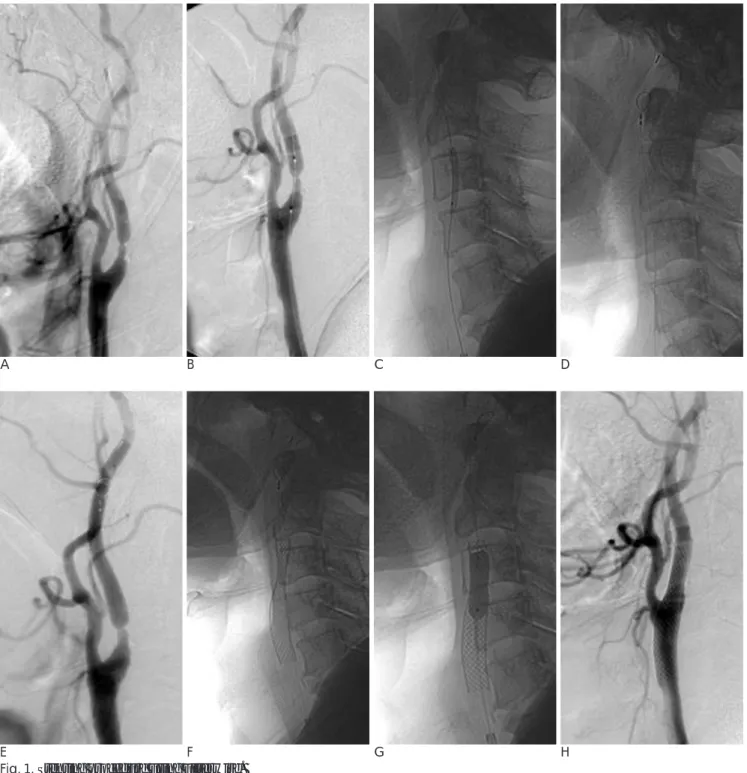
A B C D
E F G H
Fig. 1. Stenting procedure using Filterwire.
A. Angiography shows severe stenosis in left internal carotid artery.
B, C. Predilatation with low profile balloon is performed.
D, E. Filterwire and delivery sheath is passed across the lesion and Filterwire is deployed at distal internal carotid artery.
F, G. Stent is deployed and dilated.
H. Filterwire is removed and completion angiography is performed.
(Table 2), 시술 전 평균 협착은 89%(70-100%) 그리고, 시 술직후 평균 협착은 거의 0%로, 100%의 기술적인 성공을 이 루었다.
24예에서 자가팽창형 스텐트가 사용되었으며, 한 예에서는 풍선팽창형 스텐트를 설치하였다. 자가팽창형 스텐트 설치술 시에는 Filterwire의 진행을 원활하게 하고, 원위부 색전합병 증을 줄이기 위해 2.5 mm 관상동맥용 풍선을 이용하여 전확 장술(predilatation)을 시행하였다.
한 예에서 술후 중증 뇌졸중(major stroke)이 발생하였으 나 그외 30일 추적관찰에서 뇌졸중이나 심근경색 또는 사망 등은 없었다. 중증 뇌졸중이 발생하였던 한 예는 좌측 내경동 맥의 완전 폐색과 양측 추골동맥의 중증 협착이 있고 좌측 내 경동맥의 혈류는 후교통동맥을 통한 측부순환을 받고 있었던
환자로 시술과 관련된 원위부 색전합병증에 의한 것이 아니 라 스텐트 설치술 후 발생한 저혈압에 의한 저관류로 반대측 중뇌동맥 분포부에 뇌졸중이 발생하였다.
Filterwire는 25예 모두 잘 진행되고 성공적으로 설치되었 으며, 8예에서 Filterwire와 관련된 원위부 내경동맥의 일과성 연축이 있었으나, 곧 회복되었고 신경학적 증상을 남기지 않 았다. 14예에서 Filterwire에 눈으로 보이는 색전물질이 있었 으며 그 중 11예는 노란색 물질, 2예는 빨간색 부스러기 (debris), 1예는 동맥경화성 반(plaque)이었다. 병리조직학적 분석에서 이러한 색전물질은 섬유소, 혈소판, 석회화 물질, 그 리고 혈액성분으로 둘러싸인 콜레스테롤 열(cleft)과 지방 액 포(vacuole)로 구성되어 있었다.
술 후 4예에서 확산강조 자기공명영상에서 술전 영상에서
A B
Fig. 2. Images obtained in a 72-year- old man with severe stenosis of left carotid artery.
A. Left lateral angiogram shows about 90% stenosis of the left internal carotid artery.
B. Left lateral angiogram shows the re- sult after stent implantation. The tran- sient vasospasm distal to the stent is noted (arrow).
C, D. Compared with preprocedural image (C), postprocedural diffusion- weighted MR image (D) shows two new lesions (<5 mm) in left basal gan- glia, but these lesions were clinically silent.
C D
는 보이지 않던 새로운 색전병변이 관찰되었으나(Fig. 2), 임 상적인 증상은 4예 모두 없었다. 4예 중 2예는 내경동맥이 급 성 완전 폐색 되었던 환자였으며, 1예는 방사선치료에 기인 하는 중증협착이 있었던 환자였다.
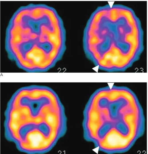
13예에서 시술 전, 후에 단일광자방출 전산화 단층촬영 (SPECT)를 시행하였는데, 13예 모두에서 시술 전과 비교하 여 시술 후 뇌혈류(CBP; cerebral blood perfusion)나 뇌혈 관반응성(CVR; cerebral vasoreactivity)이 회복되는 소견을 보였다(Fig. 3).
고 찰
경동맥내막 절제술(CEA)은 현재까지 두개강외 경동맥 폐 쇄성 질환에 대한 표준치료법으로 알려졌다. 그러나 경동맥 스텐트 설치술(CAS)이 동맥내막 절제술을 대체할 수 있는 치 료법으로 최근 새로이 부각되고 있다. 여러 연구에서 기술적 인 성공률, 시술관 관련된 이환율 (morbidity)과 사망률 (mortality), 재협착율(re-stenosis rate)등이 경동맥내막 절 제술과 비슷하다고 보고되고 있으며, 시술관련 뇌졸중 발생률 과 사망률은 약 6% 정도로 경동맥내막 절제술과 비슷한 것 으로 보고되고 있다. 그러나, Jordan 등(18)은 스텐트 설치술
(CAS)이 경동맥내막 절제술(CEA)에 비해 조기 경증 뇌졸중 발생률(6.6% vs. 0.6%)과 30일 내 뇌졸중 발생률과 사망률 (9.7% vs. 0.9%)이 높은 것으로 보고 하였으며 이는 대부분 술중 색전 합병증에 의한 것이었다. 그래서 술중 색전 합병증 을 막기 위한 다양한 색전방지기구가 현재까지 연구되고 있 다.
Theron 등(19)이 최초로 풍선을 이용한 색전방지기구를 고 안하여 색전 합병증이 감소하였다고 보고 한 이후로, 풍선 또 는 여과장치와 같은 다양한 색전방지기구가 연구되어 임상실 험이 시행되고 있다. Kastrup 등(20)은 경동맥 스텐트 설치 술의 30일 이내의 경증 뇌졸중, 중증 뇌졸중 그리고 사망률 에 관한 전반적인 고찰을 하였는데, 색전방지기구를 사용한 환자군에서 30일내 모든 합병증 발생률은 1.8%로 색전방지 기구를 사용하지 않은 환자군의 5.5%에 비해 현저히 낮았다 고 보고하고 있다. 또한, Cremonesi 등(21)은 색전방지기구 를 이용한 442명의 환자에게서의 30일내 합병증 발생률이 약 3.4%였다고 보고하였다. 이런 연구들은 색전방지기구의 사용 이 경동맥 스텐트 설치술에 있어 급성 합병증을 줄이는데 아 주 효과적이라는 것을 보여주고 있다.
본 연구에서는 1예의 증증 뇌졸중이 발생하였는데(1/25, 4%), 이는 색전 합병증이나 Filterwire와는 무관하게 반대측
A
Fig. 3. SPECT image of 75-year-old man with severe stenosis of right inter- nal carotid artery.
A. Pre-procedural images reveal de- creased blood flow in right internal cerebral artery territory (arrowheads).
B. Post-procedural images show mar- ked recovery of regional blood flow (arrowheads).
B
중뇌동맥 분포영역에 발생하였으며, 경동맥 압수용체에 대한 직접적인 자극으로 인한 스텐트 설치술 후 저혈압(post-CAS hypotension)에 기인한 것이었다(22, 23). 환자는 스텐트 설 치술 직후, 출혈 등이 없었으며 적절한 약물투여와 체액 보충 에도, 수축기 혈압이 90 mmHg이하로 측정되었다. 확산강조 자기공명 영상에서 중뇌동맥 분포영역에 새로운 고신호강도 의 병변이 관찰되었다. 이 환자는 이미 반대측 내경동맥이 막 혀 있었으며, 후교통동맥을 통해 측부순환을 받고 있었는데, 뇌관류의 저하로 말미암아 측부순환을 통한 혈류가 감소하면 서 반대측 중뇌동맥 영역의 뇌경색이 온 것으로 추정되었다.
Filterwire를 사용한 최근의 연구들(24, 25)은 일부 일과성 허혈증이 발생한 것을 제외하고는 30일 내 색전 합병증이 거 의 발생하지 않은 것으로 보고하고 있다. 저자들의 연구에서 도 모든 환자가 중증의 협착이 있었음에도 동측의 색전 합병 증은 발생하지 않았다.
지금까지 30일 이내의 뇌졸중 발생률과 사망률에서 풍선차 단기구 시술군와 여과장치 시술군 사이에 의미 있는 차이는 없는 것으로 알려져 오고 있다. 풍선차단기구는 병변을 지나 기에 적절한 굵기가 있으나, 수 분 동안 상행하는 혈류가 완 전히 차단되기 때문에 일부 환자는 사용할 수 없다. 반면에 여과장치는 더 굵고 강직하지만, 사용이 단순하며 상행혈류를 잘 유지하며 시술중 혈관내경을 잘 볼 수 있기 때문에 더욱 정확한 위치에 스텐트를 설치할 수 있는 장점이 있다(8).
Filterwire와 같은 여과장치에 있는 구멍을 통해서도 색전 물질이 빠져나갈 가능성이 있다. 그러나 이전의 연구들(26, 27)은 색전과 임상적 후유증 간에 상관관계가 없다고 보고하 고 있으며, 대다수의 색전 물질은 200 μm미만의 혈소판응집 이었으며 예후가 좋았다고 보고하고 있다. 여과장치를 사용하 더라도 여과장치를 펼칠 때와 접을 때에는 불완전한 색전방 어가 이루어질 수 있으며(25), 여과장치가 협착부위를 가로 질러 진행할 때와 풍선 전확장술(predilatation)을 시행할 때 에도 원위부로의 색전이 일어날 가능성이 있다.
시술 후 확산강조 자기공명영상에서 새로이 고강도신호의 병변은 4예에서 관찰되었으나(4/25, 16%), 임상적인 증상은 없었다. 이전의 확산자기공명영상을 이용한 연구(15)와 비교 할 때, Filterwire를 사용한 본 연구에서는 모든 예에서 중증 이상의 경동맥 협착이 있었지만 색전을 나타내는 고강도신호 의 새로운 병변은 감소한 것으로 나타났다(16% vs. 29%).
13예에서 시술전후 뇌관류를 포함하는 단일광자방출 전산 화 단층촬영을 시행하였으며 13예 모두에서 술전과 비교하여 술후에 뇌관류(CBP) 또는 뇌혈관반응성(CVR)이 정상화되는 소견을 보였다.
혈관내막손상, 일과성 혈관연축 또는 혈류장애와 같은 여과 장치와 관련된 합병증에 대한 연구가 보고되고 있다(21, 28).
Cremonesi 등(21)은 두개강내 동맥의 일과성 혈관연축과 많 은 양의 색전 절편이 의한 내경동맥의 혈류장애(13.1%)를 보 고하고 있다. 저자들의 연구에서는 8예에서 여과장치 바스켓 이 놓였던 원위부 내경동맥의 주위 혈관벽에 일과성 혈관연 축이 있었으나, 8예 모두 신경학적 증상을 남기지 않고 회복
되었다.
저자들의 Filterwire 바스켓에 걸린 색전 물질들은 조직병 리학적 분석 결과 섬유소, 혈소판, 석회화 물질, 그리고 혈액 성분으로 둘러싸인 콜레스테롤 열 (cleft)과 지방 액포 (vacuole)로 구성되어 있었다. 이는 이전의 타 연구결과(4, 8, 11)와 같았다.
이번 연구는 비교적 적은 환자군을 대상으로 분석하였으며, 적절한 대조군이 없다는 제한점이 있다. 또한, 술전 시행한 자 기공명영상의 시간간격이 일정하지 않으며, 다수의 환자가 외 부병원에서 자기공명영상을 시행한 후 의뢰되어 영상연쇄 (sequence)와 영상절편(slice)이 다양하게 시행되어서 술후의 새로운 병변에 대한 파악에 제한이 있었다.
결론적으로, Filterwire 색전방지기구를 이용한 경동맥 스텐 트 설치술은 기술적으로 적용가능하고, 안전하며, 시술과 관 련된 색전 합병증을 줄이는데 매우 효과적이었다. 다른 색전 방지기구, 그리고 경동맥내막 절제술과의 비교평가를 위해서 는 대단위 환자군을 대상으로 하는 장기간의 추적연구가 필 요할 것으로 생각된다.
참 고 문 헌
1. Lin PH, Bush RL, Lumsden AB. Carotid artery stenting: current status and future directions. Vasc Endovascular Surg 2003;37:315- 322
2. Castriota F, Cremonesi A, Manetti R, Liso A, Oshola K, Ricci E, et al. Impact of cerebral protection devices on early outcome of carotid stenting. J Endovasc Ther 2002;9(6):786-792
3. Roubin GS, New G, Iyer SS, Vitel JJ, Al-Mubarak N, Liu MW, Yadav J, et al. Immediate and late clinical outcomes of carotid artery stenting in patients with symptomatic and asymptomatic carotid artery stenosis: 5-year prospective analysis. Circulation 2001;103:532-537
4. Whitlow PL, Lylyk P, Londero H, Mendiz OA, Mathias K, Jaeger H, et al. Carotid artery stenting protected with an emboli contain- ment system. Stroke 2002;33(5):1308-1314
5. Vitek JJ, Roubin GS, Al-Mubarak N, New G, Iyer SS. Carotid artery stenting: technical considerations. AJNR Am J Neuroradiol 2000;21(9): 1736-1743
6. Endovascular versus surgical treatment in patients with carotid stenosis in the Carotid and Vertebral Artery Transluminal Angioplasty Study (CAVATAS): a randomized trial. Lancet 2001;357:1729-1737
7. Alberts MJ. Results of a multicenter prospective randomized trial of carotid artery stenting versus carotid endarterectomy. Stroke 2001;32:325
8. Reimers B, Corvaja N, Moshiri S, Sacca S, Albiero R, Di Mario C, et al. Cerebral protection with filter devices during carotid artery stenting. Circulation 2001;104:12-15
9. Muller-Hulsbeck S, Husler EJ, Schaffner SR, Jahnke T, Glass C, Wenke R, et al. An in vitro analysis of a carotid artery stent with a protective porous membrane. J Vasc Interv Radiol 2004;15:1295- 1305
10. Sievert H, Rabe K. Role of distal protection during carotid stenting.
J Interv Cardiol 2002;15:499-504
11. Lin PH, Bush RL, Lubbe DF, Cox MM, Zhou W, McCoy SA, et al.
Carotid artery stenting with routine cerebral protection in high-
risk patients. Am J Surg 2004;188:644-652
12. van Heesewijk HP, Vos JA, Louwerse ES, Van Den Berg JC, Overtoom TT, Ernst SM, et al. Carotid PTA and Stenting Collaborative Research Group. New brain lesions at MR imaging after carotid angioplasty and stent placement. Radiology 2002;224:361-365
13. Jordan WD Jr, Voellinger DC, Doblar DD, Plyushcheva NP, Fisher WS, McDowell HA. Microemboli detected by transcranial Doppler monitoring in patients during carotid angioplasty versus carotid endarterectomy. Cardiovascular Surg 1999;7:33-38
14. Jaeger HJ, Mathias KD, Drescher R, Hauth E, Bockisch G, Demirel E, et al. Diffusion-weighted MR imaging after angioplasty or angio- plasty plus stenting of arteries supplying the brain. AJNR Am J Neuroradiol 2001;22:1251-1259
15. Jaeger HJ, Mathias KD, Hauth E, Drescher R, Gissler HM, Hennigs S, et al. Cerebral ischemia detected with diffusion-weight- ed MR imaging after stent implantation in the carotid artery. AJNR Am J Neuroradiol 2002;23:200-207
16. Yadav J. Stenting and Angioplasty with Protection in Patients at High Risk for Endarterectomy (SAPPHIRE). Chicago, III: American Heart Association; 2002
17. Mas JL, Chatellier G, Beyssen B; EVA-3S Investigators. Carotid an- gioplasty and stenting with and without cerebral protection: clini- cal alert from the endarterectomy versus angioplasty in patients with symptomatic severe carotid stenosis (EVA-3S) trial. Stroke 2004;35:18-20
18. Jordan WD Jr, Schroeder PT, Fisher WS, McDowell HA. A com- parison of angioplasty with stenting versus endarterectomy for the treatment of carotid artery stenosis. Ann Vasc Surg 1997;11:2-8 19. Theron JG, Payelle GG, Coskun O, Huet HF, Guimaraens L.
Carotid artery stenosis: treatment with protected balloon angio- plasty and stent placement. Radiology 1996;201:627-636
20. Kastrup A, Groschel K, Krapf H, Brehm BR, Dichgans J, Schulz JB.
et al. Early outcome of carotid angioplasty and stenting with and without cerebral protection devices. Stroke 2003;34:813-819 21. Cremonesi A, Manetti R, Setacci F, Setacci C, Castriota F.
Protected carotid stenting: clinical advantages and complications of embolic protection devices in 442 consecutive patients. Stroke 2003;34:1941-1943
22. Dangas G, Laird JR Jr, Satler LF, Mehran R, Mintz GS, Larrain G, et al. Postprocedural hypotension after carotid artery stent place- ment: predictors and short- and long-term clinical outcomes.
Radiology 2000;215:677-683
23. Mangin L, Medigue C, Merle JC, Macquin-Mavier I, Duvaldestin P, Monti A, et al. Cardiac autonomic control during balloon carotid angioplasty and stenting. Can J Physiol Pharmacol 2003;81:944-951 24. Bosiers M, Peeters P, Verbist J, Schroe H, Deloose K, Lauwers G,
et al. Belgian experience with FilterWire EX in the prevention of embolic events during carotid stenting. J Endovasc Ther 2003;10:695-701
25. Grube E, Colombo A, Hauptmann E, Londero H, Reifart N, Gerckens U, et al. Initial multicenter experience with a novel distal protection filter during carotid artery stent implantation. Catheter Cardiovasc Interv 2003;58:139-146
26. Crawley F, Clifton A, Buckenham T, Loosemore T, Taylor RS, Brown MM. Comparison of hemodynamic cerebral ischemia and microembolic signals detected during carotid angioplasty. Stroke 1997;28:2460-2464
27. Bladin CF, Bingham L, Grigg L, Yapanis AG, Gerraty R, Davis SM.
Transcranial Doppler detection of microemboli during percuta- neous transluminal angioplasty. Stroke 1998;29:2367-2370 28. Cardaioli P, Giordan M, Panfili M, Chioin R. Complication with an
embolic protection device during carotid angioplasty. Catheter Cardiovasc Interv 2004;62:234-236
J Korean Radiol Soc 2007;56:119-126
Address reprint requests to : Tae Hong Lee, M.D., Department of Diagnostic Radiology, Pusan National University College of Medicine 1-10, Ami-dong, Seo-gu, Busan 602-739, Korea.
Tel. 82-51-240-7354 Fax. 82-51-244-7534 E-mail: [email protected]
Results of Carotid Artery Stenting using Filter Wire in 24 Consecutive Symptomatic Patients with Severe Stenosis
1Hyun Wook Choi, M.D., Tae Hong Lee, M.D., Hak Jin Kim, M.D., Chang Won Kim, M.D., Suk Kim, M.D., Ki Seok Choo, M.D., Suk Hong Lee, M.D.
1Department of Diagnostic Radiology, Pusan National University College of Medicine
Purpose: The aims of this study were to analyze the results of carotid artery stenting using distal protection with FilterWire, and evaluate the effectiveness of FilterWire for distal embolic protection.
Materials and Methods: Between June and December in 2004, elective carotid artery stenting using FilterWire was attempted in 25 lesions of 24 consecutive patients. All patients were symptomatic, with recurrent tran- sient ischemic attacks (TIA) or a stroke. The cerebral ischemic lesions of embolic origin were evaluated before and after the procedure using magnetic resonance imaging, including diffusion-weighted images. Both pre- and post-procedural 99mcTc-ECD SPECT were performed to assess the cerebral blood flows. Any visible de- bris within the FilterWire was sent for histological/cytological analyses.
Results: Technical success was achieved all 25 cases. The mean pre-procedural stenosis was 89% (range 70- 100%), and that immediately after stent placement was nearly 0%. With the exception of only one major stroke (1/25, 4%), no periprocedural complications were encountered. On the diffusion weighted images, new lesions were observed in four patients (4/25, 16%), but these were clinically silent. FilterWire-related transient spasm occurred in eight of the 25 procedures (32%).
Conclusion: Carotid artery stenting, with FireWire distal protection, seems technically feasible, safe and effec- tive in preventing procedural related embolic complications.
Index words :Carotid arteries, stenosis or occlusion Arteries, transluminal angioplasty Carotid arteries
Stents and prostheses