• 검색 결과가 없습니다.

Routine Shunting is Safe and Reliable for Cerebral Perfusion during Carotid Endarterectomy in Symptomatic Carotid Stenosis

N/A
N/A
Protected

Academic year: 2021

Share "Routine Shunting is Safe and Reliable for Cerebral Perfusion during Carotid Endarterectomy in Symptomatic Carotid Stenosis"

Copied!
6
0
0

로드 중.... (전체 텍스트 보기)

전체 글

(1)

http://dx.doi.org/10.5090/kjtcs.2012.45.2.95 ISSN: 2233-601X (Print) ISSN: 2093-6516 (Online)

Department of Thoracic and Cardiovascular Surgery, Division of Neurology, Chonbuk National University Hospital, Chonbuk National University Medical School

Received: August 22, 2011, Revised: September 14, 2011, Accepted: October 16, 2011

Corresponding author: Jong Bum Choi, Department of Thoracic and Cardiovascular Surgery, Chonbuk National University Hospital, Chonbuk National University Medical School, 20 Geonji-ro, Deokjin-gu, Jeonju 561-712, Korea

(Tel) 82-63-250-1486 (Fax) 82-63-250-1480 (E-mail) jobchoi@chonbuk.ac.kr

C

The Korean Society for Thoracic and Cardiovascular Surgery. 2012. All right reserved.

CC
This is an open access article distributed under the terms of the Creative Commons Attribution Non-Commercial License (http://creative- commons.org/licenses/by-nc/3.0) which permits unrestricted non-commercial use, distribution, and reproduction in any medium, provided the original work is properly cited.

Routine Shunting is Safe and Reliable for Cerebral Perfusion during Carotid Endarterectomy in Symptomatic Carotid Stenosis

Tae Yun Kim, M.D., Jong Bum Choi, M.D., Kyung Hwa Kim, M.D., Min Ho Kim, M.D., Byoung-Soo Shin, M.D., Hyun Kyu Park, M.D.

Background: The purpose of this report is to describe the perioperative outcomes of standard carotid endarter- ectomy (CEA) with general anesthesia, routine shunting, and tissue patching in symptomatic carotid stenoses.

Materials and Methods: Between October 2007 and July 2011, 22 patients with symptomatic carotid stenosis (male/female, 19/3; mean age, 67.2±9.4 years) underwent a combined total of 23 CEAs using a standardized technique. The strict surgical protocol included general anesthesia and standard carotid bifurcation endarterectomy with routine shunting. The 8-French Pruitt-Inahara shunt was used in all the patients. Results: During the ischemic time, the shunts were inserted within 2.5 minutes, and 5 patients (22.7%) revealed ischemic cerebral signals (flat wave) in electroencephalographic monitoring but recovered soon after insertion of the shunt. The mean shunting time for CEA was 59.1±10.3 minutes. There was no perioperative mortality or even minor stroke. All patients woke up in the operating room or the operative care room before being moved to the ward. One patient had difficulty swallowing due to hypoglossal nerve palsy, but had completely recovered by 1 month postsurgery. Conclusion:

Routine shunting is suggested to be a safe and reliable method of brain perfusion and protection during CEA in symptomatic carotid stenoses.

Key words: 1. Carotid arteries 2. Endarterectomy

3. Surgical procedures, operative 4. Shunts

INTRODUCTION

Carotid endarterectomy (CEA) is better than medical ther- apy in treating symptomatic carotid stenosis of more than 50% and preventing major strokes [1,2], but the surgical team needs to evaluate the occurence of perioperative neurologic complications including major and minor strokes, especially in symptomatic carotid stenosis.

Carotid artery surgery raises some problems such as patient selection, determining the safest and most reliable technique (standard versus eversion CEA, direct suture versus patch an- gioplasty), the use of general or locoregional anesthesia, in- tra-procedural cerebral protection, and whether or not to use a shunt and, if so, the kind of shunt to use.

Recently, with the development of cerebral protection de-

vices for carotid artery stenting [3], the number of surgical

(2)

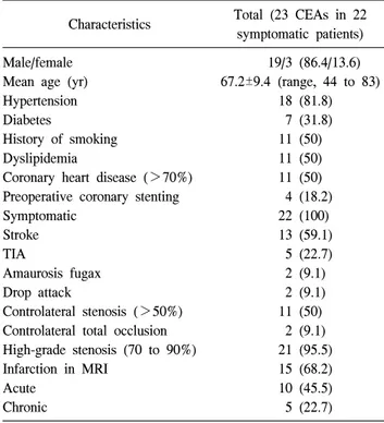
Table 1. Characteristics of the patients undergoing carotid endar- terectomy

Characteristics Total (23 CEAs in 22 symptomatic patients) Male/female

Mean age (yr) Hypertension Diabetes

History of smoking Dyslipidemia

Coronary heart disease (>70%) Preoperative coronary stenting Symptomatic

Stroke TIA

Amaurosis fugax Drop attack

Controlateral stenosis (>50%) Controlateral total occlusion High-grade stenosis (70 to 90%) Infarction in MRI

Acute Chronic

19/3 (86.4/13.6) 67.2±9.4 (range, 44 to 83)

18 (81.8) 7 (31.8) 11 (50) 11 (50) 11 (50) 4 (18.2)

22 (100) 13 (59.1) 5 (22.7) 2 (9.1) 2 (9.1) 11 (50) 2 (9.1) 21 (95.5) 15 (68.2) 10 (45.5) 5 (22.7) Values are presented as mean±standard deviation or number (%).

CEA=carotid endarterectomy; TIA=transient ischemic attack;

MRI=magnetic resonance imaging.

Fig. 1. (A) Preoperative four-vessel angiogram and (B) postoperative co- mputed tomography carotid angio- gram. The carotid stenosis (white ar- row) was repaired with endarterecto- my and patch angioplasty (white ar- rowhead).

cases has been decreasing and most of them include the symptomatic cases that are unsuitable for stenting. The main issue that surgeons face in surgical therapy of carotid artery stenosis is determining the safety of the surgical methods for treating symptomatic carotid stenosis.

The aim of this report is to describe the perioperative and early outcomes of standard CEA performed under general an- esthesia with routine shunting and patch angioplasty to show

that routine shunting is a safe and reliable method of cerebral perfusion and protection during CEA, especially in most pa- tients in whom a stenting procedure would be inappropriate.

MATERIALS AND METHODS

Between October 2007 and July 2011, 22 patients with symptomatic carotid stenosis (male/female, 19/3; mean age, 67.2±9.4 years) underwent a combined total of 23 CEAs us- ing a standardized technique. Eleven patients (50%) had a significant coronary artery disease; four of them (18.2%) un- derwent preoperative coronary artery stenting, and the remain- ing seven patients (31.8%) did not undergo revascularization.

The indication for surgery was one of the three following criteria: a high-grade (range, 70 to 99%) carotid stenosis as revealed by a duplex scan evaluated by a neurologist, more than 50% of carotid stenosis associated with a large aneur- ysm, or vulnerable ulceration in the symptomatic patients. All patients underwent a magnetic resonance imaging (MRI) and magnetic resonance angiography (MRA) to evaluate the intra- cerebral lesions and intra- and extra-cerebral vascular lesions.

Four-vessel angiography or 3-dimensional (3D) computed to- mography (CT) carotid angiography was then used to accu- rately evaluate the extent of the carotid stenosis. Twenty pa- tients (90.9%) had received combination therapy of anti-plate- let agents (aspirin 80 mg/day and clopidogrel 75 mg/day) be- fore surgery. Electroencephalography (EEG) was performed to continuously monitor intraoperative cerebral perfusion during CEA with shunting. The patients’ characteristics are shown in Table 1.

We recorded the total shunting time taken for CEA. On the

(3)

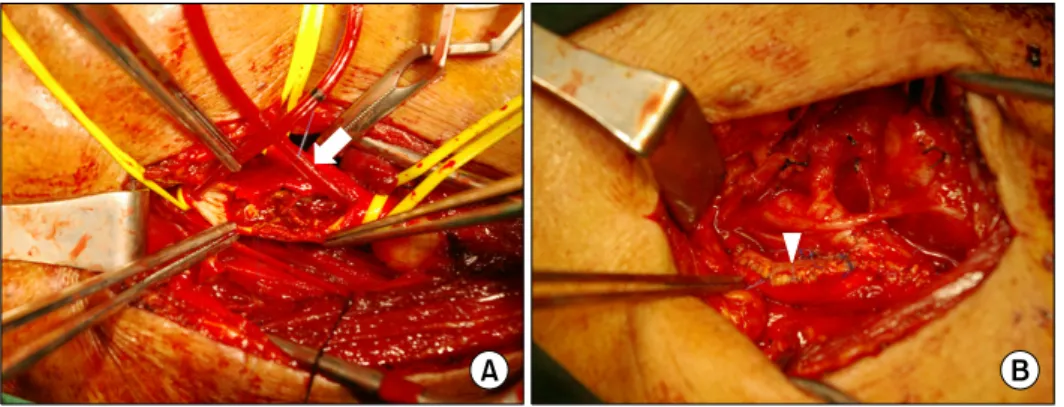
Fig. 2. Operative view. (A) A shunt (white arrow) was placed to maintain cerebral flow during carotid endarte- rectomy. (B) A bovine pericardial pa- tch (white arrowhead) was used to close the arteriotomy with a patch angioplasty technique.

Table 2. Reasons for which patients (n=16) were not appropriate candidates for carotid artery stenting

Contraindications Number (%)

Inaccessibility for stenting Friable thrombus Large aneurysm Deep ulceration

Severe controlateral lesion Calcification

4 (17.4) 2 (8.7) 2 (true, 1; false, 1) (8.7)

2 (8.7) 5 (21.7)

1 (4.3) third to fifth postoperative day, 3D CT carotid angiography

was performed to evaluate the patch-augmented arterial lumen in the carotid artery (Fig. 1). In the perioperative data, con- tinuous variables were expressed as mean±standard deviation (SD).

Operative management

All of the operations were performed by following a strict protocol involving general anesthesia, routine shunting (Pruitt- Inahara carotid shunt with T-port; LeMaitre Vascular Inc., Burlington, MA, USA) (Fig. 2A) and an autologous greater saphenous vein patch or a bovine pericardial strip (Vascu- Guard peripheral vascular patch; Synovis Life Technologies Inc., St. Paul, MN, USA) (Fig. 2B).

The carotid artery stenosis was approached through a longi- tudinal incision parallel to the medial border of the sternoclei- domastoid muscle, with intravenous heparin (5,000 units) be- ing administered before clamping. The common carotid artery (CCA) and the internal carotid artery (ICA) were fully dis- sected with minimal touching of the stenotic lesion site, which was predicted in the preoperative angiographic films (Fig. 1A). Two vascular loops were doubly passed around the CCA and ICA, respectively. The carotid body was gently in- jected with 1% xylocaine. At that time, the hypoglossal nerve was identified and exposed in the operating field in order to avoid touching the nerve during surgery. The CCA and ICA were temporarily clamped, avoiding the intraluminal athero- sclerotic lesion. The ICA was incised in the distal intact arte- rial wall, and the distal end of the shunt (closed with a small Kelly clamp) was inserted into the distal ICA and fixed with a 0.5 mL saline balloon and double ligation of the vessel

loop. The shunt was filled with blood that was retrograde flown from the ICA and clamped. The proximal end of the shunt was inserted into the CCA and fixed with a 1.5 mL balloon and double ligation of the vessel loop. Two clamps on the shunt were released in turn to evacuate air through the central arm of the shunt, and then free flow was established from the CCA to the ICA by releasing the clamps. A longi- tudinal incision was made from the distal shunt site to the proximal shunt site; the intraluminal atheroma was exposed.

After the endarterectomy, the arteriotomy was closed using

an autologous saphenous vein patch or a bovine pericardial

strip of 9.0 mm width (Vascu-Guard patch) and 6/0 poly-

propylene sutures (Fig. 2B). The shunt was removed before

the completion of the suture. The artery was then briefly

clamped proximally and distally to the patch angioplasty por-

tion, and the suture was finally completed. In cases with a

bleeding tendency, a small Penrose strip drain was placed

through a separate small incision opening and removed on

postoperative day two.

(4)

Table 3. Surgical findings and procedures

Characteristics Number (%)

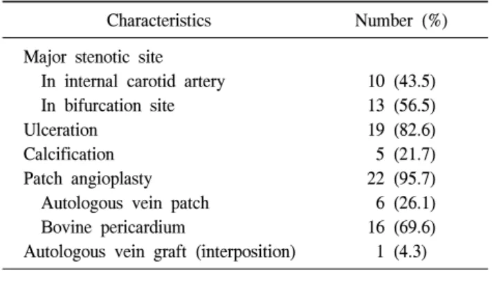
Major stenotic site In internal carotid artery In bifurcation site Ulceration

Calcification Patch angioplasty Autologous vein patch Bovine pericardium

Autologous vein graft (interposition)

10 (43.5) 13 (56.5) 19 (82.6) 5 (21.7) 22 (95.7) 6 (26.1) 16 (69.6) 1 (4.3)

Table 4. Postoperative mortality and morbidity

Operative results Number (%)

Hospital mortality Major or minor stroke

Swallowing difficulty for 1 month (major hypoglossal nerve palsy) Re-exploration for wound hematoma

0 0 1 (4.3)

1 (4.3)

RESULTS

Sixteen patients (72.7%) were unsuitable for carotid artery stenting (CAS) (Table 2). Two patients had previous carotid stenting in the controlateral carotid lesion, but the endarter- ectomy side was inappropriate for carotid stenting due to the marked angle at the bifurcation of the CCA and eccentric stenosis due to calcification. One patient with two carotid stenoses underwent CEAs in two week intervals.

The 8-French Pruitt-Inahara shunt was the only shunt used and was easily inserted in all the cases. Its insertion could be performed within 2.5 minutes. During the short clamping time for insertion of the shunt, five patients (22.7%) revealed a sudden onset of flat cerebral signals on the EEG, but the ischemic signals disappeared soon after the shunting was performed.

The mean shunting time for endarterectomy and patch an- gioplasty was 59.1±10.3 minutes (range, 45 to 71 minutes).

The mean intraluminal pressure in the distal ICA after clamp- ing was 42.8±14.5 mmHg (range, 27 to 70 mmHg), which was measured in 11 patients who did not show ischemic cer- ebral signals on electroencephalography during insertion of the shunt.

All patients except one with a false aneurysm had patch angioplasty at both bifurcation and internal carotid artery sites (Table 3).

There was no postoperative mortality or even minor stroke.

Postoperatively, one patient had difficulty swallowing due to a left hypoglossal nerve palsy, which was possibly compli- cated by marked traction. The complication was treated 30 days after Levin tube feeding (Table 4). One other patient

had a temporary minor nerve palsy of the hypoglossal nerve, but recovered before discharge.

All patients were extubated in the operating room or in the postoperative recovery room before being moved to the ward.

DISCUSSION

Shunting during CEA remains controversial. The elective application of temporary shunting was advocated by Thom- pson et al. [4] in the early 1960s. The practice of intraopera- tive cerebral monitoring varies among surgeons and insti- tutions depending on the surgeons’ preferences, technical re- sources, and the fact that no single method is superior in all aspects [5]. Some surgeons have used it routinely, such as Thompson [6], some have used it selectively based on the as- sessment of the cerebral collateral circulation, and a few have stated that they have rarely or never used it [7,8]. Shunting is performed only to prevent ischemic stroke, and it may be in- herently risky for embolic stroke if performed poorly. Intra- operative cerebral ischemia is a relatively uncommon cause of intraoperative stroke [8]. This would support the argument of the routine non-shunters, who argue that because cerebral is- chemia is a rare cause of stroke and shunting may do more harm than good, then even selective shunting is never justi- fied. However, there is no denying that when cerebral ische- mia does occur, it can lead to perioperative stroke [9,10].

Intraoperative EEG is a reliable method for evaluating routine

shunting in CEA. Some authors have reported good post-

operative results with selective use of shunting for CEA

[11,12]. If the shunt is prepared only when a cerebral ische-

mic signal is identified on EEG, the cerebral ischemic time

will be prolonged. However, routine stenting can prevent pro-

longed cerebral ischemic time because it is an ordinary and

comfortable procedure for surgeons who always use the meth-

(5)

od for CEA. Routine shunting needs to be comfortable in cases with a long operation time, especially as in our cases with a mean shunting time of 59.1 minutes. We worried about our relatively long operation time, but we were com- fortable using the shunt for preventing cerebral ischemia. The benefit is realin the case that showed an ischemic cerebral signal late on EEG during CEA [13]. Our cases had longer operating (shunting) time than other cases that have been re- ported [14], but even minor stroke did not occur, although all the patients were symptomatic and many had severe lesions that were inappropriate for stenting. Recently, the number of surgical methods for carotid artery stenosis has decreased with the development of stenting devices. The infrequent op- erations and the increase of complex cases were causes of the extended operation time. The other causes that extended the operation time were long arteriotomy for seeking intact arte- rial lumen in which to insert the shunt and augmentation us- ing a long pericardial patch.

The etiology of intraoperative stroke may be ischemic or embolic. Most perioperative strokes were due to technical er- rors made during CEA; the strokes were related to difficulties in the shunt placement. This was more common in the pa- tients with controlateral carotid artery occlusion or pre- operative stroke. Some authors have demonstrated worse re- sults when shunting was used in selective patients with ence- phalic ischemic signs during various cerebral protection sys- tems [15,16]. All of these authors have suggested that neuro- logical complications are directly attributable to intraluminal shunting. The complications are related to the atherosclerotic lesion in the insertion site of the shunt because emboli de- bridement passes through the shunt from the atherosclerotic lesion. This complication can be reduced if the proximal and distal ends of the intraluminal atherosclerotic lesion are accu- rately defined in the angiographic films. In order to reduce that complication, four-vessel angiography or 3D CT angiog- raphy and a duplex scan is required for diagnosis. We per- formed the angiography to define the intraluminal extent of the atherosclerotic lesion in addition to the duplex scan and MRI/MRA in all cases. That allows surgeons the ability to predict convenient sites for inserting the shunt before a long arteriotomy for CEA is performed.

There are some reasons for believing that the shunt com-

plications that have been reported in the literature may have been due to the placement difficulties that resulted from a surgeon who performed a hurried and imprecise surgery and also did not routinely perform the procedure. This could be particularly likely when a patient who is awake under locore- gional anesthesia manifests dramatic ischemic symptoms such as coma or convulsions [14]. In this subgroup of patients, the reduced ischemic time of carotid shunting may lead to better results for postoperative neurologic complications.

Hypoglossal nerve dysfunction, manifested by ipsilateral tongue weakness and deviation to the affected side with pro- trusion and difficulty masticating, is the most frequent cranial deficit that has been documented in most reports [8,17,18].

We think that the only method for avoiding stretch trauma from excessive traction is to identify and expose the nerve in the operating field and not to touch it during operation.

Identification of the nerve can be safely done after the carotid arteries have been dissected rather than during the division of the muscles. We have used the identification and protection technique ever since we experienced a nerve injury in a case.

It has recently been reported that CAS with the use of an emboli-protection device is not inferior to CEA (cumulative stroke and death 1%−2.6% in the endarterectomy arm) [19-21]. The CAS procedure is currently a popular technique for carotid stenosis and is more commonly performed by inter- ventional cardiologists or radiologists in some centers, includ- ing ours. Two-thirds of our cases had relative and absolute contraindications to CAS such as inaccessibility, friable throm- bi, combined aneurysm, severe controlateral stenosis, long le- sion, or calcification. Two other cases underwent a previous contralateral CAS with unsatisfactory outcomes and as a result needed to undergo a surgical operation for ipsilateral carotid lesion. While endarterectomies are infrequently performed due to the popularity of CAS, routine stenting and intraoperative EEG monitoring needs to be a safe and reliable method to prevent perioperative complications, especially in the patients who have relative and absolute contraindications to CAS.

We are aware that shunting is not always necessary.

However, when routinely performed, the insertion maneuvers

become simple and safe, and the intraoperative cerebral mon-

itoring helps surgeons to be more comfortable during CEA,

thus minimizing complications.

(6)

REFERENCES

1. Clinical alert: benefit of carotid endarterectomy for patients with high-grade stenosis of the internal carotid artery.

National Institute of Neurological Disorders and Stroke Stroke and Trauma Division. North American Symptomatic Carotid Endarterectomy Trial (NASCET) investigators. Stro- ke 1991;22:816-7.

2. Barnett HJ, Taylor DW, Eliasziw M, et al. Benefit of carotid endarterectomy in patients with symptomatic moderate or se- vere stenosis. North American Symptomatic Carotid Endar- terectomy Trial Collaborators. N Engl J Med 1998;339:

1415-25.

3. Wholey MH, Al-Mubarek N, Wholey MH. Updated review of the global carotid artery stent registry. Catheter Cardio- vasc Interv 2003;60:259-66.

4. Thompson JE, Austin DJ, Patman PD. Endarterectomy of the totally occluded carotid artery for stroke: results in 100 operations. Arch Surg 1967;95:791-801.

5. MRC European Carotid Surgery Trial: interim results for symptomatic patients with severe (70-99%) or with mild (0- 29%) carotid stenosis. European Carotid Surgery Trialists' Collaborative Group. Lancet 1991;337:1235-43.

6. Thompson JE. Carotid surgery: the past is prologue. The John Homans lecture. J Vasc Surg 1997;25:131-40.

7. Steed DL, Peitzman AB, Grundy BL, Webster MW. Causes of stroke in carotid endarterectomy. Surgery 1982;92:634-41.

8. Whitney EG, Brophy CM, Kahn EM, Whitney DG. Inade- quate cerebral perfusion is an unlikely cause of perioper- ative stroke. Ann Vasc Surg 1997;11:109-14.

9. McDowell HA Jr, Gross GM, Halsey JH. Carotid endarter- ectomy monitored with transcranial Doppler. Ann Surg 1992;215:514-8.

10. Riles TS, Imparato AM, Jacobowitz GR, et al. The cause of perioperative stroke after carotid endarterectomy. J Vasc Surg 1994;19:206-14.

11. Choi IS, Park JC, Chung KC, Jang DI. Clinical experiences of carotid endarterectomy for carotid stenosis. Korean J Thorac Cardiovasc Surg 1999;32:1087-92.

12. Kim DH, Yi IH, Youn HC, et al. Surgical treatment for carotid artery stenosis. Korean J Thorac Cardiovasc Surg 2006;39:815-21.

13. Lee CH, Kim JS, Kim KB, Chae H. Surgical management of coronary artery disease combined with carotid artery stenosis: a report of two cases. Korean J Thorac Cardiovasc Surg 1995;28:876-80.

14. Bellosta R, Luzzani L, Carugati C, Talarico M, Sarcina A.

Routine shunting is a safe and reliable method of cerebral protection during carotid endarterectomy. Ann Vasc Surg 2006;20:482-7.

15. Davies MJ, Mooney PH, Scott DA, Silbert BS, Cook RJ.

Neurologic changes during carotid endarterectomy under cervical block predict a high risk of postoperative stroke.

Anesthesiology 1993;78:829-33.

16. Frawley JE, Hicks RG, Beaudoin M, Woodey R. Hemodyna- mic ischemic stroke during carotid endarterectomy: an ap- praisal of risk and cerebral protection. J Vasc Surg 1997;

25:611-9.

17. Schauber MD, Fontenelle LJ, Solomon JW, Hanson TL.

Cranial/cervical nerve dysfunction after carotid endarterec- tomy. J Vasc Surg 1997;25:481-7.

18. Ballotta E, Da Giau G, Renon L, et al. Cranial and cervical nerve injuries after carotid endarterectomy: a prospective study. Surgery 1999;125:85-91.

19. Shah DM, Darling RC 3rd, Chang BB, et al. Carotid endar- terectomy by eversion technique: its safety and durability.

Ann Surg 1998;228:471-8.

20. Ecker RD, Pichelmann MA, Meissner I, Meyer FB. Durabil- ity of carotid endarterectomy. Stroke 2003;34:2941-4.

21. Hertzer NR, O’Hara PJ, Mascha EJ, Krajewski LP, Sullivan

TM, Beven EG. Early outcome assessment for 2228 consec-

utive carotid endarterectomy procedures: the Cleveland Cli-

nic experience from 1989 to 1995. J Vasc Surg 1997;26:1-10.

수치

Table 1. Characteristics of the patients undergoing carotid endar- endar-terectomy
Fig. 2. Operative view. (A) A shunt  (white arrow) was placed to maintain  cerebral flow during carotid  endarte-rectomy
Table 4. Postoperative mortality and morbidity

참조

관련 문서

Temperature sensor Angioplasty for canine common carotid artery. 0.6

Environmental engineering is the solution of problems of environmental sanitation, notably in the provision of:.. • Safe, palatable, and ample

Taylor Series

According to above, liquid based cytology is considered to be adequate for routine oral examination or diagnostic tool for oral dysplasia and carcinoma

In this thesis, this method choosing the node having reliable RSSI values based on the reliability of the signal is proposed for the... more accurate

<Hwang Tae-euljeon> is a Korean translation, and the Korean version is mostly translated, and the sentences appear deformed during the

The locations of aneurysms were middle cerebral artery in 15 patients, cerebral artery in 15 patients, cerebral artery in 15 patients, cerebral artery in

Also, the quasi-infraversion is larger for the inferior safe insertion limit direction (Fig. In practical design of a screw guiding device, targeted safe angles can