Received on June 13, 2013. Revised on June 24, 2013. Accepted on June 26, 2013.
CC This is an open access article distributed under the terms of the Creative Commons Attribution Non-Commercial License (http://creativecommons.org/licenses/by-nc/3.0) which permits unrestricted non-commercial use, distribu- tion, and reproduction in any medium, provided the original work is properly cited.
*Corresponding Author. Seung-Hyo Lee, Cellular Immunology Laboratory, Graduate School of Medical Science and Engineering, Korea Advanced Institute of Science and Technology, 291, Daehak-ro, Yuseong-gu, Daejeon, Korea. Tel:
82-42-350-4235; Fax: 82-42-350-4240; E-mail: [email protected]
#These authors contributed equally to this work.
Keywords: Rheumatoid arthritis (RA), Citrullinated fibrinogen (cFBG), CD4 T cell, HLA-DR4, Interferon-γ(IFN-γγγ), Interleukin-17A (IL-17A)
Abbreviations: RA, rheumatoid arthritis; CD, cluster of differentiation; PPD, purified protein derivative; ACPA, anti-cit- rullinated protein antibodies; CCP, cyclic citrullinated peptide; cFBG, citrullinated fibrinogen; Cit, citrulline; HLA, human leukocyte antigen; PBMC, peripheral blood mononuclear cell; CFSE, carboxyfluorescein succinimidyl ester
Role of Citrullinated Fibrinogen Peptides in the Activation of CD4 T Cells from Patients with Rheumatoid Arthritis
Kihyuk Shin1#, SeokChan Hong1#, Eun-Hye Choi1, Mi-Kyoung Lim2, Seung-Cheol Shim2, Ji-Hyeon Ju3 and Seung-Hyo Lee1*
1Graduate School of Medical Science and Engineering, Biomedical Research Center, KAIST Institute for the BioCentury, Korea Advanced Institute of Science and Technology, Daejeon 305-701, 2Division of Rheumatology, Department of Internal Medicine, Eulji Medi-Bio Research Institute, Eulji University, Daejeon 302-799, 3The Center for Rheumatic Diseases, Kangnam St. Mary’s Hospital, and Rheumatism Research Center, College of Medicine, The Catholic University, Seoul 137-701, Korea
This study was conducted to determine whether CD4 T cell re- sponses to citrullinated fibrinogen occur in patients with rheu- matoid arthritis (RA), especially in HLA-DR4-positive subjects.
Whole peripheral blood mononuclear cells (PBMCs) of RA pa- tients and control subjects were stimulated with citrullinated fi- brinogen peptides, and T-cell production of proliferation and proinflammatory cytokines, such as interferon-γ(IFN-γ) and interleukin-17A (IL-17A), were measured. In addition, CD4 T cells from RA patients were stimulated with the citrullinated fi- brinogen peptide, Fib-α R84Cit, identified as a DRB1*0401-re- stricted T cell epitope in HLA-DR4 transgenic mice, and the de- gree of T cell activation was examined similarly. No proliferative responses to the citrullinated fibrinogen peptides were ob- served in whole PBMCs or CD4 T cells from RA patients.
Furthermore, no increased production of IFN-γor IL-17A was found in whole PBMCs or CD4 T cells stimulated with the cit- rullinated fibrinogen peptides, although these cells responded to recall antigen, a mixture of tetanus toxoid, purified protein derivative (PPD) from Mycobacterium tuberculosis, and Can- dida albicans. The results of this study indicate that anti-citrul- line immunity in RA patients may be mediated by fibrinogen be- cause there is no evidence of CD4 T cell-mediated immune re- sponses to citrullinated fibrinogen peptides.
[Immune Network 2013;13(4):116-122]
INTRODUCTION
Rheumatoid arthritis (RA) is a chronic inflammatory poly- arthritis characterized by cartilage destruction and bone erosion. Although RA has long been considered an auto- immune disease, there is no consensus regarding critical auto- antigen involved in the pathogenesis of the disease (1,2).
Recently, there is increasing evidence to support that im- munity to citrullinated protein i.e., to the peptide post-transla- tionally modified by the conversion of arginine to citrulline, is highly specific and even pathogenic for RA (3,4). Anticit- rullinated protein antibodies (ACPA), such as anti-cyclic cit- rullinated peptide (CCP) antibodies, are useful serological markers for the diagnosis and prediction of development of RA (1,2). Recent studies have shown that the presence of an- ti-CCP antibodies correlates with the presence of specific hu- man leukocyte antigen (HLA)-DRB1 alleles, known as“shared epitope”genes (3,5). Citrullinated peptides can bind with a higher affinity to major histocompatibility complex (MHC) containing the shared epitope and thereby lead to activation of CD4 T cells (6). This may provide a link between immune responses to citrullinated protein and CD4 T cells in the con-
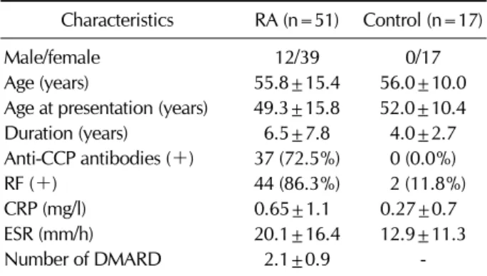
Table I. The demographic and clinical characteristics of the study population
Characteristics RA (n=51) Control (n=17)
Male/female 12/39 0/17
Age (years) 55.8±15.4 56.0±10.0
Age at presentation (years) 49.3±15.8 52.0±10.4
Duration (years) 6.5±7.8 4.0±2.7
Anti-CCP antibodies (+) 37 (72.5%) 0 (0.0%)
RF (+) 44 (86.3%) 2 (11.8%)
CRP (mg/l) 0.65±1.1 0.27±0.7
ESR (mm/h) 20.1±16.4 12.9±11.3
Number of DMARD 2.1±0.9 -
Data are mean±SD. Anti-CCP antibodies (+): (>5 U/ml), RF (+): (>18 IU/ml). CCP, cyclic citrullinated peptide; CRP, C reactive protein; ESR, Erythrocyte sedimentation rate.
text of certain HLA molecules.
Several proteins, such as type II collagen, proteoglycan and fibrinogen, have been proposed as potential auto-anti- gens in RA model mice and RA patients (7). Fibrinogen, which has been shown to be citrullinated in synovial tissues of inflamed joints, is one of the most extensively studied auto-anti- gens in RA. A recent study has reported that citrullinated fibri- nogen (cFBG) was detected in synovial fluids and may be a potential auto-antigen for ACPA in patients with RA (8).
Indeed, experimental arthritis can be induced in HLA-DR4-IE transgenic mice which carry the RA-associated shared epit- ope, such as HLA-DRB1*0401, after immunization with cFBG (9). Furthermore, this study demonstrated the HLA-DR4-res- tricted T cell epitope within cFBG. Although there is relatively strong evidence to support that cFBG acts as an auto-antigen in RA-induced animals, CD4 T cell-mediated immune re- sponses to cFBG have not yet been fully investigated in RA patients.
In this study, we investigated the possibility that cFBG pep- tides could be autoantigens in RA patients, and especially cFBG peptides (Fib-α R84Cit) within cFBG targeted by T cells in HLA-DR4-IE transgenic mice could be CD4 T-cell epitopes restricted to the susceptible HLA-DR allele in RA patients.
MATERIALS AND METHODS Study subjects
All RA patients (n=51) who fulfilled the 1987 revised criteria for the classification of RA proposed by the American College of Rheumatology (formerly, the American Rheumatism Asso- ciation) (10) were included in this study. The control group (n=17) consisted of patients with osteoarthritis, Behcet’s dis- ease, or other inflammatory diseases (e.g., fibromyalgia and systemic lupus erythematosus). Demographic and clinical characteristics of the subjects are summarized in Table I. The study was approved by the Institutional Review Board of Eulji University Hospital and Kangnam St. Mary’s Hospital, and written informed consent was obtained from each participant (approval No. 08-11).
Anti-CCP antibody and HLA-DRB1 genotyping Anti-CCP antibody levels were assessed using the DIASTAT anti-CCP kit (MBL Co., Nagoya, Japan). According to the manufacturer’s instruction, anti-CCP antibody was considered positive when its absorbance was higher than the cutoff value (5 U/ml). HLA-DRB1 genotyping was performed by the
PCR-SSP (sequence specific primer) method (11). Each tube contained a primer mix consisting of allele- or group-specific primer pairs as well as a positive control primer that matched the non-allelic sequences. Specific amplification of the HLA- DRB1 gene was performed using 20 specific pairs of primers for HLA-DRB1.
Peptides
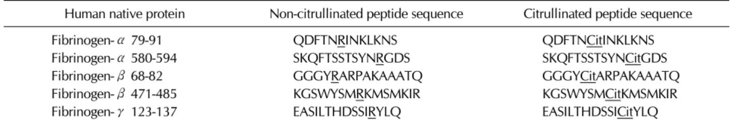
Citrullinated and non-citrullinated fibrinogen peptides were chemically synthesized (Peptron, Daejeon, Korea). High-per- formance liquid chromatography showed the purity of pep- tides is over 90%. Peptide sequences are shown in Table II, and the underlined arginine residue was substituted with citrulline.
Cell purification and culture
Peripheral blood mononuclear cells (PBMCs) were isolated from whole blood samples by density-gradient centrifugation using lymphocyte separation medium (PAA Laboratories, Linz, Austria). PBMCs (1×10⁶) were stimulated with either non-citrullinated or cFBG peptides (1, 10, and 30μg/ml for each) in RPMI 1640 medium supplemented with penicillin G (50 U/ml), streptomycin (50μg/ml), L-glutamine (2 mM), and 10% fetal bovine serum (WelGene, Seoul, Korea) at 37oC in a humidified atmosphere of 5% CO2. For recall antigen re- sponses, tetanus toxoid (5μg/ml; Calbiochem, San Diego, CA, USA), purified protein derivative (PPD) from Mycobacte- rium tuberculosis (5μg/ml; Statens Serum Institute, Copenha- gen, Denmark), and Candida albicans (10μg/ml; Greer La- boratories, Lenoir, NC, USA) were used.
Table II. Sequences of fibrinogen peptides used in the study
Human native protein Non-citrullinated peptide sequence Citrullinated peptide sequence
Fibrinogen-α 79-91 QDFTNRINKLKNS QDFTNCitINKLKNS
Fibrinogen-α 580-594 SKQFTSSTSYNRGDS SKQFTSSTSYNCitGDS
Fibrinogen-β 68-82 GGGYRARPAKAAATQ GGGYCitARPAKAAATQ
Fibrinogen-β 471-485 KGSWYSMRKMSMKIR KGSWYSMCitKMSMKIR
Fibrinogen-γ 123-137 EASILTHDSSIRYLQ EASILTHDSSICitYLQ
Arginine (R) converted to citrulline (Cit) was underlined.
For Fib-α R84Cit stimulation, CD4 T cells and CD14-pos- itive monocytes were positively isolated from PBMCs using magnetic cell sorting (autoMACSpro, Miltenyi Biotec Inc., Germany). CD4 T cells (5×10⁵) were stimulated in the pres- ence or absence of 50μg/ml non-citrullinated or cFBG pep- tides. CD14-positive cells (5×10⁴) used as antigen-presenting cells (APCs) were irradiated in a dose of 3,000 rads.
In vitro proliferation and cytokine measurement For cell proliferation, carboxyfluorescein succinimidyl ester (CFSE) labeling was performed (Invitrogen, Carlsbad, CA, USA). Briefly, whole PBMCs were labeled with 3μM CFSE before stimulation. After 4 days of culture, the levels of CFSE dilution indicating cell proliferation were measured within CD4 T cells by FACS analysis.
For Fib-α R84Cit peptide stimulation, cell pellets were col- lected, and proliferation was determined by using the BrdU proliferation ELISA assay kit (Roche diagnostics, Germany) ac- cording to the manufacturer’s instruction. Briefly, the BrdU labeling solution was added to the wells and incubated for additional 24 hours. The amount of BrdU uptake was sub- sequently determined by fixation and incubation with an an- ti-BrdU antibody conjugated with peroxidase (POD), followed by colorimetric detection.
Supernatants from the wells were removed, and the levels of IFN-γ and IL-17A were measured using a standard sandwich ELISA according to the manufacturer's instructions (eBioscience, San Diego, CA, USA).
Statistical analysis
Statistical analysis (unpaired t test) was performed using GraphPad Prism Software V5.0 (GraphPad Software, San Diego, CA, USA). A p-value of <0.05 was considered statisti- cally significant.
RESULTS
Effect of cFBG peptide stimulation on PBMCs from RA patients
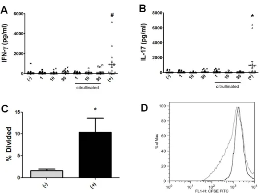
To determine whether antigen-specific T cell responses can be elicited by cFBG peptides, we first searched HLA DRB1*0401-restricted peptides from α, β and γ chains of fibrinogen by using a web- based program (Immune epitope database, National Institute of Allergy and Infectious Diseases [NIAID]). Peptides were selected according to their predicted affinity for DRB1*0401 as in a previous study (12). Because citrulline (Cit) is not accounted for in the predictive algorithm, the value of glutamine (Q) was substituted for arginine (R) when identifying T cell epitope candidates (Q has a same ter- minal side chain group with Cit), and each epitope sequences are shown in Table II. For cFBG peptide-specific stimulation, we combined 5 high-peptides at various concentrations and added 1 million PBMCs isolated from RA patients. After 4 days of culture, the levels of IFN-γ and IL-17 were measured by ELISA, which were not enhanced (Fig. 1A and B). For T-cell proliferation, we utilized CFSE labeling in which whole PBMCs were stained with CFSE and the degree of CFSE, dilu- tion indicating cell proliferation was measured with FACS within the CD4 T-cell population. Similar to cytokine pro- duction, we could not detect T-cell proliferation stimulated with cFBG peptides (Fig. 1C and D). To check for T-cell acti- vation, we performed T-cell proliferation and cytokine pro- duction assay in response to recall antigen which is a combi- nation of tetanus toxoid, M. tuberculosis-PPD, and C.
albicans. We detected CD4 T cells from RA patients that re- sponded well to a panel of recall antigen (Fig. 1). For exam- ple, T cells responded to the recall antigen to produce IFN-γ (Fig. 1A), IL-17 (Fig. 1B) and to proliferate well (Fig. 1C and D). Therefore, these findings indicated that CD4 T cells from RA patients may be not specific for cFBG peptides, although they respond well to the recall antigen.
Figure 1. Immune responses of PBMCs from RA patients to cFBG peptides.
PBMCs from RA patients (n=17) were stimulated with either non-citrullinated or citrullinated fibrinogen (cFBG) peptides at indicated concentrations (μg/ml). IFN-γ secretion was quan- tified from culture supernatant of peptide-stimulated PBMCs (A). Si- milarly, IL-17 secretion was quanti- fied from the supernatant (B). Cell proliferation was evaluated with a dilution of CFSE (C). Representative FACS results are shown (D) (un-sti- mulation in black [% division: 0.31]
and recall antigen stimulation in gray line [% division: 16.5]). #p
<0.01 compared with non-stimula- tion (−). *p<0.05 compared with non-stimulation. (+) represents a mixture of recall antigens.
Role of DRB1*0401-restricted cFBG peptide in sti- mulation of CD4 T cells
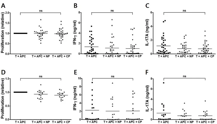
Next, we investigated the possibility that a specific cFBG pep- tide, Fib-α R84Cit, which is identified as a DRB1*0401-re- stricted T cell epitope in DR4 transgenic mice (9), can be rec- ognized by CD4 T cells from RA patients. Thus, CD4 T cells from RA patients were incubated with either a non-citrulli- nated (Fib-α 79-91) or a cFBF peptide (Fib-α R84Cit), in the presence of CD14-positive monocytes as APCs. Significant proliferative responses to the cFBG peptide were not observed after in vitro stimulation with both Fib-α 79-91 and Fib-α R84Cit (Fig. 2A). We also examined cytokine production by stimulation with cFBG peptide. Neither IFN-γ nor IL-17A in- duction was observed in T cells stimulated with Fib-α R84Cit peptide (Fig. 2B and C). Similar to the results from RA pa- tients, proliferation and cytokine production were not ob- served from CD4 T cells from control subjects (Fig. 2D∼F).
These results indicate that CD4 T cells may not respond to Fib-α R84Cit peptide regardless of the presence of RA.
Anti-CCP antibodies are useful serological markers for the diagnosis of RA, and immune responses to citrullinated protein could be confined to RA patients who have a high titer of an- ti-CCP antibodies (3). Because we also used the DRB1*0401-re- stricted T-cell epitope within the cFBG to assess CD4 T
cell-mediated immune responses, we assessed T-cell respon- ses to anti-CCP antibody in HLA-DR4-positive patients. Howe- ver, neither proliferative response nor IFN-γ and IL-17A se- cretion was observed when CD4 T cells were stimulated with the citrullinated peptide even in anti-CCP antibody-positive RA patients carrying HLA-DRB1*04 (Fig. 3). Again, to ensure that failure to detect significant responses in our study was not attributed to improper assay, we examined T-cell pro- liferation and cytokine production in response to the recall antigen. We were able to detect CD4 T cells from RA patients and healthy controls that responded well to the panel of re- call antigens, including tetanus toxoid, M. tuberculosis-PPD, and C. albicans (data not shown). Therefore, these results demonstrated that significant proliferation and cytokine re- sponses may not be induced by cFBG peptide stimulation of CD4 T cells from RA patients, although these CD4 T cells re- spond well to common recall antigens.
DISCUSSION
In this study, we determined whether T-cell epitope within cFBG could act as an auto-antigen in RA. We detected neither T-cell proliferative response nor Th1 (IFN-γ) response by stimulation with cFBG peptides. Furthermore, Th17 (IL-17A)
Figure 3. CD4 T-cell responses of PBMCs from anti-CCP Ab/HLA-DRB1*04-positive RA patients to Fib-α R84Cit peptide. CD4 T cells from anti-CCP Ab/HLA-DRB1*04-positive RA patients (n=13) were stimulated with non-citrullinated (NP) and citrullinated (CP) fibrinogen peptides in the presence of APCs. Cell proliferation was quantified by the incorporation of BrdU (A). IFN-γ (B) and IL-17A (C) secretion was quantified from each peptide-stimulated culture supernatant by ELISA. ns=not significant.
Figure 2. CD4 T-cell responses of PBMCs from RA patients and control subjects to Fib-α R84Cit peptide. CD4 T cells from RA patients (n=34) were stimulated with non-citrullinated (NP) and citrullinated (CP) fibrinogen peptides in the presence of APCs. Cell proliferation was quantified by the incorporation of BrdU. The degree of cell proliferation was compared between stimulation and non-stimulation, and values represent the degree of well proliferation in individual samples (A). IFN-γ (B) and IL-17A (C) secretion was quantified from each peptide-stimulated culture supernatant by ELISA. Similarly, T-cell proliferation (D), and IFN-γ (E) and IL-17A (F) production of CD4 T cells from control subjects (n=17) are shown. ns=not significant.
response against cFBG peptides, which may be more in- timately associated with RA, was not enhanced in RA patients, either.
Genetic studies have shown that certain HLA-class II al- leles, "shared epitopes" are the most important risk factor for RA (3). In addition, it has been shown that HLA-DRB1 shared
epitopes only confer susceptibility to anti-CCP and antibody- positive RA and that they contribute to the production of ACPA (5,13). However, we failed to observe any significant responses in CD4 T cells from anti-CCP and HLA-DR4-positive RA patients.
It is not clear why our findings are different from those of an HLA-DR4 transgenic mouse RA model (9). The diversity and/or heterogeneity of human RA is a good possibility.
Thus, it seems likely that multiple peptides, instead of a single peptide, successfully activate multiclonic cFBG-specific T cells. Indeed, fibrinogen has several sites that can be citrulli- nated (14). However, in our study, multiple cFBG peptide stimulation clearly showed that T cells were not activated.
Furthermore, it should be pointed out that immunization with cFBG protein, which is citrullinated by peptidylarginine de- aminase, induces arthritis in only less than half of cases even in HLA-DR4 transgenic mice (9). It has also been shown that immunization with Fib-α R84Cit peptide itself does not in- duce arthritis even if T-cell responses to Fib-α R84Cit are evident. In addition, recent studies have demonstrated that T cell-mediated immune responses are detected in response to different citrullinated proteins, such as citrullinated vi- mentin and/or citrullinated aggrecan in RA patients (15,16).
Those studies also stated that the degree of immune re- sponses to citrullinated proteins is not significant.
The difference in the T-cell receptor (TcR) repertoire be- tween species is another possibility. A previous study has documented that cFBG peptide or Fib-α R84Cit is identified as DRB1*0401- restricted T-cell epitope in DR4 transgenic mice (9). However, it is apparent that the TcR repertoire of RA patients differs from that of HLA-DR4 transgenic mice and that T cells from RA patients may respond to citrullinated peptides other than Fib-α R84Cit.
In summary, our study demonstrates that T-cell responses to cFBG peptides may not be present in RA patients, suggest- ing that anti-citrulline immunity in RA patients is unlikely to be mediated by citrullinated fibrinogen.
ACKNOWLEDGEMENTS
This work was supported by a grant from the Korea Health- care Technology R&D Project, Ministry for Health, Welfare &
Family Affairs, the Republic of Korea (A101151), the Korea Health 21 R&D Project, by the Ministry of Health and Welfare (01-PJ3-PG6-01GN09-003), and Basic Science Research Pro- gram through the National Research Foundation of Korea (NRF)
funded by the Ministry of Education (NRF-2013R1A1A2010714).
CONFLICTS OF INTEREST
The authors have no financial conflict of interest.
REFERENCES
1. Klareskog, L., A. I. Catrina, and S. Paget. 2009. Rheumatoid arthritis. Lancet 373: 659-672.
2. Imboden, J. B. 2009. The immunopathogenesis of rheuma- toid arthritis. Annu. Rev. Pathol. 4: 417-434.
3. Klareskog, L., J. Ronnelid, K. Lundberg, L. Padyukov, and L. Alfredsson. 2008. Immunity to citrullinated proteins in rheumatoid arthritis. Annu. Rev. Immunol. 26: 651-675.
4. Kuhn, K. A., L. Kulik, B. Tomooka, K. J. Braschler, W. P.
Arend, W. H. Robinson, and V. M. Holers. 2006. Antibodies against citrullinated proteins enhance tissue injury in ex- perimental autoimmune arthritis. J. Clin. Invest. 116: 961- 973.
5. van der Helm-van Mil, A. H., K. N. Verpoort, F. C.
Breedveld, T. W. Huizinga, R. E. Toes, and R. R. de Vries.
2006. The HLA-DRB1 shared epitope alleles are primarily a risk factor for anti-cyclic citrullinated peptide antibodies and are not an independent risk factor for development of rheu- matoid arthritis. Arthritis. Rheum. 54: 1117-1121.
6. Hill, J. A., S. Southwood, A. Sette, A. M. Jevnikar, D. A. Bell, and E. Cairns. 2003. Cutting edge: the conversion of arginine to citrulline allows for a high-affinity peptide interaction with the rheumatoid arthritis-associated HLA-DRB1*0401 MHC class II molecule. J. Immunol. 171: 538-541.
7. Wegner, N., K. Lundberg, A. Kinloch, B. Fisher, V. Malm- ström, M. Feldmann, and P. J. Venables. 2010 . Autoimmuni- ty to specific citrullinated proteins gives the first clues to the etiology of rheumatoid arthritis. Immunol. Rev. 233: 34-54.
8. Takizawa, Y., A. Suzuki, T. Sawada, M. Ohsaka, T. Inoue, R. Yamada, and K. Yamamoto. 2006. Citrullinated fibrinogen detected as a soluble citrullinated autoantigen in rheumatoid arthritis synovial fluids. Ann. Rheum. Dis. 65: 1013-1020.
9. Hill, J. A., D. A. Bell, W. Brintnell, D. Yue, B. Wehrli, A.
M. Jevnikar, D. M. Lee, W. Hueber, W. H. Robinson, and E. Cairns. 2008. Arthritis induced by posttranslationally modi- fied (citrullinated) fibrinogen in DR4-IE transgenic mice. J.
Exp. Med. 205: 967-979.
10. Arnett, F. C., S. M. Edworthy, D. A. Bloch, D. J. Mcshane, J. F. Fries, N. S. Cooper, L. A. Healey, S. R. Kaplan, M. H.
Liang, H. S. Luthra, T. A. Medsger Jr, D. M. Mitchell, D. H.
Neustadt, R. S. Pinals, J. G. Schaller, J. T. Sharp, R. L. Wil- der, and G. G. Hunder. 1988. The American Rheumatism Association 1987 revised criteria for the classification of rheumatoid arthritis. Arthritis. Rheum. 31: 315-324.
11. Jordan, F., A. J. McWhinnie, S. Turner, N. Gavira, A. A.
Calvert, S. A. Cleaver, R. H. Holman, J. M. Goldman, and J. A. Madrigal. 1995. Comparison of HLA-DRB1 typing by DNA-RFLP, PCR-SSO and PCR-SSP methods and their applica- tion in providing matched unrelated donors for bone marrow
transplantation. Tissue. Antigens. 45: 103-110.
12. Hammer, J., E. Bono, F. Gallazzi, C. Belunis, Z. Nagy, and F. Sinigaglia. 1994. Precise prediction of major histocompati- bility complex class II-peptide interaction based on peptide side chain scanning. J. Exp. Med. 180: 2353-2358.
13. Huizinga, T. W., C. I. Amos, A. H. van der Helm-van Mil, W. Chen, F. A. van Gaalen, D. Jawaheer, G. M. Schreuder, M. Wener, F. C. Breedveld, N. Ahmad, R. F. Lum, R. R. de Vries, P. K. Gregersen, R. E. Toes, and L. A. Criswell. 2005.
Refining the complex rheumatoid arthritis phenotype based on specificity of the HLA-DRB1 shared epitope for antibodies to citrullinated proteins. Arthritis. Rheum. 52: 3433-3438.
14. Nakayama-Hamada, M., A. Suzuki, K. Kubota, T. Takazawa, M. Ohsaka, R. Kawaida, M. Ono, A. Kasuya, H. Furukawa,
R. Yamada, and K. Yamamoto. 2005. Comparison of enzy- matic properties between hPADI2 and hPADI4. Biochem.
Biophys. Res. Commun. 327: 192-200.
15. Feitsma, A. L., E. I. van der Voort, K. L. Franken, H. el Bannoudi, B. G. Elferink, J. W. Drijfhout, T. W. Huizinga, R. R. de Vries, R. E. Toes, and A. Ioan-Facsinay. 2010.
Identification of citrullinated vimentin peptides as T cell epito- pes in HLA-DR4-positive patients with rheumatoid arthritis.
Arthritis. Rheum. 62: 117-125.
16. von Delwig, A., J. Locke, J. H. Robinson, and W. F. Ng.
2010. Response of Th17 cells to a citrullinated arthritogenic aggrecan peptide in patients with rheumatoid arthritis.
Arthritis. Rheum. 62: 143-149.