Correspondence to: Jong-Seok Lim, Laboratory of Cell Biology, Korea Research Institute of Bioscience and Biotechnology, Yusung P.O. Box 115, Daejeon 305-600, Korea. (Tel) +82- 42-860-4193, (Fax) +82-42-860-4593, (E-mail) jslim@kribb.re.kr This work was supported in part by grants from MOHW (02- PJ1-PG10-21206-0002) and the Ministry of Science and Techno- logy (M1-0016-00-0027), Korea.
188
Immune Network
Introduction
Dendritic cells (DCs) are highly effective antigen- presenting cells with the unique capability of inducing primary immune responses against tumor-associated antigens (1). DCs pulsed with synthetic tumor-derived MHC class I-restricted peptides, tumor lysates or tumor cell-derived RNA induced tumor-specific T
Activation by Necrotic Tumor Cell-loaded Dendritic Cells in Therapeutic Vaccinations
Aeyung Kim1, Kwang Dong Kim1, Seung-Chul Choi1, Moon-Jin Jeong2, Hee Gu Lee1, Yong-Kyung Choe1, Sang-Gi Paik3 and Jong-Seok Lim1
1Laboratory of Cell Biology, Korea Research Institute of Bioscience and Biotechnology, Daejeon,
2Department of Oral Histology, College of Dentistry, Chosun University, Gwangju, 3Department of Biology, Chungnam National University, Daejeon, Korea
ABSTRACT
Background: Immunization of dendritic cells (DCs) pulsed with tumor antigen can activate tumor-specific cytotoxic T lymphocytes (CTL) that are responsible for protection and regression. In this study, we examined whether the uptake of necrotic tumor cells could modulate DC phenotypes and whether the immunization of necrotic tumor cell-loaded DCs could elicit efficient tumor specific immune responses followed by a regression of established tumor burdens. Methods: We prepared necrotic tumor cell-pulsed DCs for the therapeutic vaccination and investigated their phenotypic characteristics, the immune responses induced by these DCs, and therapeutic vaccine efficacy against colon carcinoma in vivo. Several parameters including phagocytosis of tumor cells, surface antigen expression, chemokine receptor expression, IL-12 production, and NK as well as CTL activation were assessed to characterize the immune response. Results: DCs derived from mouse bone marrow efficiently phagocytosed necrotic tumor cells and after the uptake, they produced remarkably increased levels of IL-12. A decreased CCR1 and increased CCR7 expression on DCs was also observed after the tumor uptake, suggesting that antigen uptake could induce DC maturation.
Furthermore, co-culturing of DCs with NK cells in vitro enhanced IL-12 production in DCs and IFN-γ production in NK cells, which was significantly dependent on IL-12 production and cell-to-cell contact. Immunization of necrotic tumor cell-loaded DCs induced cytotoxic T lymphocytes as well as NK activation, and protected mice against subsequent tumor challenge. In addition, intratumoral or contra-lateral immunization of these DCs not only inhibited the growth of established tumors, but also eradicated tumors in more than 60% of tumor-bearing mice. Conclusion: Our data indicate that production of IL-12, chemokine receptor expression and NK as well as CTL activation may serve as major parameters in assessing the effect of tumor cell-pulsed DC vaccine.
Therefore, DCs loaded with necrotic tumor cells offer a rational strategy to treat tumors and eventually lead to prolonged survival. (Immune Network 2003;3(3):188-200) Key Words: Dendritic cells (DC), IL-12, NK cells, cytotoxic T lymphocytes (CTL),
IFN-γ
Abbreviations: DC, dendritic cells; CCR, CC chemokine receptor; NKT, natural killer T; MIP-1α, macrophage-inflammatory protein-1α; SLC, secondary lymphoid-tissue chemokine.
cell activation, host-protective and therapeutic anti- tumor immunity in mice and humans (2-5). The use of tumor-derived antigens in the form of tumor cell lysates or whole tumor cells has advantages in com- parison with vaccinations against a single antigen, because so far only a few human tumor-associated antigens have been identified and immunity against a single antigen may be ineffective in tumors with he- terogenous cell populations. In addition, the high polymorphism of the human HLA system has pre- sented problems in identifying and applying tumor- associated markers to be used as vaccines for cancer immunotherapy. In contrast, given that the induction of stronger cytotoxic T lymphocytes (CTL) responses appears to be a major goal of current cancer vaccine strategies, lysates-loaded DCs containing multiple known as well as unknown antigens that can be pre- sented to T cells by both MHC class I- and class II-pathways provide the potential to induce efficient antitumor immune responses (6).
Recently, it has been shown that phagocytosis of apoptotic/necrotic tumor cells induced the matur- ation of DCs (7,8). DC maturation is characterized by down-regulation of antigen acquisition, increased expression of MHC and co-stimulatory molecules, production of proinflammatory cytokines, and altered expression of chemokine receptors (1,9). As they mature, DCs migrate to the T-cell areas of lymphoid organs, where antigen is presented to naive CD4+ and CD8+ T cells. Although a number of reports de- monstrated that apoptotic/necrotic tumor cells might be a good source of tumor antigens for presentation to DCs in vitro and in vivo (7,10-12), the causal relation between the phenotypic characteristics of DCs that phagocytosed tumor cells, the immune responses induced by these DCs and their therapeutic vaccine efficacy against an established tumor have not been well studied. Importantly, it seems clearly not enough to phagocytose killed tumor cells to induce immune stimulatory signals leading to the breaking of tol- erance to tumor antigens in vivo. Furthermore, there is a great need in controlled clinical trials for the establishment of key experimental parameters of the immune response by DC-based tumor vaccine, especially DCs loaded with necrotic tumor cells.
We have previously reported that vaccination of DC co-cultured with tumor cells in mice elicits an efficient protective antitumor activity against a chal- lenge by colon cancer CT-26 cells (13). In this study, we prepared necrotic tumor cell-pulsed DCs for the therapeutic vaccination and investigated their pheno- typic characteristics, the immune responses induced by these DCs, and therapeutic vaccine efficacy against colon carcinoma in vivo. Several parameters including phagocytosis of tumor cells, surface antigen expres-
sion, chemokine receptor expression, IL-12 produc- tion, and NK as well as CTL activation were assessed to characterize the immune response. Our data in- dicate that production of IL-12, chemokine receptor expression and NK as well as CTL activation may serve as major parameters to assess the effect of tumor cell-pulsed DC vaccine.
Materials and Methods
Mice and tumor cell lines. Five-to six-week-old female Balb/c mice purchased from the Genetic Resources Center, KRIBB (Daejeon, Korea) were maintained under specific pathogen-free conditions until the experiments. The experiments employing the mice were performed in accordance with institutional guidelines. CT-26 cells were obtained from Seoul National University Hospital (Seoul, Korea). CT-26/
NP cells, stable cell line of CT-26 cells transfected with influenza type A NP, were kindly provided from Dr. Y. C. Sung at Pohang University of Science and Technology (Pohang, Korea). Renca, renal adenocar- cinoma cell line was obtained from Samsung Medical Center (Seoul, Korea). All tumor cells were cultured in RPMI containing 10% heat inactivated FBS, 100 U/ml penicillin, 100 μg/ml streptomycin, and 2 mM L-glutamine (Gibco BRL, Grand Island, NY, USA) at 37oC in 5% CO2. They were routinely checked for the absence of Mycoplasma contamination by using ELISA detection kit (Roche Diagnostics GmbH, Mannheim, Germany).
Generation of dendritic cells from mouse bone marrow cells.
Dendritic cells were generated from bone marrow cells by following Dr. Inaba's method with slight mo- dification (14). In brief, bone marrow cells obtained from femurs and tibias of Balb/c mice were incubat- ed with an antibody cocktail containing J1j.10 (anti- CD90), J11d (anti-CD11b), 3.168 (anti-CD8), GK1.5 (anti-CD4), RA3-3A1 (anti-B220), and M5/114.15.2 (anti-I-Ab,d,q & I-Ed,k) at 4oC for 1 h. They were washed twice with 10% FBS RPMI and treated with rabbit complement (Low-ToxR-M, Cedarlane, Onta- rio, Canada) according to the manufacturer's instruc- tion. To eliminate dead cells, cells were subjected to density centrifugation on Histopaque 1077 (Sigma, Saint Louis, MO, USA), and then washed twice with RPMI 1640 medium without serum. They (5×10⁵ cell/well) were further incubated in culture medium supplemented with 10 ng/ml recombinant murine GM-CSF and IL-4 (Endogen, Woburn, MA, USA) in 24-well plates (Costar, Cambridge, USA). On days 2 and 4, non-adherent cells were discarded and culture media were replaced with fresh DC medium. On day 7, the non-adherent cells that had acquired typical dendritic cell morphology as identified on the phase contrast microscope were harvested by a gentle
swirling, and used in the subsequent experiments.
Induction of tumor necrosis and uptake of necrotic tumor cells.
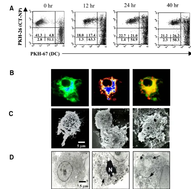
Tumor necrosis was induced by exposing the cells to one cycle of rapid freezing (liquid nitrogen) and thawing at 37oC in serumfree medium. Upon treat- ment all the cells became trypan blue positive. During incubation, these necrotic cells were degraded into fragments. Supernatants were obtained from necrotic cells by spinning at 1,500 rpm for 10 min. To analyze uptake of necrotic tumor cells by DCs, tumor cells were dyed red with PKH-26 and DCs were dyed green with PKH-67 (Sigma) according to the manu- facturer's protocol. Briefly, tumor cells and DCs were incubated with 2×10⁻⁶ M PKH-26 and PKH-67 at room temperature for 5 min, respectively, then stopped with the same volume of fetal bovine serum, and rinsed extensively with culture medium. PKH-26- labeled tumor cells were subjected to necrosis as described above, and then co-cultured with PKH-67- labeled DCs at the ratio of 1:1 for 12∼40 h at 4oC or 37oC. Phagocytosis of necrotic cells by DCs was defined by the percentage of double positive cells by flow cytometry. For fluorescence images, coverslips containing DCs stained with PKH-67 were mounted on glass slides after pulsing PKH-26-labeled necrotic tumor cells for 24 h and subjected to a confocal laser scanning microscope (Leica Laser Technik GmbH, Heidelberg, Germany). Phagocytosis of necrotic tu- mor cells was also examined in a JEOL JEM CXII transmission electron microscope at an accelerating voltage of 80 kV and a scanning electron microscope (ISI-SS40) at an accelerating voltage of 10 kV.
Flow cytometric analysis of cell surface antigens. For the analysis of surface molecules, 1∼5×10⁵ DCs or DCs pulsed with necrotic tumor cells were incubated at 4oC for 30 min using the following FITC-conjugated monoclonal antibodies (PharMingen, San Diego, CA, USA): anti-CD80 (B7-1; 16-10A1), anti-CD86 (B7-2;
GL-1), and anti-CD40 (MH 40.3). After washing twice with phosphate buffered saline, cells were analyzed by using a FACSCalibur (Becton Dickinson, Mountain View, CA, USA).
Determination of IL-12 p70 and IFN-γ by ELISA. Nec- rotic tumor cells were co-cultured with DCs at the ratio of 1:1 for 12∼32 h. For determining IL-12 p70 or IFN-γ concentration, culture supernatants were collected after co-culturing necrotic tumor cell and DCs, or contact of pulsed-DCs and NK cells in the absence or presence of anti-IL-12 neutralizing antibody C17.8 (Endogen). After incubation, culture supernatants were collected, and then subjected to IL-12 p70 or IFN-γ ELISA kit (Endogen). Briefly, 50μl of standard or culture supernatants was in- cubated in anti-mouse IL-12 p70 or IFN-γ anti- body pre-coated well plates for 1 h. After biotin-
labeled antibody was added, the plates were incubated for 1 h, and then incubated with streptavidin-HRP for 30 min. TMB substrate solution was added and the plates were developed in the dark for 30 min.
The reaction was stopped by adding 100μl of 0.18 M sulfuric acid, and the absorbance was read at 450 nm using an ELISA reader. All steps were performed at room temperature. The relationship between absor- bance and cytokine concentration was linear between 0 and 1,000 pg/ml, with correlation coefficients > 0.999, consistently.
mRNA expression analysis for chemokine receptors and cytokines using RT-PCR. Total RNA was isolated from each sample by the acid guanidinium thiocyanate phe- nol chloroform extraction method. cDNA was syn- thesized from 10μg total RNA using ProSTARTM kit (Stratagene, La Jolla, CA, USA), and used as the template for PCR (CCR1 and CCR7; 95oC for 1 min, 57oC for 1 min, and 72oC for 1 min, IL-12 p35, p40 and IFN-γ; 94oC for 1 min, 55oC for 1 min, and 72oC for 1 min) using specific primers. To ensure the quality of the procedure, RT-PCR was performed on the same samples using specific primers for β-actin.
PCR products were harvested and resolved on a 1.2%
agarose gel containing ethidium bromide. They were quantified with Quantity One software supplemented with Gel Doc 2000 system (Biorad, Hercules, CA, USA). To compare relative expression, relative values were calculated as follows; (mean concentration×area) of CCR1, CCR7 or IFN-γ/(mean concentration×
area) of β-actin. The sequence of each primer was as follows: mCCR1 (forward: 5'-TCTAGTGTTCAT- CATTGGAGTGGTG; reverse: 5'-GACGCACGGC- TTTGACCTTCTTCTC), mCCR7 (forward: 5'-ACA- GCGGCCTCCAGAAGAAGAGCGC; reverse: 5'-T- GACGTCATAGGGCAATGTTGAGCTG), mIL-12 p35 (forward: 5'-TCGATCATGAAGACATCACA;
reverse: 5'-GATTCAGAGACTGCATCAGC), mIL-12 p40 (forward: 5'-ATGTGTCCTCAGAAGCTAACC;
reverse: 5'-CCAAATTCCATTTTCCTTCT), and mIFN- γ (forward: 5'-AACGCTACACACTGCATCT; reverse:
5'-TGCTCATTGTAATGCTTGG).
In vitro chemotaxis assay. An in vitro chemotaxis assay was performed in 24-well transwell culture chamber (Costar) with 5.0μm pore size as described pre- viously (15). Briefly, after DCs were co-cultured with necrotic tumor cells at the ratio of 1:1 for 24 h, cells (5×10⁵) were added to the top chamber in assay medium at a final volume of 100μl, and recom- binant chemokines (MIP-1α and MIP-3β; R & D Systems, Inc, Minneapolis, MN, USA) were diluted in the bottom chamber with assay medium to a final volume of 600μl at appropriate concentrations. After the plates were incubated at 37oC in 5% CO2 for 4 h, the cells in the bottom chamber were collected.
The migrating cells were stained with trypan blue and counted five times. Each assay was performed in triplicate.
Determination of cytotoxic activity. Groups of Balb/c mice at 6∼8 wks age were immunized subcutaneously (s.c.) in the right flank with 0.5∼1×10⁶ DCs, ne- crotic tumor cell-loaded DCs, necrotic tumor cells, or PBS. To examine CTL activity, splenocytes were har- vested, stimulated in vitro with irradiated DCs pulsed with necrotic tumor cells for 3∼4 days, and then used as effector cells. As effector cells for NK assay, splenocytes incubated for 12 h in the presence of IL-2 (10 ng/ml) were used. The target cells, such as CT-NP, CT-26, Renca, and YAC-1 were incubated with Na51CrO4 (100μCi per 1 2×10⁶, NEN- DuPont) for 90 min at 37oC with shaking every 15 min. Radioisotope-labeled target cells were combined with various numbers of effector cells in 96-well U-bottomed culture plates (Corning, NY, USA) in triplicate. Wells containing only culture medium and target cells were served as the spontaneous 51Cr release control, whereas those containing 1% SDS and target cells were used as the maximum release.
After incubation for 4 h at 37oC in 5% CO2, su- pernatants (100μl) were collected from each well, and the 51Cr release was measured in a gamma counter (Wallac Inc., Gaithersburg, MD, USA). The percentage of specific lysis was calculated as follows:
(cpm of test - cpm of spontaneous release)/(cpm of maximum release - cpm of spontaneous release)×
100. Data shown are the mean of triplicate cultures.
Immunization of mice. To measure protective immunity, Balb/c mice were immunized s.c. with 0.5∼1×10⁶ DCs, necrotic tumor cell-loaded DCs, or necrotic tumor cells in the right flank. Control mice were immunized with the same volume of PBS. Seven days after immunization, mice were inoculated s.c. with 2
×105 viable tumor cells. To measure therapeutic effect, 2×10⁵ viable tumor cells were injected s.c. in the right flank. Vaccination was performed intra- tumorally or contra-laterally in the left flank twice on day 3 when tumor was noticeably developed, and on day 10. The size of tumors was measured in two perpendicular dimensions with a vernier caliper twice a week.
Statistical analysis. Data are expressed as mean±SD.
Statistical analysis was performed using the Student t-test, two-sided. Differences were considered statis- tically significant with P<0.05.
Results
Uptake of necrotic tumor cells by DC. We first examined whether DC could efficiently phagocytose necrotic tumor cells induced by a freeze-thawing by using flow cytometric analysis of fluorescence-labeled cells. After
necrosis induction, all tumor cells were positively stained with trypan blue, and fragmented into small debris during 24 h incubation. When PKH-67-labeled DCs obtained from the bone marrow of syngeneic Balb/c mice were co-cultured with PKH-26-labeled necrotic tumor cells at the ratio of 1:1 at 37oC, phagocytosis increased as the incubation time was prolonged (Fig. 1A). When the incubation was performed on ice for 24 h to discriminate uptake of necrotic tumor cells from non-specific binding, the phagocytosis percentage was the same as the one from spontaneous uptake (data not shown). The uptake of necrotic cells appeared to occur relatively fast since a significant enhancement of double posi- tive cells (4.8% vs. 17.4%) was already observed after the incubation for 12 h. Optimum phagocytosis was consistently observed after 24 h (∼30% of DCs), and the relative percentage of phagocytosis was scarcely changed at higher ratios of DC and tumor cell numbers (data not shown). Therefore, the co- culturing condition at the ratio of 1:1 for 24 h was used for subsequent experiments. This finding was further confirmed by confocal microscopic image analysis, showing that necrotic tumor cells were phagocytosed into the cytoplasm of DCs (Fig. 1B).
Tumor cell fragments could be identified in DCs that had phagocytosed dye-labeled necrotic tumor cells (Fig. 1B, middle). Characteristic morphology of DCs with surface microvilli and long fillopodia was ob- served by scanning or transmission electron micro- scopy (Fig. 1C and 1D, left). After the physical contact with necrotic tumor cells in the tissue culture, micovilli on DCs displayed interaction with tumor debris at the initial phase of phagocytosis (Fig. 1C, middle). During the phagocytic processes, uptake of cell debris by micovilli and several phagocytotic cups on DC surfaces were found (Fig. 1C, right). In the cytoplasmic area of DCs, engulfment of necrotic cells that showed a collapsed nucleus or cell fragments was shown by transmission electron microscopy (Fig. 1D, middle and right). These results indicate that DCs are able to phagocytose necrotic tumor cells through the recognition of specific molecules on necrotic cells.
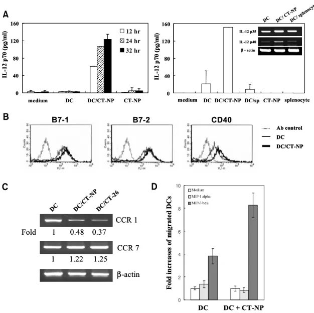
Uptake of necrotic tumor cells induces phenotypical change of DCs. Next, we investigated IL-12 production of DCs pulsed with necrotic tumor cells. Tumor cells were subjected to necrosis, and supernatant and/or pellets were separated by a centrifugation to test their respective effects on IL-12 production by pulsed DCs. IL-12 p70 production was induced by an addition of pellets of necrotic tumors that are absent from the supernatant (data not shown). However, in the presence of supernatant it was markedly increased by necrotic tumor cells in a time-dependent manner, indicating that maximal IL-12 production by DCs
required not only a soluble form of some tumor components, but also the uptake of necrotic cell pellets (Fig. 2A, left). This effect was not due to the contamination of endotoxin because tumor cells were confirmed to be endotoxin-free by the Limulus amo- ecyte lysate assay (BioWhittaker) and the treatment of polimyxin-B in the same culture did not affect IL-12 production. In addition, phagocytosis of necrotic tumor cells including their supernatant was not dif- ferent from that of the necrotic cell pellets (data not shown). Therefore, necrotic tumor cells including
their supernatant were used for further experiments.
The next question was whether the induction of IL-12 production is associated with tumor-related components. We prepared necrotic cells of syngeneic splenocytes and added them to the DC culture to compare with necrotic tumor cells. In necrotic tumor cell-pulsed DCs, IL-12 production was repeatedly observed, whereas after uptake of necrotic splen- ocytes DCs were not able to produce IL-12 (Fig. 2A, right). RT-PCR analysis revealed that IL-12 p40 mRNA could be induced in DCs pulsed with necrotic
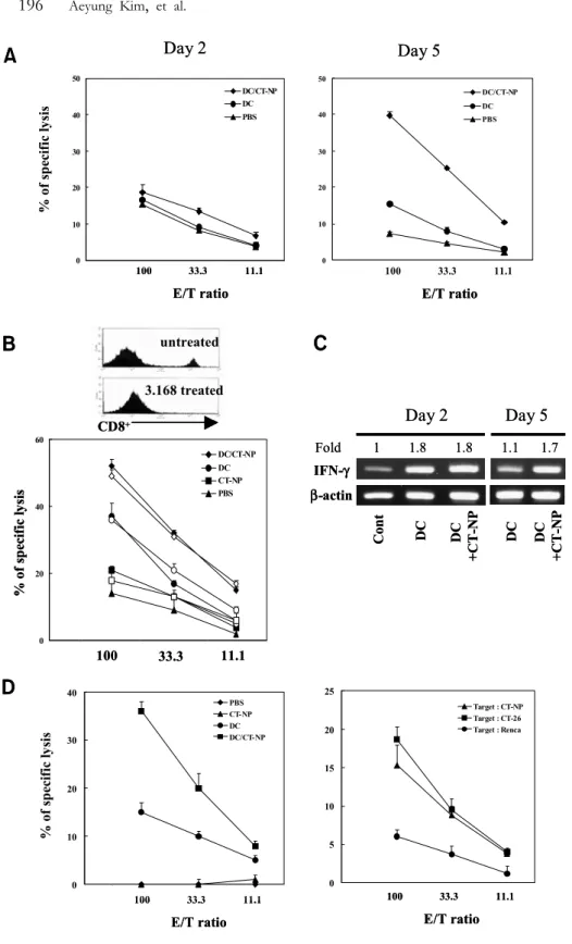
Figure 1. Uptake of necrotic CT-NP tumor cells by DCs. A. PKH-26-labeled necrotic CT-NP and PKH-67-labeled DCs were co-cultured at 37oC for 0, 12, 24, and 40 h at the ratio of 1:1. Cells were analyzed by flow cytometer where double positive cells indicate uptake of the necrotic CT-NP by DCs. The uptake of necrotic CT-NP was not detectable when the incubation was performed on ice. Data are representative of a minimum of three independent experiments. B. DCs co-cultured with necrotic CT-NP were examined by confocal microscopy. left; PKH-67-labeled DC, middle; PKH-26-labeled CT-NP fragments in DCs. Merging of two images (right) shows that necrotic CT-NP fragments have been internalized into DCs. C and D. Ultra-structural observation of DCs (left), and DCs co-cultured with necrotic CT-NP for 24 h (middle, right) by SEM and TEM. Arrows show engulfed tumor cells (N; nucleus of necrotic tumor cell) or tumor fragments.
PKH-26 (CT-NP)
41.3 4.8
2.8 51.1 18.0 17.4
1.3 63.3 22.7 21.0
1.4 54.9 21.2 26.2 2.3 50.3
A
B
7.5 µm 5 µm
C
D
0 hr 12 hr 24 hr 40 hr
PKH-67 (DC)
N PKH-26 (CT-NP)
41.3 4.8
2.8 51.1 18.0 17.4
1.3 63.3 22.7 21.0
1.4 54.9 21.2 26.2 2.3 50.3
A
B
7.5 µm 5 µm
C
D
0 hr 12 hr 24 hr 40 hr
PKH-67 (DC)
N
tumor cells, but not with necrotic normal splenocytes (Fig. 2A, right, inset).
IL-12 production is a phenotype typical of mature or activated DCs, with high levels of MHC class I and class II expression as well as other markers,
including B7.1 and B7.2. Therefore, we investigated whether pulsing with necrotic tumor cells would affect the expression of surface antigens involved in antigen presentation and T cell stimulation. As shown in Fig. 2B, FACS analysis indicated that DCs have
Figure 2. Uptake of necrotic tumor cells induces DC maturation. A. Production of IL-12p70 after the uptake of necrotic tumor cells.
DCs were co-cultured with necrotic CT-NP at the ratio of 1:1. After incubation for 12, 24 and 32 h, culture supernatants were collected, and then subjected to IL-12 p70 ELISA. These data are representative of a minimum of three experiments with similar results (left).
DCs were co-cultured with necrotic tumor cells or necrotic splenocytes (sp) for 12 h at the ratio of 1:1 (right). RT-PCR for IL-12 gene expression was performed and prepared cDNAs were amplified with specific primers for IL-12 p35 and p40 for 28 cycles (inset).
B. Expression of co-stimulatory molecules on DCs after uptake of necrotic CT-NP cells. After incubation for 24 h, the DCs were stained with FITC-conjugated anti-B7-1, B7-2, or CD40 mAb, and then analyzed by using flow cytometer. C. Changes of CCR expression on DCs after the uptake of necrotic tumor cells. DCs were co-cultured with necrotic CT-NP or CT-26 for 24 h. RT was performed and prepared cDNAs were amplified with specific primers for CCR1 and CCR7. The relative values of PCR products were calculated as follows; (mean intensity X band area) of CCR1 or CCR7/(mean intensity X band area) of β-actin. These data are representative of three (B) or two experiments (C) with similar results. D. Migration of DCs or tumor pulsed DCs. DCs co-cultured with necrotic tumor cells respond well to CCR7 ligand, MIP-3β, but not to the CCR1 ligand, MIP-1α. DCs or tumor-pulsed DCs were harvested and their responses to MIP-1α (30 ng/ml) and MIP-3β (10 ng/ml) were determined in migration assay as described in “Materials and Methods”. Results are expressed as the ratio of chemokine-responded DCs to spontaneously migrated DCs. Data shown are the mean of triplicate cultures.
B
β-actin CCR 1
CCR 7
C
0 40 80 120 160
medium DC DC/CT-NP CT-NP
IL-12 p70 (pg/ml) 12 hr
24 hr 32 hr
A
0 40 80 120 160
medium DC DC/CT-NP DC/sp CT-NP splenocyte
IL-12 p70 (pg/ml)
DC DC/ CT-NP DC/ splenocyte IL-12 p35
IL-12 p40 β- actin
B7-1 B7-2 CD40
Ab control DC DC/CT-NP
DC DC/CT-NP DC/CT-26
Fold 1 0.48 0.37 1 1.22 1.25
0 2 4 6 8
10 Medium
MIP-1 alpha MIP-3 beta
DC DC + CT-NP
Fold increases of migrated DCs
D B
β-actin CCR 1
CCR 7
C
0 40 80 120 160
medium DC DC/CT-NP CT-NP
IL-12 p70 (pg/ml) 12 hr
24 hr 32 hr
A
0 40 80 120 160
medium DC DC/CT-NP DC/sp CT-NP splenocyte
IL-12 p70 (pg/ml)
DC DC/ CT-NP DC/ splenocyte IL-12 p35
IL-12 p40 β- actin
B7-1 B7-2 CD40
Ab control DC DC/CT-NP Ab control DC DC/CT-NP
DC DC/CT-NP DC/CT-26
Fold 1 0.48 0.37 1 1.22 1.25
0 2 4 6 8
10 Medium
MIP-1 alpha MIP-3 beta
DC DC + CT-NP
Fold increases of migrated DCs
D
relatively high levels of B7.1, B7.2 and CD40 ex- pression. When these cells were incubated with ne- crotic tumor cells, we consistently observed a slight up-regulation of antigen expression. A moderate increase in the level of MHC molecules was also induced (data not shown). These data suggest that necrotic tumor cells are able to mature DCs with respect to IL-12 production as well as increased surface expression of costimulatory molecules.
Chemokine receptor expression on DCs has been shown previously to correlate with cell's maturation status and in vivo homing ability (16). The function
of DCs to migrate first to the site of inflammation, and then to the draining lymph nodes can be explained in terms of a switch in the expression of chemokine receptors. To assess the impact of ex- posure to necrotic tumor cells on chemokine receptor gene expression by DCs, we performed RT-PCR to evaluate the transcription of mRNA encoding the CCR1 and CCR7. RT-PCR analysis using mRNA extracted from DCs revealed that expression of CCR1 was significantly reduced by co-culturing with necrotic CT-NP tumor cells compared with that in untreated DCs, whereas CCR7 mRNA expression
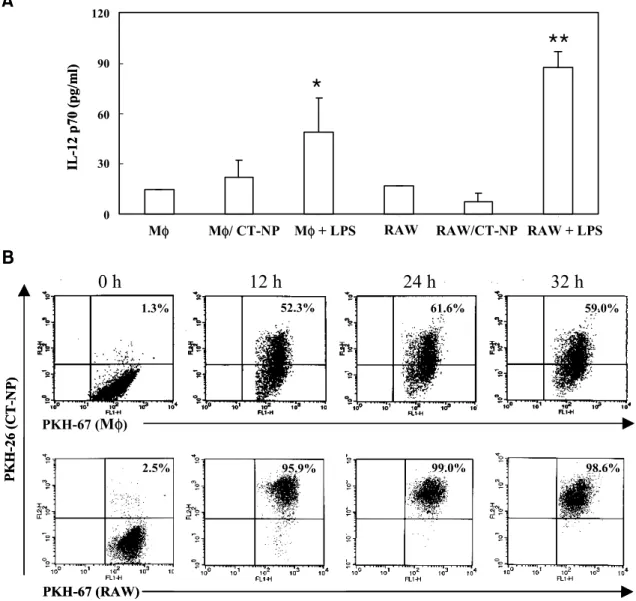
Figure 3. Peritoneal macrophages and RAW cells efficiently phagocytose necrotic tumor cells, but they are not active in producing IL-12. A. Peritoneal macrophages or RAW cells were co-cultured with necrotic tumor cells for 12 h at the ratio of 1:1. Thereafter, culture supernatants were collected, and examined for IL-12 p70 production by ELISA. Macrophages and RAW cells stimulated with LPS (1 μg/ml) were used as positive control. Data represent one of two experiments, each of which produced similar results (*P<0.05;
**P<0.01; Student's t test). B. PKH-26-labeled (FL2) and PKH-67-labeled (FL1) cells were co-cultured at the ratio of 1:1 for 0, 12, 24, and 32 h. Cells were gated on the FL1 positive cells where FL2 positive cell population represents necrotic cells phagocytosed by macrophages or RAW cells.
A
0 30 60 90 120
1 2 3 4 5 6
Mφ Mφ/ CT-NP Mφ+ LPS RAW RAW/CT-NP RAW + LPS
IL-12 p70 (pg/ml)
*
**
1.3% 52.3% 61.6% 59.0%
2.5% 95.9% 99.0% 98.6%
PKH-67 (RAW) PKH-67 (Mφ)
PKH-26 (CT-NP)
B
0 h 12 h 24 h 32 h
A
0 30 60 90 120
1 2 3 4 5 6
Mφ Mφ/ CT-NP Mφ+ LPS RAW RAW/CT-NP RAW + LPS
IL-12 p70 (pg/ml)
*
**
1.3% 52.3% 61.6% 59.0%
2.5% 95.9% 99.0% 98.6%
PKH-67 (RAW) PKH-67 (Mφ)
PKH-26 (CT-NP)
B
0 h 12 h 24 h 32 h
was slightly increased (Fig. 2C). In fact, migration assay showed that tumor-pulsed DC had an enhanced ability to migrate responding to CCR7 ligand, MIP-3β (Fig. 2D). Thus, in terms of CCR1 expression and responsiveness to CCR7 ligand at least, pulsing with necrotic tumor cells induced a mature phenotype of DCs. These changes were also evident in DCs ex- posed to parent CT-26 cells, indicating that expres- sion of NP protein in cells does not have any differ- ential effect on CCR gene expression by DCs.
Pulsing with necrotic tumor cells does not induce IL-12 production in macrophages. We next questioned whether pulsing with tumor cells could induce enhanced IL-12 production in macrophages. When other antigen-pre- senting cells including peritoneal macrophages and RAW 264.7 cell line were tested, we observed mark- edly increased production of IL-12 by LPS stimul- ation, but not any significant increase of IL-12 pro- duction by co-cultures with necrotic tumor cells (Fig.
3A). In order to examine the possibility that the low level of IL-12 production was due to the defect of phagocytic activity, PKH-67-labeled antigen-present- ing cells were mixed with PKH-26-labeled necrotic cells and then analyzed by flow cytometry. As shown in Fig. 3B, the phagocytosis of necrotic tumor cells by primary macrophages and the macrophage cell line occurred more efficiently than the one by DCs. Thus, we concluded that IL-12 production in antigen- presenting cells does not correlate with phagocytic activity and the increased IL-12 production after contact with necrotic tumor cells is a characteristic feature of DCs.
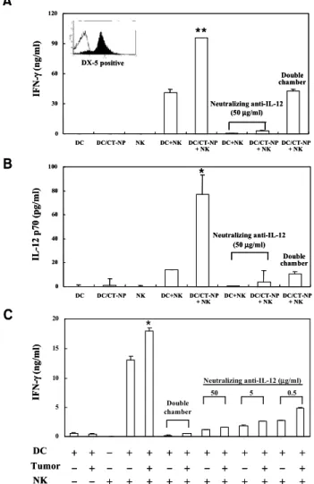
Induction of NK cell activity by vaccination with necrotic tumor cell-loaded DCs. It was recently reported by our group and others that murine DCs are capable of modul- ating innate immunity by stimulating NK cells thro- ugh cell-to-cell contact (13,17). Thus we examined NK cell stimulation by tumor-pulsed DCs in two different experiments; determining IFN-γ produc- tion after co-culturing with purified splenic NK cells in vitro and NK cell activity after DC vaccination in vivo. As shown in Fig. 4A, NK cells purified with DX-5 beads or DCs alone were not able to produce IFN-γ, whereas co-culturing them exhibited signifi- cant production of IFN-γ. However, DCs pulsed with necrotic tumor cells induced a much larger IFN- γ secretion than non-treated DCs. IFN-γ produc- tion was not observed in the culture supernatant from tumor cell-pulsed DCs, suggesting that at least DCs pulsed with necrotic tumor cells are not producers of IFN-γ. In addition, although we did not detect IL-12 after the washing of tumor-pulsed DC, the co-culture of DCs with NK cells induced IL-12 production that was significantly inhibited by a transwell culture (Fig. 4B). IFN-γ secretion was
confirmed again in co-culture of DCs and NK cells (Fig. 4C), which was blocked by a transwell culture or neutralizing anti-IL-12 antibody. These results show that NK cells can be stimulated to produce IFN-γ by tumor-pulsed DCs via cell-to-cell contact as well as soluble factor such as IL-12. Our results, however, could not rule out the possibility that after contact with NK cells, DCs might be converted to IFN-γ producers.
In order to examine the NK cell activity of
Figure 4. Co-culture of DCs with NK cells enhances IL-12 production and subsequent IFN-γ production. NK cells were positively selected with MACS (purity>88%). DCs (4×10⁵) or DCs pretreated with necrotic tumor cells for 24 h were thoroughly washed and co-cultured with purified splenic NK cells (2×10⁶) under various conditions in 24-well plates for 48 h.
IL-12 (B) and IFN-γ (A and C) in the culture supernatant were determined by ELISA. For IL-12 depletion, anti-IL-12 neu- tralizing antibody (50μg/ml) was added in cultures. In some ex- periments, transwell plates (pore size: 0.4μm, Costar, Cam- bridge, MA) were used to block cell-to-cell contact. Data are representative of more than three experiments (*, P<0.05 com- pared with DC + NK; **, P<0.01 compared with DC + NK).
0 30 60 90 120
DC DC/CT-NP NK DC+NK DC/CT-NP
+ NK DC+NK DC/CT-NP + NK DC/CT-NP
+ NK
0 20 40 60 80 100
DC DC/CT-NP NK DC+NK DC/CT-NP
+ NK DC+NK DC/CT-NP + NK DC/CT-NP
+ NK
IFN-γ(ng/ml)IL-12 p70 (pg/ml)
Neutralizing anti-IL-12 (50 µg/ml)
B A
DX-5 positive
0 5 10 15 20
1 2 3 4 5 6 7 8 9 1 0 1 1 1 2 1 3
C
IFN-γ(ng/ml)
DC Tumor
NK
Neutralizing anti-IL-12 (µg/ml) Double
chamber
Neutralizing anti-IL-12 (50 µg/ml)
+ + − + + + + + + + + + +
− + − − + − + − + − + − +
− − + + + + + + + + + + +
50 5 0.5
Double chamber
Double chamber
**
*
*
splenocytes in vivo, 51Cr release assay was performed 2 or 5 days after DC vaccination using NK-sensitive YAC-1 cells as targets. As expected, DCs pulsed with necrotic tumor cells markedly induced NK cell- mediated cytolysis after a short-term restimulation of vaccinated splenocytes with IL-2 in vitro (Fig. 5A).
NK cell activity was not due to contamination of
cytotoxic T cells because target cell killing was not influenced by a depletion of CD8-posive T cells using specific mAb and complement (Fig. 5B). Similar to NK cell activity, vaccination with tumor-pulsed DCs influenced INF-γ mRNA expression in splenocytes, which was still elevated 5 days after vaccination when compared to vaccination with DC alone (Fig. 5C).
Figure 5. Activation of NK cells and induction of tumor-specific CTL after immunization with necrotic tumor cell-loaded DCs. A. Balb/c mice were subcutaneously immuni- zed and splenocytes were harvested on days 2 and 5, cultured for 12 h in the presence of IL-2 (10 ng/ml), and used as effector cells against
51Cr-labeled YAC-1 cells. B. To examine NK activity after depletion of CD8+ T cell, splenocytes from each group were treated with anti- CD8 mAb (3.168) and rabbit com- plement, and then used as effector cells. Data are representative of two experiments with similar results. C.
Splenocytes from immunized mice were harvested and total RNA was prepared. RT was performed and prepared cDNA were amplified with specific primers for IFN-γ. The relative values of PCR products were calculated as described in Fig. 2. D.
Mice were subcutaneously immuni- zed with PBS, necrotic tumor cells, DCs, or DCs pulsed with necrotic tumor cells. One week after immu- nization, splenocytes were harvest- ed, cultured for 3 days with irradi- ated DC pulsed with necrotic CT- NP, and used as effector cells for CTL assay. Target cells labeled with 100μCi of Na51CrO4 were incu- bated with graded numbers of ef- fector cells in round-bottomed 96 well plates. After 4 h, the super- natants were harvested and counted in a gamma counter. CTL activity against CT-NP, CT-26, or Renca target cells was examined by using the same effector cells harvested from splenocytes of DC/CT-NP immunized mice. These data are re- presentative of a minimum of three experiments with similar results.
0 10 20 30 40 50
0 0 .5 1 1 .5 2 2.5 3 3 .5
DC/CT-NP DC PBS
0 10 20 30 40 50
0 0.5 1 1 .5 2 2 .5 3 3 .5
DC/CT-NP DC PBS
% of specific lysis
A
B
Day 2 Day 5
100 33.3 11.1
E/T ratio
D
0 5 10 15 20 25
0 0.5 1 1 .5 2 2 .5 3 3 .5
Target : CT-NP Target : CT-26 Target : Renca
0 10 20 30 40
0 0.5 1 1 .5 2 2 .5 3 3 .5
PBS CT-NP DC DC/CT-NP
100 33.3 11.1
E/T ratio
100 33.3 11.1
E/T ratio
100 33.3 11.1
E/T ratio
% of specific lysis
IFN-γ β-actin
Day 2 Day 5
Cont DC DC +CT-NP DC DC +CT-NP
Fold 1 1.8 1.8 1.1 1.7
100 33.3 11.1
% of specific lysis
CD8+
untreated 3.168 treated
0 20 40 60
0 0 .5 1 1 .5 2 2 .5 3 3.5
DC/CT-NP DC CT-NP PBS
C
0 10 20 30 40 50
0 0 .5 1 1 .5 2 2.5 3 3 .5
DC/CT-NP DC PBS
0 10 20 30 40 50
0 0.5 1 1 .5 2 2 .5 3 3 .5
DC/CT-NP DC PBS
% of specific lysis
A
B
Day 2 Day 5
100 33.3 11.1
E/T ratio
D
0 5 10 15 20 25
0 0.5 1 1 .5 2 2 .5 3 3 .5
Target : CT-NP Target : CT-26 Target : Renca
0 10 20 30 40
0 0.5 1 1 .5 2 2 .5 3 3 .5
PBS CT-NP DC DC/CT-NP
100 33.3 11.1
E/T ratio
100 33.3 11.1
E/T ratio
100 33.3 11.1
E/T ratio
% of specific lysis
IFN-γ β-actin
Day 2 Day 5
Cont DC DC +CT-NP DC DC +CT-NP
Fold 1 1.8 1.8 1.1 1.7
100 33.3 11.1
% of specific lysis
CD8+
untreated 3.168 treated
untreated 3.168 treated
0 20 40 60
0 0 .5 1 1 .5 2 2 .5 3 3.5
DC/CT-NP DC CT-NP PBS
C
However, consistent with IFN-γ production, vac- cination with DC alone exhibited to a certain extent enhanced NK cell activity though it was significantly lower compared to the vaccination with tumor-pulsed DCs. Together, these results suggest that necrotic tumor cell-loaded DCs stimulate NK cells to produce IFN-γ as well as induce NK cell activation to efficiently kill target cells.
Induction of tumor-specific CTL by vaccination with necrotic tumor cell-loaded DCs. We further examined the induction of CTL activity against tumor cells after vaccination with necrotic tumor cell-loaded DCs. The spleen cells harvested from vaccinated mice were restimulated in vitro with DCs pulsed with necrotic tumor cells and their cytolytic activities were deter- mined by 51Cr release assay. As shown in Fig. 5D, vaccination with necrotic tumor cells alone did not induce cytolytic activity against CT-NP target tumor cells. However, vaccination with necrotic tumor cell-loaded DCs induced markedly enhanced cyto- toxic activities that were almost completely inhibited by an in vitro treatment with CD8-specific mAb and complement, suggesting cytotoxic T lymphocyte-med- iated target killing (data not shown). It was notable that similar to NK activity induction, moderate cyto- lytic activity was also observed after vaccination with DCs alone. When cytotoxic activities were tested against different target cells, the cytolysis of syngeneic Renca cells was marginal, whereas cytotoxic activities against parent CT-26 tumor cells were not signi- ficantly different from those against CT-NP cells (Fig.
5D, right). These results indicate that vaccination of tumor cell-pulsed DCs is able to induce efficient tumor-specific cytotoxic T cell responses.
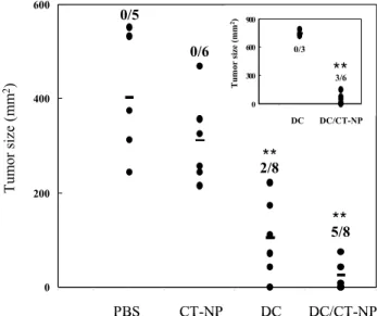
Therapeutic effect of vaccination with necrotic tumor cell-loaded DCs. To investigate the in vivo efficacy of immu- nization with necrotic tumor cell-loaded DCs, both protective and therapeutic vaccinations were perform- ed. When necrotic tumor cell-loaded DCs as a pro- phylactic vaccine were injected s.c. 7 days before tumor challenge at the same injection site, we consis- tently observed tumor protection. In addition, even after rechallenging both CT-NP and parent CT-26 cells, tumor formation was not observed and this protective immunity lasted for at least 4 months of observation (data not shown). To evaluate the thera- peutic efficacy of necrotic tumor cell-loaded DCs, viable 2×10⁵ tumor cells were inoculated in the right flank. After 3 days, when tumors were measurable, mice were immunized intratumorally with PBS buffer, necrotic tumor cells, DCs or tumor cell-loaded DCs.
A second immunization was done 7 days after the first one. As shown in Fig. 6, vaccination with necrotic tumor cell-loaded DCs significantly increased the number of tumor-free mice and reduced tumor
size compared with any other vaccinations when tumor size was determined on day 30 after first im- munization. Importantly, a substantial reduction of tumor size was also observed in mice treated with DCs alone. Thus, we questioned whether DCs ad- ministered by intratumoral injection might behave like tumor cell-pulsed DCs in vivo. To test this, therapeutic vaccination was done in the opposite flank to block encounter of DCs and tumor cells. While a significant therapeutic effect was repeatedly obtained by the contra-lateral subcutaneous injection of necrotic tu- mor cell-loaded DCs, tumors with a similar growth rate to the control tumors were formed in mice injected contra-laterally with DCs alone (Fig. 6, inset).
These results indicate that intratumoral injection of DC caused DC to uptake tumor cells in vivo similar to tumor-pulsed DCs and resulted in the partial inhibition of tumor growth. Furthermore, when vi- able tumor cells were rechallenged on the opposite left flank in completely cured mice, tumors did not form over a 30- to 40-day period, indicating that the therapeutic vaccination induced long-lasting immune memory responses against tumor cells (data not shown).
Figure 6. Inhibition of tumor growth by immunization with necrotic tumor cell-loaded DCs. Balb/c mice were challenged subcutaneously with viable 2×10⁵ tumor cells on the right flank.
After 3 days, when tumor size was measurable (≥9 mm2), mice were treated intratumorally with tumor cell-loaded DCs as indi- cated. Two groups of mice were treated contra-laterally on the left flank (inset). Seven days later, the treatment was repeated in the same manner. Tumor size was evaluated in two perpendicular dimensions with calipers on day 30 after first immunization. The numbers indicate the ratio of tumor-free mice to treated mice.
Data obtained from intratumoral experiments are mixed from three independent experiments with two or three mice per group.
(**, P<0.001 compared with PBS control or DC alone).
0 200 400 600
0 0.5 1 1 .5 2 2 .5 3 3 .5 4 4 .5
PBS CT-NP DC
Tumor size (mm2)
0/5
0/6
2/8
5/8
DC/CT-NP
0 300 600 900
0 0 .5 1 1 .5 2 2 .5
Tumor size (mm2)
0/3 3/6
DC DC/CT-NP
**
**
**
0 200 400 600
0 0.5 1 1 .5 2 2 .5 3 3 .5 4 4 .5
PBS CT-NP DC
Tumor size (mm2)
0/5
0/6
2/8
5/8
DC/CT-NP
0 300 600 900
0 0 .5 1 1 .5 2 2 .5
Tumor size (mm2)
0/3 3/6
DC DC/CT-NP
**
**
**
Discussion
Although a number of strategies for cancer therapy using DC-based vaccine have previously been report- ed, the most efficient method of vaccine preparation suitable for individual tumor types still remains to be explored. Especially, the establishment of key para- meters on the immune response by DC vaccine, load- ed ex vivo with well-characterized tumor antigens, their peptides or tumor materials, is of crucial impor- tance and necessary for the precise prediction and evaluation of vaccine efficacy. In this study, we in- vestigated the uptake of necrotic tumor cells by dendritic cells (DCs) by means of flow cytometric, confocal and electron microscopic analysis. Although the relative ratio of tumor uptake in DCs was lower than that in primary macrophages or the macrophage cell line, they were capable of picking up necrotic tumor cells effectively. Most notably, while macro- phages could not be activated to produce hetero- dimeric cytokine IL-12, DCs pulsed with necrotic tumor cells produced markedly increased amount of IL-12 or IL-12 p40 transcript. It seems highly unlikely that the enhanced IL-12 production in DCs is due to infection of tumor cell lines as recently demon- strated (18), because Mycoplasma contamination of them was routinely checked and, more importantly, even after a treatment of them with cyproxin, IL-12 secretion in necrotic tumor-pulsed DCs was detected to a similar extent (data not shown).
IL-12 is a central component of the cellular im- mune response as a surrogate marker for type 1 T cell response and used as an indicator of DC acti- vation (19). Increased IL-12 production by DCs has been suggested as a crucial step in the process of CTL priming (20,21). IL-12 induces IFN-γ secretion by activated T cells, NK cells and NKT cells and is required for optimal adaptive and innate responses against tumors (22,23). Indeed, IL-12 and IL-18 have been reported to promote NK cell cytotoxicity through up-regulation of NK cell-derived IFN-γ production (24,25). Our observation that DCs pulsed with necrotic tumor cells produce a significant amount of IL-12, and stimulate NK cell cytolytic activity and IFN-γ secretion is consistent with this concept. However, IL-12 production may not be enough to result in NK cell cytotoxicity and IFN-γ production, because DCs showing low levels of IL-12 production also have the ability to induce increased production of IFN-γ in NK cells and the IFN-γ production is not induced by culture supernatants of tumor-pulsed DCs. In fact, tumor cell-pulsed DCs prepared for immunization in vivo appeared to be a weak IL-12 producer after washing of DCs for injection as shown in Fig. 4B. Thus antigen-pulsed
DC and NK interaction is more important for IL-12 production and subsequent NK cell activation than necrotic tumor cell pulsing itself, and supposed to be relevant in the control and the amplification of the antitumor immune response. Moreover, immuniza- tion with DC alone could induce NK cell cytoto- xicity, albeit to a lesser extent. It is important that while contralateral injection of tumor-pulsed DCs induces a significant therapeutic effect, DCs alone do not show any tumor suppressive effect, indicating that although DCs alone can, at least in part, activate NK cells, they are not sufficient for the induction of systemic antitumor immune responses.
It was recently reported that murine or human DCs were capable of modulating the innate immunity by stimulating NK cells through cell-to-cell contact (26,28). However, the requirement of DC-derived IL-12 during the development of cellular immunity is somewhat controversial since DC-derived IL-12 is not required for the generation of protective immu- nity against melanoma and treatment of DCs with anti-IL-12 antibody does not block the enhancement of NK cell-mediated cytolysis by DCs (27,28).
Grufman et al. indeed demonstrated that it is indeed possible to generate good CTL responses by immu- nizing B6 mice with IL-12-deficient DC (23). They suggested that either DC-derived IL-12 may still be required, but the inoculated DC may be engulfed and presented by host APC or MHC class I-peptide complexes may be transferred from the inoculated cells to host APC, which are able to produce IL-12.
Alternatively, neighboring cells may provide IL-12 required for optimal responses. In the present study, we demonstrated that intratumoral injection of DCs alone had a minor but substantial effect on the inhibition of tumor formation. Thus, the results showing that the ability of DCs to produce IL-12 correlates with NK cytotoxicity, IFN-γ production and tumor-specific T cell cytotoxicity are consistent with this notion. Recent work from our group has shown that NK cell depletion in vivo correlates closely with the weakness of DC vaccine-induced protective immunity and tumor rejection (13). In another thera- peutic setting using the B16 model, immunization with the DC vaccine could inhibit lung metastasis of tumor cells, whereas the depletion of NK cells before DC vaccination abrogated the therapeutic effect (manuscript in preparation). Therefore, one might expect that DCs might acquire nominal tumor antigens and stimulate tumor-specific CTLs by phagocytosis of dying tumor cells at the tumor site, presenting them in both MHC class I and class II molecules (29-32). Taking this into consideration, we speculate that IL-12 production in tumor-pulsed DCs or DCs administered into tumor sites after contact