Copyright © 2011 by Th e Korean Orthopaedic Association
Th is is an Open Access article distributed under the terms of the Creative Commons Attribution Non-Commercial License (http://creativecommons.org/licenses/by-nc/3.0) which permits unrestricted non-commercial use, distribution, and reproduction in any medium, provided the original work is properly cited.
Clinics in Orthopedic Surgery • pISSN 2005-291X eISSN 2005-4408
Clinical Results of Technique for Double Bundle Anterior Cruciate Ligament Reconstruction Using
Hybrid Femoral Fixation and Retroscrew
Doo-Sup Kim, MD, Chang-Ho Yi, MD, Hoi-Jung Chung, MD, Yeu-Seung Yoon, MD
Department of Orthopedic Surgery, Yonsei University Wonju College of Medicine, Wonju, Korea
Received March 1, 2011; Accepted September 17, 2011 Correspondence to: Yeu-Seung Yoon, MD
Department of Orthopedics, Yonsei University Wonju College of Medicine, 162 Ilsan-dong, Wonju 220-701, Korea
Tel: +82-33-741-1357, Fax: +82-33-746-4326 E-mail: [email protected]
In spite of many excellent outcomes with arthroscopic an- terior cruciate ligament (ACL) reconstruction, problems remain with this procedure; rotation instability and physi- cal disabilities are not always completely resolved.1,2) Rota- tion stability has been achieved by tunneling the femur at the 10-o'clock position or by double bundle ACL recon-
Background: Anatomic anterior cruciate ligament (ACL) reconstruction has been presented as a means to more accurately re- store the native anatomy of this ligament. This article describes a new method that uses a double bundle to perform ACL recon- struction and to evaluate the clinical outcome.
Methods: Grafts are tibialis anterior tendon allograft for anteromedial bundle (AMB) and hamstring tendon autograft without detachment of the tibial insertion for posterolateral bundle (PLB). This technique creates 2 tunnels in both the femur and tibia.
Femoral fi xation was done by hybrid fi xation using Endobutton and Rigidfi x for AMB and by biointerference screw for PLB. Tibial fi xations are done by Retroscrew for AMB and by native insertion of hamstring tendon for PLB. Both bundles are independently and differently tensioned. We performed ACL reconstruction in 63 patients using our new technique. Among them, 47 participated in this study. The patients were followed up with clinical examination, Lysholm scales and International Knee Documentation Committee (IKDC) scoring system and radiological examination with a minimum 12 month follow-up duration.
Results: Signifi cant improvement was seen on Lachman test and pivot-shift test between preoperative and last follow-up. Only one of participants had fl exion contracture about 5 degrees at last follow-up. In anterior drawer test by KT-1000, authors found im- provement from average 8.3 mm (range, 4 to 18 mm) preoperatively to average 1.4 mm (range, 0 to 6 mm) at last follow-up. Aver- age Lysholm score of all patients was 72.7 ± 8.8 (range, 54 to 79) preoperatively and signifi cant improvement was seen, score was 92.2 ± 5.3 (range, 74 to 97; p < 0.05) at last follow-up. Also IKDC score was normal in 35 cases, near normal in 11 cases, abnormal in 1 case at last follow-up.
Conclusions: Our new double bundle ACL reconstruction technique used hybrid fi xation and Retroscrew had favorable outcomes.
Keywords: Anterior cruciate ligament reconstruction, Double bundle, Anatomical, Hybrid fi xation, Retroscrew
struction.3-8) However, there has been a great deal of de- bate as to the ability of the double bundle technique with respect to the selection of grafts, fixation methods, and techniques for tightening each bundle.4,5,8,9) Complications of this technique have included failure of graft fixation and excessive tunnel dilation due to an increase in tunnel numbers and adversity when an operation has required revision.
Endobutton (Smith & Nephew, Andover, MA, USA) was introduced and commonly used as a femoral fi xation method to overcome limitations in length and thickness of the autograft.5) However, Endobutton has had some problems in the graft healing in the bony tunnel due to
shortening of graft ed tendons, as well as excessive dilation of tunnels for the receipt of Endobuttons.10) For this rea- son, we minimized graft motion by fi xating the graft with a bio-absorbable transfemoral pin, Rigidfix pin (Mitek, Johnson and Johnson, Raynham, MA, USA). In order to restore the native tibial insertion of ACL, the Retroscrew (Arthrex, Naples, FL, USA) was designed and introduced.
It was proven to facilitate optimum joint line fixation at the level of the intercondylar fl oor for soft tissue autograft s and allograft s to maximize graft stiff ness, fi xation strength, abrasion resistance, and anatomical placement.11) To prove its advantages in the clinical setting and in clinical out- comes, we used the Retroscrew as a tibial fixation to the articular outlet of the tibial tunnel for anteromedial (AM) bundle.
In addition, for further fi xation to the distal portion, the gracillis and semi-tendinosus tendons, without detach- ment of the tibial insertion, were used for the posterolat- eral (PL) bundle, and a bio-absorbable interference screw, Intrafix (Mitek, Johnson and Johnson), was also applied to the articular outlet of the PL femoral tunnel in order to minimize graft motion. Th us, we designed a clinical study to investigate the clinical outcomes of this technique.
METHODS
Study Subjects
From May 2005 to May 2007, 63 patients underwent anterior cruciate ligament reconstruction with our new technique using the double bundle. We excluded patients with combined injuries of the medial or lateral collateral ligaments and posterior cruciate ligament. Patient with a meniscus injury more than and equal to grade 3 in mag- netic resonance imaging (MRI)12) were excluded as well as patient who had undergone revisional surgery. We per- formed a conventional single bundle reconstruction for osteoporotic patients (bone mineral density < −3.0), these was excluded also. However, there were no patients ex- cluded as a result of loss to follow-up. In total, 47 patients were enrolled into this prospective study. Th e mean time of follow-up was 18.7 months (range, 12 to 23 months);
there were 46 males and 1 female; there was an average age of 23.8 years (range, 19 to 38 years) and 38 operations oc- curred on right knees, while 9 operations occurred on left knees. Th e time period from trauma to operation was an average of 6.8 months (range, 1 to 22 months). Th e reason for anterior cruciate ligament reconstruction included the following: 42 patients reported sports injury; 26 reported jumping injuries and 12 reported running injuries when playing soccer, 2 reported jumping injuries when playing
basketball, 2 injured during recreational activities, and 5 patients reported having falling down as a result of their occupation.
In 28 cases, we found meniscal injury under ar- throscopy: 18 were medial meniscus injuries, 5 were lat- eral meniscus, and 5 cases were bilateral. For treatment of combined meniscal injury, we sutured torn meniscus in 13 medial meniscus injuries, 3 lateral meniscus injuries; we did menisectomy in 10 medial meniscus injuries, 6 lateral meniscus. In one case of lateral meniscus injury, we per- formed a meniscal transplantation using an allograft aft er 6 months aft er total menisectomy.
Methods
We performed the Lachman test and the pivot-shift test prior to operation and at last follow-up. In addition, we used the Lysholm score and International Knee Documen- tation Committee (IKDC) for clinical outcome evaluation.
For the evaluation of instability, we used the KT-1000 (Medmetric Co., San Diego, CA, USA) arthrometer. A second-look arthroscopy was performed in 28 cases. Th e indication for second-look arthroscopy was those persons wanted to remove the screw for tibial fi xation of the graft 1 year postoperative (average, 13.2 months; range, 12 to 18 month).
We classified graft tension by pulling the middle portion of the graft with probe at 80o fl exion of knee joint.
If the graft pulled less than 3 mm on the basis of lateral femoral condyle, it was considered ‘Normal’. If the graft pulled 4 to 5 mm, it was considered ‘Partial Relaxation’.
If the graft was torn or pulled over 5 mm, it considered a ‘Failure’.13) In addition, we classifi ed synovial formation into 3 categories. When the graft was covered with syno- vial membrane completely, we made a judgment of ‘Good’.
When the synovial membrane was thin or insufficient compared with the posterior cruciate ligament, it was con- sidered ‘Half ’. In cases of minimal synovialization and vis- ible strands of graft , we considered it ‘Pale’.13)
Stastical Analysis
In evaluation of continuous variables, we used the paired t-test in evaluation of knee scores, results of KT-1000 arthrometers and the Wilcoxon signed-rank test in evalu- ation of the Lachman test, degree of synovialization, and pivot-shift test. Spearson correlation analysis was used in the evaluation of follow-up periods and in the degree of graft tension that occurred in second-look operations. Th e statistical program SPSS ver. 11.0 (SPSS Inc., Chicago, IL, USA) was for statistical analyses. Th e level of signifi cance was set at a p-value less than 0.05.
Surgical Procedure
Tibialis anterior tendon allograft is obtained for the AM bundle, and the gracillis and semi-tendinosus tendon, without detachment of the tibial insertion, are used for the PL bundle. The tibialis allograft is double-looped, 8 mm in thickness and ≥ 120 mm in total length, so that additional fi xation to the tibia. Endobutton is suspended at the looped portion of the allograft in relation to the length of a femoral tunnel. Whipstitch sutures with No. 2 Ethibond were placed 3 cm below the suspended site, and No. 1 Ethibond (Ethicon, Somerville, NJ, USA) was passed through the 2 holes of the Endobutton (Fig. 1A). Th e ham- string tendon graft is harvested cautiously with an open
tendon stripper, in order to prevent the insertion site from detaching from the tibia. Gracillis and semi-tendinosus tendons are sutured to form 1 bundle that measures 6 mm in thickness and ≥ 120 mm in total length (Fig. 1B).
Whipstitch sutures with No. 2 Ethibond were placed 3 cm below the femoral end of the graft , and 5-0 Ethibond su- tures were placed in the femoral end of the graft .
Aft er minimal notchplasty, the tibia is fi rst reamed for the PL bundle with the knee held in 90 degrees of fl ex- ion. An ACL tibial guide (Acufex Micro Surgical, Mans- fi eld, MA, USA) set a 45 degrees is introduced through the anteromedial portal. Th e tunnel is reamed with consider- ation of the graft size aft er the intra-articular side is placed
Fig. 1. (A) The tibialis allograft is pre- pared (a, 30 mm for the femoral tunnel; b, 30 mm for the intra-articular space; c, 40 mm for the tibial tunnel). Mersilene tape of the Endobutton is suspended at the looped portion of the allograft. Whipstitch sutures with No. 2 Ethibond are placed 3 cm below the suspended site. (B) Anterolateral view of the operative right knee: The hamstring tendon autograft was harvested and prepared. The gra- cillis and semi-tendinosus tendons are stripped from the femoral side without detachment of the tibial insertion site (left) (a, 40 mm for the tibial tunnel; b, 20 mm for the intra-articular space; c, 30 mm for the femoral tunnel). The portion for tibial tunnel of graft was prepared with Ethibond (right).
Fig. 2. (A) Tibial tunnel for posterolateral (PL) bundle and anteromedial (AM) bundle was seen on arthroscopic view. (B) Anterior cruciate ligament (ACL) footprint of PL bundle (arrow) was seen on intercondylar notch of femoral condyle (left) and femoral tunnel of the PL bundle was guided at remnant of the ACL footprint using outside-in technique (right).
5 mm anterior to the posterior cruciate ligament, just in the anteromedial portion of the posterior horn of the lat- eral meniscus, and in the posterolateral portion of ACL footprint (Fig. 2A).
Th e tibial tunnel for the AM bundle was made at the extra-articular portion, which was placed 1.5 cm above the upper margin of the pes anserius and 1 cm posterior to the medial margin of the tibial tubercle. An ACL tibial drill guide set at 40o is placed 7 mm anterior to the PL bundle, while the intra-artucular starting point was maintained in the anteromedial portion of ACL footprint (Fig. 2A). Th e tibal tunnel was reamed over a guide pin according to the diameter of graft and a transtibial technique that was used.
Th e femoral tunnel for the AM bundle was reamed proxi- mal to and posterior to femoral ACL footprint. Aft er a 6 mm off set guide was introduced through the tibial tunnel into the intra-articular space and was placed at the 10- to
10:30-o'clock position and 6 mm anterior to the posterior margin of the intercondylar notch in the case of a right knee. When the target position was reached, the tunnel was reamed with a 4.5 mm Endobutton cannulated drill over the guide pin, and the tunnel length was measured.
The femoral tunnel was reamed with consideration of a diameter of 8 mm that allows for Endobutton rotation, a length of 30 mm that allows for fi xing a Rigidfi x pin, and the diameter of the graft .
The femoral tunnel was reamed for the PL bundle with an ACL guide by the outside-in technique. After introducing an ACL guide through the anterolateral por- tal, the intra-articular side on medial wall of the lateral femoral condyle was placed 5-7 mm above to the lateral meniscus posterior horn at 90 degree knee flexion. The guide pin was positioned towards the intra-articular side of the bundle in the just posterior and proximal portion
Fig. 3. (A) Wire loop is introduced through the posterolateral (PL) femoral tunnel via outside-in technique (left) and a femoral Ethibond suture of the graft for the PL bundle is suspended to a wire loop that is pulled out of the PL tibial tunnel and the graft for the PL bundle is introduced through the femoral tunnel via a wire loop (right). (B) Lateral view of operative right knee: a Rigidfi x fi xation guide system is introduced into the anteromedial femoral tunnel through a transtibial approach. The guide sheaths are carefully positioned in the lateral epicondylar area 1 cm above the PL femoral tunnel through the skin incision for the fi xation of the bundle in order to prevent the graft from wrapping around the sheath. (C) Anteromedial (AM) bundle was fi xed with Retroscrew at tibial articular side (left) and with 1 spiked washer screw outside the tibia. Hamstring autograft for PL bundle was seen at medial side of AM bundle with no fi xation (arrow) (right). (D) Final view of the construct at 90° of fl exion showing the PL bundle crossing the AM bundle from the back.
of the lateral femoral epicondyle. Th e tunnel was reamed along the guide pin, and the distance between the 2 femo- ral tunnels was maintained at ≥ 4 mm. Th e wire loop was pulled out of the posterolateral tibial tunnel after being introduced into the intra-articular space from the lateral femoral condyle.
A femoral Ethibond suture of the graft for the PL bundle was suspended to a wire loop that was pulled out of the PL tibial tunnel and the graft for the PL bundle was introduced through the femoral tunnel via a wire loop (Fig.
3A). After a tension-measuring instrument is connected to the PL bundle, cyclic loading was applied to the graft 20 times, at a tension of 15-20 lbs. Next, a bio-absorbable interference screw, Intrafix was introduced through the anteromedial portal into the intra-articular surface and fi xed to the femur with the knee held in 10-20 degrees of flexion. Before the AM bundle was introduced through the tunnel, a Retroscrew was prepared for insertion. Aft er introducing a No. 5 fiberwire through the anteromedial portal into the intra-articular space and pulling it out of the anteromedial tibial tunnel to the extra-articular space, a cannula was introduced through the antermedial portal, and the suture was tucked in the cannula. Thereafter, a guide pin was introduced through the tunnel for the AM bundle by the transtibial approach. The No. 5 Ethibond suture in the AM bundle was tied to the eyelet of the guide pin and the graft for AM bundle was introduced through the tibial tunnel to the femoral tunnel by pulling out the No. 5 Ethibond suture. Aft er the Endobutton was fl ipped, 1 Rigidfi x bioabsorbable pin (length, 42 mm; diameter, 3.3 mm; Rigid Fix, Ethicon, Depuy Mitek Division, Norder- stedt, Germany) was fi xed to the proximal portion of the sheath by preparing the site in a manner so to prevent the graft from wrapping around the sheath (Fig. 3B); it’s the tight fi xation was confi rmed by pulling the graft . Another Rigidfi x pin was applied to the distal portion. Aft er cyclic loading was applied about 20 times at 15-20 lbs of tension, with the knee held at 60-70 degrees of flexion, a suture was connected to a Retroscrew and tucked in the intra- articular space with a pituitary rongeur. Th e suture outside the tibial tunnel was fixed with a cannulated Retroscrew driver, while it was advanced retrograde in the tunnel, then the AM bundle outside the tibia was fi xed with 2 staples or with a 6.5 mm spiked washer screw (Fig. 3C).
Postoperative Rehabilitation
After skin closure, cylinder splint was applied with full extension of the knee joint. Splint was maintained until subsiding of postoperative pain approximately 2 days. Af- ter the closed drain removed, continuous passive range of
motion exercise was started. For recovery of range of mo- tion by 90o fl exion at postoperative 4 weeks, partial weight bearing ambulation with crutch was permitted at postop- erative 1 day and hinged knee brace with limited motion was applied at postoperative 2 days with increasing exer- cise intensity and maintained at 4 weeks postoperative.
Aft er 6 weeks of partial weight bearing, full weight bearing was permitted at 7 weeks postoperative and patients were educated on being completely postoperative at 3 months.
We had the patients to start muscle strengthening exer- cises aft er disappearance of postoperative pain; bicycling exercise was allowed at postoperative 2 months; swimming was allowed at 3 months postoperative; and light exercise, such as jogging, was allowed at 6 months postoperative;
every sports activity was allowed at 9 months postopera- tive.
RESULTS
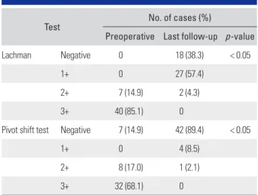
Significant improvement was seen on Lachman test and pivot-shift test between preoperative and last follow-up (Table 1). At last follow-up, negative in pivot-shift test was seen in 42 cases. In range of motion, there was preopera- tive fl exion contracture between 5 to 10 degrees in 8 cases.
In one case, fl exion contracture remained of about 5 de- grees at last follow-up.
We checked the diff erence from contralateral side in anterior drawer test by KT-1000 before operation and at last follow-up (Table 2). We found improvement from an average 8.3 mm (range, 4 to 18 mm) preoperative to aver- age 1.4 mm (range, 0 to 6 mm) at last follow-up. At last follow-up, a diff erence of less than 3 mm was seen in 42 cases. In one case, a 6 mm diff erence was seen.
Table 1. Results of Lachman and Pivot-Shift Test
Test
No. of cases (%)
Preoperative Last follow-up p -value
Lachman Negative 0 18 (38.3) < 0.05
1+ 0 27 (57.4)
2+ 7 (14.9) 2 (4.3)
3+ 40 (85.1) 0
Pivot shift test Negative 7 (14.9) 42 (89.4) < 0.05
1+ 0 4 (8.5)
2+ 8 (17.0) 1 (2.1)
3+ 32 (68.1) 0
Average Lysholm score of whole patients was 72.7
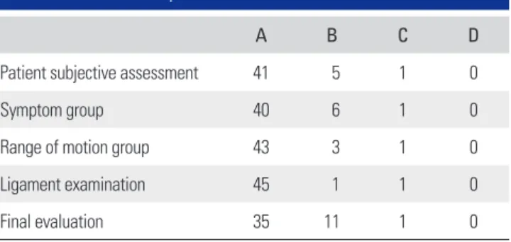
± 8.8 (range, 54 to 79) preoperative. At last follow-up, sig- nifi cant improvement was seen, score was 92.2 ± 5.3 (range, 74 to 97; p < 0.05). At last follow-up, IKDC score was nor- mal (A) in 35 cases, near normal (B) in 11 cases, abnormal (C) in 1 case. There was no patient that was considered severely abnormal (D) (Table 3).
In the 28 cases that required a second-look opera- tion, graft tension was checked at 80o flexion.13) The an- teromedial bundle wasnormal in 22 cases, while partial relaxation was reported in 6 cases. Th ere were no cases of failure. Th e posterolateral bundle was normal in 16 cases, partially relaxation in 8 cases, and failure was seen in 4 cases. Th ere was no correlation between follow-up periods and degree of graft tension in anteromedial and postero- lateral bundles (p = 0.48, 0.31).
Synovial membrane formation of graft was reported as good in 18 cases, half in 9 cases, pale in 1 case in antero- medial bundle. The posterolateral bundlewas good in 16 cases, half in 8 cases, and pale in 4 cases. However, there was no significant correlation between graft tension and clinical results (p > 0.05).
Intraoperative complication was seen in 5 cases during the fl ipping of Endobutton device. We confi rmed fi xation of Endobutton by pulling graft aft er fl ipping En- dobutton. In cases where the skin retracted when the graft pulled, we checked the radiograph during operation. Th e Endobutton had not been fully attached to the femur due to impingement in muscles, so we put additional incisions to release impinged muscles and repositioned the Endo- button. Complications after the operation were reported in 13 cases. Sensory changes at the donor site occurred in 12 cases, superfi cial infection was reported in 1 case. Th ere was no deep infection. Among 28 cases taken second-look operation, partial rupture was seen in the posterolateral
bundle in 4 cases and debridement was performed in these cases. Despite partial rupture, satisfactory clinical results were shown.
DISCUSSION
In double bundle ACL reconstruction, there are contro- versies and weaknesses. One of them is taking much time for graft incorporation into bone due to an increase in the number of tunnels. Th erefore, we did not detach the tibial insertion of the hamstring tendon autograft in order to obtain a more durable fi xation of the distal portion of the graft or to shorten incorporation time into bone with a more viable graft .14) Also, we were able to reduce discom- fort in the tibial area during postoperative rehabilitation due to excessive volume of the graft in the anteromedial portion of the tibia aft er tibial fi xation by using the con- ventional technique, since fixation of the PL bundle was not needed in our technique (Fig. 4).14) But, there were limitations, such as lengthened fixation length causing tunnel widening.
Edwards et al.15) have reported that the most excel- lent outcome can be obtained when 2 tunnels are used for the tibia and the femur each, respectively. Th us, we used 2 tibial and 2 femoral tunnels, because the single tibial tunnel technique can be diffi cult in the restoration of the native shape of the ACL. But the increase in the number of tunnels may lead to some disadvantages, such as diffi culty in the operative technique and more frequent occurrence of dilation of tunnels. Siebold et al.16) found that MRI taken at the 1-year follow-up revealed remarkable dilation of the 2 femoral tunnels when 2 tibial tunnels are broken and communicated in double bundle ACL reconstruction using 4 tunnels. Authors were able to separate 2 tibial tun- nels by leaving the tibial cortical bone between them when reaming the tibial tunnel.
The choice of grafts is inevitable issue in discus- Table 2. Results of KT-1000 Manual Maximum Side-to-Side Di-
fferences
Differences (mm)
No. of cases (%) Preoperative Last follow-up
< 3 0 45 (95.7)
3-5 2 (4.3) 2 (4.3)
6-10 38 (80.8) 0
> 10 7 (14.9) 0
Mean (mm) ± SD 8.3 ± 2.3 1.4 ± 1.2
SD: standard deviation.
Table 3. Last Follow-up IKDC Grade
A B C D
Patient subjective assessment 41 5 1 0
Symptom group 40 6 1 0
Range of motion group 43 3 1 0
Ligament examination 45 1 1 0
Final evaluation 35 11 1 0
IKDC: International Knee Documentation Committee.
sion of our technique. Many graft options are available for ACL reconstruction, including diff erent autograft and allograft tissues. Autograft s include bone-patellar tendon- bone composites, combined semitendinosus and gracilis hamstring tendons, and quadriceps tendon. Allograft op- tions include the same types of tendons harvested from donors, in addition to Achilles and tibialis tendons. In studies deal with graft choice recently, allograft s proved its durability and strength in single-bundle technique17) and double bundle technique,18) and through comparison with autografts.19) Though risk of graft failure was higher in hamstring tendon than bone-patellar tendon-bone graft s, the hamstring tendon has shown favorable clinical results.
For this reason, we thought there would be no diffi culties using allograft s in double-bundle reconstruction.20) But, in aspect of interaction between autograft and allograft, we could not predict the types ofside eff ects. Studies discussed this subject have to be done in near future.
Endobutton is also one of subjects in controversy.
Although several authors have recently used Endobut- ton to fix short grafts, this technique has difficulty to fix at femoral cortex accurately. We experienced 4 cases in which Endobutton was not able to attach to the femoral cortex because of a bottleneck between muscles. So, we recommend if skin retraction was seen when pulling graft after turning of Endobutton, intraoperative radiograph should be taken to confi rm the fi xation of Endobutton. In addition, Endobutton has led to dilation of the tunnel and the bungee cord eff ect, which is perpendicular motion of a graft due to distant fi xation of each end of the graft . To this eff ect, the authors fi xed 2 additional Rigidfi x pins in order
to strengthen Endobutton fi xation. Th ere have been many studies suggesting that 2 Rigidfi x pin possessed adequate strength to fi x a graft .21) Between them, Kousa et al.22) re- ported Rigidfix showed fixation failure at 868N at single cycle load to failure test in biomechanical analysis. How- ever, we experienced 7 cases in which the graft descended or the pin was broken while applying the cyclic load 20 times in case of fi xation with the Rigidfi x pin alone. Han et al.23) also reported breakage of Rigidfix pin at femoral tunnel aft er anterior cruciate ligament reconstruction with hamstring tendon graft . Consequently, we avoided single Rigidfi x pin fi xation. Th rough additional Rigidfi x pin fi xa- tion with Endobutton, the femoral tunnel fixation was close to the intercondylar notch, so graft motion could be minimized. And we used a Retroscrew for the tibial graft for reducing fixation length in tibial fixation close to its natural anatomy, and as natural consequence, we could achieve juxta-articular fixation as compared with ante- grade screw fixation. Also, divergence of Retroscrew can be avoided by fi xing it under arthroscopy.11) In aspects of graft tension, the theoretical advantages of Retroscrew had not been proven in other studies. In a biomechanical study by Chang et al.,24) antegrade bioscrew showed advantages compared to Retroscrew such as superior stiffness, less displacement, greater maximum load at failure in the por- cine knee. Shortages of Retroscrew as technical diffi culties, low bone density in tibial metaphysis and small diameter could be thought cause of this unfavorable result. So, more clinical study for Retroscrew has to be done to clear this debate.
Clinical studies involving double-bundle recon- struction have been available for several years. In 1999, Muneta et al.8) published the results of a 2-year follow-up in 54 patients who underwent double-bundle reconstruc- tion. They reported a “trend” toward improved anterior tibia translational stability, but no patient data or param- eters were available. Kubo et al.25) and Hara et al.26) pub- lished that their technique was a “physiologically more du- rable ACL reconstruction,” but outcome data or statistics were not described. Recently, studies with patients' data and outcomes published,9) though its double-sidedness.
Between those studies, KT-1000 or 2000 arthrometry is known as the most reliable and reproducible parameter for measurement of anterior translation available. Various results of KT-1000 or 2000 arthrometry as 1.7 ± 2.0 mm, 1.4 ± 1.4 mm, 1.6 ± 2.0 mm, 1.9 ± 1.9 mm, 1.7 ± 2.0 mm was reported by numerous authors.6,7) We found an im- provement from an average 8.3 mm (range, 4 to 18 mm) preoperative to an average 1.4 mm (range, 0 to 6 mm) at last follow-up in contrast to the unsatisfactory results of Fig. 4. Schematic view of double bundle reconstruction of anterior
cruciate ligament with hybrid femoral fi xation and Retroscrew.
the Lachman test, which had negative results on 38.3% of the time. Despite in the development of bias in comparin our results of anterior translation directly, to other authors' finding, our results are superior to other studies. We re- garded this superior outcomes comes from several advan- tages of our technique. First, viable grafts were obtained by maintaining tibial insertion of the hamstring tendon autograft. Second, AM and PL bundles were fixed to the knee joint at diff erent angles. Th ird, adequate graft length for fi xation were achieved by using the tibialis tendon al- lograft and the hamstring tendon autograft with intact tibial insertion.
Besides limitations of the study design such as the short follow-up period, small sample size and need of comparing clinical results of our technique to single bundle technique and conventional double bundle tech- nique, in our technique, there are some limitations and disadvantages by its own nature. Same as other technique, it is technically diffi cult to perform and require a learning curve. Besides technical demands, cost-effectiveness is another weak point. Allograft itself could burden patients with high cost instrument system such as Rigid fi x pin sys- tem and Retroscrew system we used in this study. If stud- ies that compare our double bundle technique to conven- tional single-bundle technique could not be done or there were no signifi cant diff erences between techniques at all, continuous application of our technique could not be rec- ommended. Even if our technique was proven superior to other double bundle techniques, clinicians must consider its cost-effectiveness. In addition, unsuccessful results of PL bundle in second-look operation is our big weakness.
In 4 cases among 28 cases that took second-look opera- tion were shown failure of PL bundle and 8 cases of partial relaxation were shown. It could eliminate advantage of double bundle reconstruction. In some point, causal fac- tors of this unfavorable result considered. Nature of PL bundle could make excessive motion of graft , which might be related to the higher non-isometric function of the PL bundle compared with the AM bundle as Siebold and Ca- faltzis27) described in study about postoperative bone tun- nel widening. Regarding the graft fixation, authors fixed the AM bundle with the knee held in 60 degrees of fl exion
and the PL bundle with the knee held in 0 to 10 degrees of flexion. Considering that 2 bundles are fixed with the knee held in 30 degrees of flexion in double bundle re- construction, it is conceivable that excessive tension may rupture the PL bundle and overconstrain knee.28) Despite our attempt not to give too much tension to PL bundle, knee fl exion angle during PL bundle fi xation with the knee held in 0 to 10 degrees of fl exion might not have been suf- fi cient for preserving PL bundle for some reasons authors didn’t recognize. Fixation method which remains native insertion of hamstring tendon can be one of the causes.
Th e authors did not perform additional fi xation because of bio-interference screw at tibial tunnel of PL bundle, which made fixation length long as described above. Excessive motion of autograft due to this long fixation length may have induced the failure of PL bundle. Femoral tunnel positioning could be one of reasons, because transtibial technique is not appropriate method to make femoral tun- nel anatomically as Giron et al.29) discovered. Th e sequence of creating 2 femoral tunnel might have affected the PL bundle failure. We created a femoral tunnel for AM bundle fi rst in this study. However, Taketomi et al.30) reported PL tunnel first technique seems to be superior to AM first technique regarding anatomic placement. Non-anatomic PL tunnel placement could be a reason of PL bundle fail- ure.
Despite these limitations, our study is significant because we attempted to apply a theoretical possibility to clinical circumstances. Th is could be a proposal for further progression of double bundle ACL reconstruction.
In conclusions, our new double bundle ACL recon- struction technique used hybrid fi xation and Retroscrew with favorable outcomes. But, this technique should be evaluated further in regards to technical difficulty, cost- eff ectiveness, and the relatively unsatisfactory results of the posterolateral bundle.
CONFLICT OF INTEREST
No potential confl ict of interest relevant to this article was reported.
REFERENCES
1. Aglietti P, Buzzi R, Zaccherotti G, De Biase P. Patellar ten- don versus doubled semitendinosus and gracilis tendons for anterior cruciate ligament reconstruction. Am J Sports Med.
1994;22(2):211-7.
2. Kim SJ, Jung KA, Song DH. Arthroscopic double-bundle anterior cruciate ligament reconstruction using autogenous quadriceps tendon. Arthroscopy. 2006;22(7):797.e1-5.
3. Loh JC, Fukuda Y, Tsuda E, Steadman RJ, Fu FH, Woo SL.
Knee stability and graft function following anterior cruciate ligament reconstruction: comparison between 11 o'clock and 10 o'clock femoral tunnel placement. 2002 Richard O'Connor Award paper. Arthroscopy. 2003;19(3):297-304.
4. Yagi M, Wong EK, Kanamori A, Debski RE, Fu FH, Woo SL. Biomechanical analysis of an anatomic anterior cruciate ligament reconstruction. Am J Sports Med. 2002;30(5):660- 6.
5. Yasuda K, Kondo E, Ichiyama H, et al. Anatomic recon- struction of the anteromedial and posterolateral bundles of the anterior cruciate ligament using hamstring tendon graft s. Arthroscopy. 2004;20(10):1015-25.
6. Aglietti P, Giron F, Cuomo P, Losco M, Mondanelli N. Sin- gle-and double-incision double-bundle ACL reconstruction.
Clin Orthop Relat Res. 2007;454:108-13.
7. Muneta T, Koga H, Mochizuki T, et al. A prospective ran- domized study of 4-strand semitendinosus tendon ante- rior cruciate ligament reconstruction comparing single- bundle and double-bundle techniques. Arthroscopy.
2007;23(6):618-28.
8. Muneta T, Sekiya I, Yagishita K, Ogiuchi T, Yamamoto H, Shinomiya K. Two-bundle reconstruction of the anterior cruciate ligament using semitendinosus tendon with endo- buttons: operative technique and preliminary results. Ar- throscopy. 1999;15(6):618-24.
9. Hamada M, Shino K, Horibe S, et al. Single- versus bi- socket anterior cruciate ligament reconstruction using autogenous multiple-stranded hamstring tendons with en- dobutton femoral fi xation: a prospective study. Arthroscopy.
2001;17(8):801-7.
10. Yamazaki S, Yasuda K, Tomita F, Minami A, Tohyama H.
Th e eff ect of graft -tunnel diameter disparity on intraosseous healing of the fl exor tendon graft in anterior cruciate liga- ment reconstruction. Am J Sports Med. 2002;30(4):498-505.
11. Morgan CD, Stein DA, Leitman EH, Kalman VR. Anatomic tibial graft fi xation using a retrograde bio-interference screw for endoscopic anterior cruciate ligament reconstruction.
Arthroscopy. 2002;18(7):E38.
12. Reicher MA, Hartzman S, Duckwiler GR, Bassett LW, An- derson LJ, Gold RH. Meniscal injuries: detection using MR imaging. Radiology. 1986;159(3):753-7.
13. Ahn JH, Cho YB, Lee JY. Second-look arthroscopy after ACL reconstruction: comparison of patellar tendon auto- grafts with hamstring tendon autografts. J Korean Orthop Assoc. 2003;38(2):159-64.
14. Kim SJ, Kim HK, Lee YT. Arthroscopic anterior cruciate lig- ament reconstruction using autogenous hamstring tendon graft without detachment of the tibial insertion. Arthros-
copy. 1997;13(5):656-60.
15. Edwards TB, Guanche CA, Petrie SG, Th omas KA. In vitro comparison of elongation of the anterior cruciate ligament and single- and dual-tunnel anterior cruciate ligament re- constructions. Orthopedics. 1999;22(6):577-84.
16. Siebold R, Dehler C, Ellert T. Prospective randomized com- parison of double-bundle versus single-bundle anterior cru- ciate ligament reconstruction. Arthroscopy. 2008;24(2):137- 45.
17. Kuhn MA, Ross G. Allografts in the treatment of an- terior cruciate ligament injuries. Sports Med Arthrosc.
2007;15(3):133-8.
18. Royalty RN, Junkin DM Jr, Johnson DL. Anatomic double-bundle revision anterior cruciate ligament sur- gery using fresh-frozen allograft tissue. Clin Sports Med.
2009;28(2):311-26.
19. Lee JH, Bae DK, Song SJ, Cho SM, Yoon KH. Comparison of clinical results and second-look arthroscopy findings aft er arthroscopic anterior cruciate ligament reconstruction using 3 diff erent types of graft s. Arthroscopy. 2010;26(1):41- 9.
20. Reinhardt KR, Hetsroni I, Marx RG. Graft selection for an- terior cruciate ligament reconstruction: a level I systematic review comparing failure rates and functional outcomes.
Orthop Clin North Am. 2010;41(2):249-62.
21. Harilainen A, Sandelin J. A prospective comparison of 3 hamstring ACL fixation devices--Rigidfix, BioScrew, and Intrafi x--randomized into 4 groups with 2 years of follow- up. Am J Sports Med. 2009;37(4):699-706.
22. Kousa P, Jarvinen TL, Vihavainen M, Kannus P, Jarvinen M.
Th e fi xation strength of six hamstring tendon graft fi xation devices in anterior cruciate ligament reconstruction. Part I:
femoral site. Am J Sports Med. 2003;31(2):174-81.
23. Han I, Kim YH, Yoo JH, Seong SC, Kim TK. Broken bioab- sorbable femoral cross-pin aft er anterior cruciate ligament reconstruction with hamstring tendon graft : a case report.
Am J Sports Med. 2005;33(11):1742-5.
24. Chang HC, Nyland J, Nawab A, Burden R, Caborn DN. Bio- mechanical comparison of the bioabsorbable RetroScrew system, BioScrew XtraLok with stress equalization ten- sioner, and 35-mm Delta Screws for tibialis anterior graft - tibial tunnel fixation in porcine tibiae. Am J Sports Med.
2005;33(7):1057-64.
25. Kubo T, Hara K, Suginoshita T, et al. Anterior cruciate liga- ment reconstruction using the double bundle method. J Orthop Surg (Hong Kong). 2000;8(2):59-63.
26. Hara K, Kubo T, Suginoshita T, Shimizu C, Hirasawa Y.
Reconstruction of the anterior cruciate ligament using a double bundle. Arthroscopy. 2000;16(8):860-4.
27. Siebold R, Cafaltzis K. Differentiation between intra- operative and postoperative bone tunnel widening and communication in double-bundle anterior cruciate liga- ment reconstruction: a prospective study. Arthroscopy.
2010;26(8):1066-73.
28. Anderson CJ, Westerhaus BD, Pietrini SD, et al. Kinematic impact of anteromedial and posterolateral bundle graft fi xa- tion angles on double-bundle anterior cruciate ligament
reconstructions. Am J Sports Med. 2010;38(8):1575-83.
29. Giron F, Cuomo P, Edwards A, Bull AM, Amis AA, Aglietti P. Double-bundle "anatomic" anterior cruciate ligament re- construction: a cadaveric study of tunnel positioning with a transtibial technique. Arthroscopy. 2007;23(1):7-13.
30. Taketomi S, Nakagawa T, Takeda H, et al. Anatomical place- ment of double femoral tunnels in anterior cruciate liga- ment reconstruction: anteromedial tunnel fi rst or postero- lateral tunnel first? Knee Surg Sports Traumatol Arthrosc.
2011;19(3):424-31.