Introduction
Tonsillar calcifications, or tonsilloliths, are rare forma- tions that have been found in the crypts of the palatal ton- sils. These calculi are composed of calcium salts such as hydroxyapatite or calcium carbonate apatite, oxalates, and other magnesium salts, and they are thought to be a result of chronic inflammation of the tonsils.1Tonsilloliths devel- op within a mass of desquamated epithelium, serum, food
debris, and bacterial colonies.2The majority are located in tonsillar tissue (69.7%), but they may also occur in the ton- sillar fossa (21.2%) and palatine tonsil (9%). Tonsilloliths range in size from a few millimeters to several centimeters.3 A review study by Ram et al reported that 26 cases of tonsilloliths appeared in the literature from 1920-2003, with the mean age of occurrence 46.2 years (age range:
16-77 years), a 2 : 1 male : female ratio and a preponderance located on the right tonsil; the heaviest and lightest tonsil- loliths were reported to weigh 42 g and 300 mg, respectively (mean weight: 14.7 g).4
While small concretions may be asymptomatic, larger tonsilloliths may present with pain and a foreign-body sensation in the throat, swelling in the tonsillar fossa, dys-
Bilateral and pseudobilateral tonsilloliths: Three dimensional imaging with cone-beam computed tomography
Melda Mısırlıoglu1, Rana Nalcaci2, Mehmet Zahit Adisen1,*, Selmi Yardımcı1
1Department of Oral and Maxillofacial Radiology, Faculty of Dentistry, Kırıkkale University, Kırıkkale, Turkey
2Department of Oral and Maxillofacial Radiology, Faculty of Dentistry, Ankara University, Ankara, Turkey
ABSTRACT
Purpose: Tonsilloliths are calcifications found in the crypts of the palatal tonsils and can be detected on routine panoramic examinations. This study was performed to highlight the benefits of cone-beam computed tomography (CBCT) in the diagnosis of tonsilloliths appearing bilaterally on panoramic radiographs.
Materials and Methods: The sample group consisted of 7 patients who had bilateral radiopaque lesions at the area of the ascending ramus on panoramic radiographs. CBCT images for every patient were obtained from both sides of the jaw to determine the exact locations of the lesions and to rule out other calcifications. The calcifications were evaluated on the CBCT images using Ez3D2009 software. Additionally, the obtained images in DICOM format were transferred to ITK SNAP 2.4.0 pc software for semiautomatic segmentation. Segmentation was performed using contrast differences between the soft tissues and calcifications on grayscale images, and the volume in mm3of the segmented three dimensional models were obtained.
Results: CBCT scans revealed that what appeared on panoramic radiographs as bilateral images were in fact unilateral lesions in 2 cases. The total volume of the calcifications ranged from 7.92 to 302.5 mm3. The patients with bilaterally multiple and large calcifications were found to be symptomatic.
Conclusion: The cases provided the evidence that tonsilloliths should be considered in the differential diagnosis of radiopaque masses involving the mandibular ramus, and they highlight the need for a CBCT scan to differentiate pseudo- or ghost images from true bilateral pathologies. (Imaging Sci Dent 2013; 43: 163-9)
KEY WORDS: Tonsillolith; Panoramic Radiography; Cone-Beam Computed Tomography
Received December 5, 2012; Revised January 14, 2013; Accepted March 15, 2013
*Correspondence to : Dr. Mehmet Zahit Adisen
Department of Oral and Maxillofacial Radiology, Faculty of Dentistry, Kırıkkale University, Kurtulus Mahallesi, 692 Sokak, No:31, 71100, Merkez/Kırıkkale, Turkey Tel) 90-318-2244927, Fax) 90-318-225-0685, E-mail) [email protected]
Copyright ⓒ 2013 by Korean Academy of Oral and Maxillofacial Radiology
This is an Open Access article distributed under the terms of the Creative Commons Attribution Non-Commercial License (http://creativecommons.org/licenses/by-nc/3.0) which permits unrestricted non-commercial use, distribution, and reproduction in any medium, provided the original work is properly cited.
Imaging Science in Dentistry∙pISSN 2233-7822 eISSN 2233-7830
phagia, otalgia, peritonsillar abscess, and halitosis.1Tonsil- loliths may be incidentally detected on panoramic radio- graphs,3 where they appear as radiopaque shadows over the middle portion of the ascending mandibular ramus.5 Such small, ill-defined radiopacities can be difficult to dif- ferentiate: depending on the relationship with the surround- ing structures in the mandibular molar-ramus region, they may be interpreted as foreign bodies, odontomas, osteomas, sialoliths, phleboliths, cysticercosis, calcified lymph nodes, carotid artery arteriosclerosis, stylohyoid ligament miner- alization, or dystrophic calcifications in acne scars. More- over, the superimposition of hard- and soft-tissue structures is common in this anatomic region, and the resulting ‘ghost’
images can mislead clinicians into interpreting a unilateral lesion as bilateral lesions.6This difficulty may be overcome with the use of computed tomography (CT).
CT is reported to be the most convenient and accurate imaging modality currently available. Through multiple slices and alternative views, surgeons can relatively easily and accurately determine the size and location of a tonsil- lolith within the surrounding inflammation, although the appearance is still not pathognomonic.7In comparison to CT, cone-beam computed tomography (CBCT) offers the advantages of significantly lower radiation exposure, rea- sonably short scanning times, and a more compact design with sufficient accuracy.8The use of the three-dimensional (3D) information provided by CBCT as a complement to the two-dimensional (2D) overviews provided by conven- tional radiographic techniques has been reported to greatly facilitate diagnosis and pre-surgery planning for a variety of dentomaxillofacial applications.9
This study was performed to highlight the benefits of CBCT in the diagnosis of tonsilloliths appearing bilaterally on panoramic radiographs.
Materials and Methods
The study was conducted with 7 subjects (3 female, 4 male) aged 31-73 selected from 3,753 patients who under- went clinical and panoramic radiographic examinations in the Department of Oral and Maxillofacial Radiology bet- ween January and November of 2012. Patients with bila- teral radiopaque lesions at the area of the ascending ramus on panoramic radiographs were selected by two oral radio- logists and one oral surgeon. Exclusion criteria for the study were 1) history of tuberculosis or any other granulo- matous diseases, 2) Gardner’s syndrome, familial polypo- sis of the colon and other diseases that involve a metabolic bone disturbance, 3) symptoms of an obstructive parotitis,
4) parotid swellings with or without pain, 5) any associa- tion with hemangioma, and 6) radiopacities with a sur- rounding radiolucent rim. All of the patients agreed to participate in the study and signed an informed consent form.
For every patient, CBCT images were obtained from both sides of the jaw in order to determine the exact loca- tions of the lesions and to rule out other calcifications in- cluding sialoliths, phleboliths, and lymph node calcifica- tions as follows. Sialoliths are usually associated with pain and swelling of the involved salivary gland and calcifica- tions in the parotid gland are located on the exterior of the mandible on coronal and axial tomographic images. Phle- boliths are usually multiple and have a more random, clus- tered distribution. They are usually associated with heman- giomas. Furthermore, the opacity of the phlebolith tends to be lamellated, while tonsilloliths are usually evenly opaque. Lymph node calcifications are usually located in the submandibular region and associated with tuberculosis or granulomatous diseases.10,11
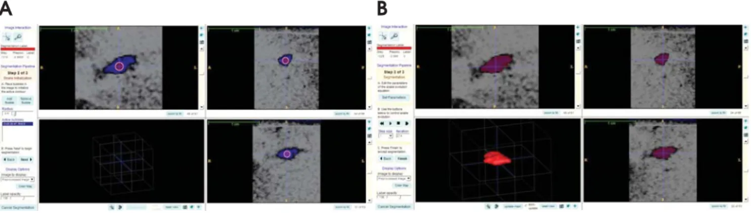
Panoramic radiographs were taken using PaxUni 3D (Vatech, Seoul, Korea) set at 60-80 kVp, 8-10 mA, and 9.7 seconds and OP 200D (Instrumentarium Dental, Tuusula, Finland) digital orthopantomograph devices set at 57-85 kVp, 2-16 mA, and 8 seconds. In all 7 cases, PaxUni 3D was also used to capture CBCT images at the following settings: 50-90 kVp, 4-10 mA, and seconds exposure time, and a 50×50 mm field of view (FOV) size. Coronal, axial, and 3D images were examined by two oral radiologists using Ez3D2009 (Vatech, Seoul, Korea) software. In order to measure calcification volumes, the images in DICOM format were transferred to ITK-SNAP 2.4.0 (Kitware, New York, USA) PC software that is used for medical image processing. Here, ITK-SNAP is an interactive image seg- mentation software developed to implement an active con- tour segmentation of structures, allowing regional segmen- tation by employing user-initialized deformable implicit surfaces that evolve to the most appropriate border bet- ween neighboring structures. Segmentation is the process by which appropriate reference points (voxels) are assigned to be part of a specific structure.12
The coronal, sagittal, and axial images were used to de- fine the calcifications in ITK-SNAP software and semi- automatic segmentation was performed by one oral radio- logist with selection of reference points on calcifications with a computer mouse.12-14 The software automatically segmented the calcifications starting from the reference points using the contrast differences between the soft tissue and calcifications on the greyscale images. Subsequent to
the segmentation, a 3D-graphical model of the volumetric object was generated by the software and the volume in mm3of the segmented 3D model was obtained (Fig. 1).
The patients with severe symptoms including dysphagia and recurrent tonsil infection were referred to the Faculty of Medicine’s Department of Otorhinolaryngology for con- sultation and one of the patients also underwent an MRI to confirm the diagnosis before surgery. The remaining patients underwent follow-up care.
Results
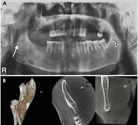
There were 4 males and 3 females who had tonsilloliths in this study. The age range of the subjects was 31-73 years (mean age: 51 years). The examination of CBCT images on coronal plane, axial, and 3D images using Ez3D2009 software showed the lesions to be located medially to each mandibular ramus and in the palatine tonsils. Irregularly shaped dense calcifications were detected in the palatal tonsils of all of the patients. In the ITK-SNAP software, bilateral calcifications were identified in 5 cases (bilateral- ly multiple in 3, unilaterally multiple in 2 cases) (Fig. 2) and a unilateral solitary calcification was identified in 2 cases (Table 1). CBCT images revealed that what appeared on panoramic radiographs as bilateral images were in fact unilateral lesions and their ghost images on the opposite side in these 2 cases (Fig. 3).
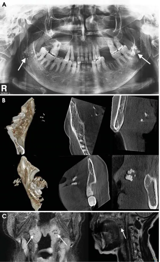
The total volume of calcifications ranged from 7.92 mm3 to 302.5 mm3. Four of the patients had symptoms includ- ing dysphagia, halitosis and tonsil infection, but the remain- ing 3 patients were asymptomatic. The patients with bilat- erally multiple and larger calcifications showed more severe symptoms (Table 1). In one of the patients, magnetic reso- nance image (MRI) results also confirmed the diagnosis of tonsilloliths by CBCT (Fig. 4).
Discussion
Although the exact etiology and pathogenesis of tonsil- loliths is unknown, most authors have believed these con- cretions to be the result of unresolved tonsillitis. Others, however, have suggested that tonsilloliths developed as a result of stasis of the saliva in the efferent ducts of the accessory salivary glands secondary to mechanical obstruc- tion arising from post-tonsillectomy scars or chronic inflam- mation.3Tonsillar concretions may produce symptoms that include non-specific chronic halitosis, irritable cough, dys- phagia, and otalgia as well as a foreign-body-like sensation and foul taste.15 Patients with tonsilloliths may also be asymptomatic, with their lesions discovered incidentally on panoramic or other skull radiographs.16
Calcifications in palatal tonsils were observed in the 7 cases in our study. Past histories did not suggest any par- ticular etiology. The absence of clinical signs and symp- toms might have originated from the small size of the cal- cifications, which remained undiagnosed until their inci-
Fig. 1.A. Selection of reference points in the area of interest is performed. B. ITK-SNAP software allows semiautomatic segmentation of calcifications.
Fig. 2.The CBCT images show a patient with multiple calcifi- cations. Every calcification can be segmented with a different color with ITK-SNAP software.
A B
dental detection during panoramic radiographic examina- tion.
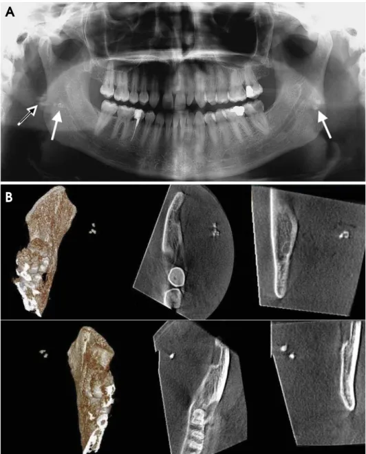
In three cases (Cases 1, 2, and 3 in Table 1), the osseous tissue density of the bilateral images, some of which over- lapped the mandibular ramus, suggested benign, intra-oss- eous lesions. However, the fact that in case 3, some of the
radiopaque images on the right side did not overlap the ramus led to a probable diagnosis of soft-tissue calcifica- tion (Fig. 5).4,6,15,17When a soft-tissue calcification is adja- cent to bone, it is sometimes difficult to determine whether the calcification is within the bone or the soft tissue. Radio- paque lesions within the mandibular ramus - such as calci-
A
B
Fig. 3.A. The panoramic radiograph shows a patient with a pseudo-bila- teral tonsillolith. The white arrow shows a unilateral calcification that overlapped the middle portion of the ascending ramus. The white and black arrow indicates a ghost image of the radiopacity on the right side appearing higher and more blurred than its source on the left side. B.
The CBCT images reveal what app- eared on panoramic radiographs as bilateral images were in fact unila- teral lesions. The 3D, axial, and coro- nal CBCT images show a unilateral mass of the right side.
Table 1.Calcification characteristics of cases
Calcification Calcification Total volume: Total volume:
Patient Age/Sex Location type at right side type at left side Signs/Symptoms
(number) (number) right side (mm3) left side (mm3)
1 61/F Bilateral Multiple (11) Multiple (5) 97.15 302.5 Recurrent tonsillitis,
dysphagia, halitosis
2 63/M Bilateral Multiple (4) Multiple (6) 22.19 87.47 Recurrent tonsillitis,
halitosis
3 33/F Bilateral Multiple (3) Multiple (3) 89.81 85.64 Recurrent tonsillitis,
dysphagia, halitosis
4 31/M Bilateral Multiple (3) Solitary 15.25 11.19 Asymptomatic
5 62/F Bilateral Solitary Multiple (3) 7.92 20.55 Asymptomatic
6 73/M Unilateral
Solitary - 16.82 - Asymptomatic
(Right tonsil)
7 34/M Unilateral - Solitary - 11.80 Halitosis
(Left tonsil)
fications in the carotid artery, lymph nodes, salivary gland, and stylohyoid ligament - may be considered in the essen- tial differential diagnosis. In panoramic images, calcifica- tions within the carotid artery appear in the soft tissue
below the angle of the mandible and between the hyoid bone and the cervical spine. Lymph node calcifications occur most often in the submandibular region, near or below the mandibular angle. The majorities (83-94%) of
Fig. 4.Case 1. Panoramic, CBCT, and MR images. A. The panoramic radiograph show bilateral calcifica- tions overlapping the middle port- ion of the ascending ramus, which suggest benign, intra-osseous lesions (white arrows). B. The 3D, axial, and coronal (both sides) CBCT images show bilateral masses in the area of the tonsils. C. The MR images of the same patient show the calcifi- cations in the tonsils (white arrows).
A
B
C
sialoliths are found in the submandibular glands, although some occur in the parotid gland (4-10%) or sublingual gland (1-7%) and can be visualized on standard occlusal projections as well as panoramic radiographs. Ossification of the stylohyoid ligament can be seen in a panoramic image to extend from the mastoid process across the pos- tero-inferior aspect of the ramus toward the hyoid bone.17-20 Tonsilloliths can be diagnosed through clinical presenta- tion, physical examination, and image studies. Enlarge- ment and hardening of the tonsil are typical findings during physical examination. On extraoral radiographs, a tonsil- lolith produces a radiopaque shadow that may be mistaken for a foreign body, tooth, prominent mandibular ramus, or maxilla, or a calcification in an artery, lymph node, sali- vary gland, or styloid ligament. Differentiation of these
calcifications can be made from a panoramic radiograph supplemented by a CT scan.17MRI may also prove valu- able in establishing a definitive diagnosis.4
The serial images provided by a CT scan make it pos- sible to identify the relationship between the lesion and the surrounding structures and to rule out diagnoses of bony tissue or calcification in vessels. MR images with ex- cellent soft tissue contrast resolution are also most helpful for identifying and localizing orofacial soft tissue lesions.
However, their high cost and relatively long imaging times limit the routine use of MRI.11 The compact design and low cost of most CBCT systems relative to conventional CT and MRI has made CBCT a valuable tool for the localization and differentiation of various radiopaque lesions.9In the 7 cases reported here, CBCT was necessary
Fig. 5.A. The panoramic radiograph of a patient shows bilateral tonsil- loliths. The white and black arrow indicates that some of the radiopaque images on the right side, which do not overlap the ramus, led to a pro- bable diagnosis of soft-tissue calci- fication. B. CBCT images revealed that they were ghost images of radio- pacities on the left side.
A
B
for a diagnosis of tonsilloliths.
In conclusion, tonsilloliths can be detected on panora- mic radiography, but differentiating these lesions from other calcifications and ghost images can be difficult. A CBCT scan is very helpful for identifying the lesions.
Asymptomatic, single, and large tonsilloliths should be removed because they can invite recurrent episodes of tonsillitis. Manual compression, curettage, or a simple incision to release the calcified body are usually successful.
However, removal of numerous, relatively small asymp- tomatic tonsilloliths can only be accomplished via a bila- teral tonsillectomy.10 In such cases, the true location of calcifications should be detected with CBCT first. Then the patient should undergo surgery to eliminate symptoms, and after the surgery, histological analysis should be made for achieving a definitive diagnosis.
References
1. Silvestre-Donat FJ, Pla-Mocholi A, Estelles-Ferriol E, Mar- tinez-Mihi V. Giant tonsillolith: report of a case. Med Oral Patol Oral Cir Bucal 2005; 10: 239-42.
2. Neville B, Damm DD, Allen CM, Bouquot J. Oral & maxillo- facial pathology. 2nd ed. Philadelphia: WB Saunders; 2002.
3. Mesolella M, Cimmino M, Di Martino M, Criscuoli G, Alb- anese L, Galli V. Tonsillolith. Case report and review of the literature. Acta Otorhinolaryngol Ital 2004; 24: 302-7.
4. Ram S, Siar CH, Ismail SM, Prepageran N. Pseudo bilateral tonsilloliths: a case report and review of the literature. Oral Surg Oral Med Oral Pathol Oral Radiol Endod 2004; 98: 110-4 5. Ozcan E, Ural A, Oktemer TK, Alpaslan G. Bilateral tonsillo-
lithiasis: a case report. Oral Surg Oral Med Oral Pathol Oral Radiol Endod 2006; 102: e17-8.
6. Sezer B, Tugsel Z, Bilgen C. An unusual tonsillolith. Oral Surg Oral Med Oral Pathol Oral Radiol Endod 2003; 95: 471-3.
7. Lo RH, Chang KP, Chu ST. Upper airway obstruction caused by bilateral giant tonsilloliths. J Chin Med Assoc 2011; 74:
329-31.
8. Poeschl PW, Schmidt N, Guevara-Rojas G, Seemann R, Ewers R, Zipko HT, et al. Comparison of cone-beam and conventional
multislice computed tomography for image-guided dental im- plant planning. Clin Oral Investig 2013; 17: 317-24.
9. Pauwels R, Beinsberger J, Stamatakis H, Tsiklakis K, Walker A, Bosmans H, et al. Comparison of spatial and contrast reso- lution for cone-beam computed tomography scanners. Oral Surg Oral Med Oral Pathol Oral Radiol 2012; 114: 127-35.
10. Guevara C, Mandel L. Panoramic radiographic demonstration of bilateral tonsilloliths. N Y State Dent J 2011; 77: 28-30.
11. White SC, Pharoah MJ. Oral Radiology: principles and inter- pretation. 5th ed. St. Louis: Mosby; 2004.
12. Yasuda CL, Costa AL, Franca M Jr, Pereira FR, Tedeschi H, de Oliveira E, et al. Postcraniotomy temporalis muscle atrophy:
a clinical, magnetic resonance imaging volumetry and electro- myographic investigation. J Orofac Pain 2010; 24: 391-7.
13. Cevidanes LH, Styner MA, Proffit WR. Image analysis and superimposition of 3-dimensional cone-beam computed tomo- graphy models. Am J Orthod Dentofacial Orthop 2006; 129:
611-8.
14. Cevidanes LH, Bailey LJ, Tucker GR Jr, Styner MA, Mol A, Phillips CL, et al. Superimposition of 3D cone-beam CT mod- els of orthognathic surgery patients. Dentomaxillofac Radiol 2005; 34: 369-75.
15. Cooper MM, Steinberg JJ, Lastra M, Antopol S. Tonsillar cal- culi. Report of a case and review of the literature. Oral Surg Oral Med Oral Pathol 1983; 55: 239-43.
16. Marshall WG, Irwin ND. Tonsilloliths. Oral Surg Oral Med Oral Pathol 1981; 51: 113.
17. de Moura MD, Madureira DF, Noman-Ferreira LC, Abdo EN, de Aguiar EG, Freire AR. Tonsillolith: a report of three clini- cal cases. Med Oral Patol Oral Cir Bucal 2007; 12: 130-3.
18. Almog DM, Horev T, Illig KA, Green RM, Carter LC. Corre- lating carotid artery stenosis detected by panoramic radiogra- phy with clinically relevant carotid artery stenosis determin- ed by duplex ultrasound. Oral Surg Oral Med Oral Pathol Oral Radiol Endod 2002; 94: 768-73.
19. Fanibunda K, Lovelock DJ. Calcified stylohyoid ligament:
unusual pressure symptoms. Dentomaxillofac Radiol 1997;
26: 249-51.
20. Ravon NA, Hollender LG, McDonald V, Persson GR. Signs of carotid calcification from dental panoramic radiographs are in agreement with Doppler sonography results. J Clin Periodontol 2003; 30: 1084-90.