- 209 -
Imaging Science in Dentistry 2019; 49: 209-12 https://doi.org/10.5624/isd.2019.49.3.209
Introduction
The clivus is a bony surface, the name of which means
“slope.” The fusion of the basisphenoidal and basiocciput bones results in the middle portion of the skull base. This fusion usually takes place before the age of 18years.1,2 Fossa navicularis is an anatomical variation of the bone in the lower part of the clivus, as well as a notch in the ba- siocciput bone. This variation has also been referred to as fossa pharyngea, large pharyngeal fossa, keyhole defect, longitudinal or transverse segmentations, fossa navicu- laris magna, and canalis basilaris medianus. Generally, fossa navicularis is an incidental finding in radiological examinations and has clearly visible cortical margins.3
Since cone-beam computed tomography(CBCT) has a lower radiation dose and higher resolution than conven- tional computed tomography(CT) scans, its usage has be- come more common in recent years. CBCT can provide a 3-dimensional morphological evaluation of dental and maxillofacial anatomy.4 Therefore, due to its widespread use, increasing interest is emerging in the anatomical fea- tures and anatomical variations of the human skull.
Dentomaxillofacial radiologists must be familiar with all the craniofacial structures and anatomical variations in the imaging field. Therefore, the anatomical variation of the fossa navicularis within the clivus must be identified and reported. In the literature, there are limited studies evaluating the prevalence and morphometric properties of the fossa navicularis.1-3,5 In this context, the aim of this study was to determine the prevalence and morphometric properties of fossa navicularis within the clivus in a Turk- ish subpopulation using CBCT.
Evaluation of morphometric features of fossa navicularis using cone-beam computed tomography in a Turkish subpopulation
Guldane Magat 1,*
1Department of Oral and Maxillofacial Radiology, Faculty of Dentistry, Necmettin Erbakan University, Konya, Turkey
ABSTRACT
Purpose: Fossa navicularis is a bone defect in the clivus. Familiarity with this anatomical variant is important because it is close to vital anatomical structures in the base of the skull. The aim of this study was to determine the prevalence and morphometric properties of fossa navicularis within the clivus in a Turkish subpopulation using cone-beam computed tomography(CBCT).
Materials and Methods: A total of 168 CBCT scans(female: 96, male: 71) were evaluated. High-quality CBCT images of patients without a syndromic condition or a history of neurological disease or surgery were included in the study. The prevalence, depth, length, and width of the fossa navicularis were performed.
Results: The prevalence of fossa navicularis was 27.5%(n=46 patients). Sex was not associated with the depth, length, or width of the fossa navicularis (P>0.05). A significant positive correlation was found between age and length of the fossa navicularis(P>0.05).
Conclusion: Fossa navicularis was found to be rare(27.5%). Anatomical variants of the skull base can also be clearly identified on CBCT images. The results of this study may be useful to radiologists, anatomists, and surgeons interested in the skull base.(Imaging Sci Dent 2019; 49: 209-12)
KEY WORDS: Anatomy; Clivus; Cone-Beam Computed Tomography; Skull Base
Copyright ⓒ 2019 by Korean Academy of Oral and Maxillofacial Radiology
This is an Open Access article distributed under the terms of the Creative Commons Attribution Non-Commercial License (http://creativecommons.org/licenses/by-nc/3.0) which permits unrestricted non-commercial use, distribution, and reproduction in any medium, provided the original work is properly cited.
Imaging Science in Dentistry·pISSN 2233-7822 eISSN 2233-7830 Received May 27, 2019; Revised July 10, 2019; Accepted July 19, 2019
*Correspondence to : Dr. Guldane Magat
Department of Oral and Maxillofacial Radiology, Faculty of Dentistry, Necmettin Erbakan University, 42050, Konya, Turkey
Tel) 90-505-945 6157, E-mail) [email protected]
Evaluation of morphometric features of fossa navicularis using cone-beam computed tomography in a Turkish subpopulation
- 210 -
Materials and Methods
A total of 168 patients(71 males, 96 females) who were admitted to the Faculty of Dentistry of Necmettin Erbakan University, Konya, Turkey between June 2016 and May 2017 and received CBCT scans for the purpose of diagno- sis and treatment were included in this study. The Necmet- tin Erbakan University Research and Ethics Committee approved this retrospective study(decision no. 2019/05).
High-quality CBCT images of patients without a syndrom- ic condition or a history of neurological disease or surgery were included in the study.
CBCT images were obtained using a Morita 3D Accu- itomo 170 device(J Morita MFG Corp., Kyoto, Japan) according to the manufacturer’s recommended protocol, using 90kVp and 5mA, a rotation time of 17.5seconds, a voxel size of 0.25mm, and a 140-×100-mm field of view.
A total of 168 CBCT images were analyzed using i-Dixel software(J Morita Manufacturing Corp., Kyoto, Japan) by the same oral and maxillofacial radiologist. The contrast and brightness of the images were adjusted using the soft- ware’s image processing tool to ensure optimum viewing.
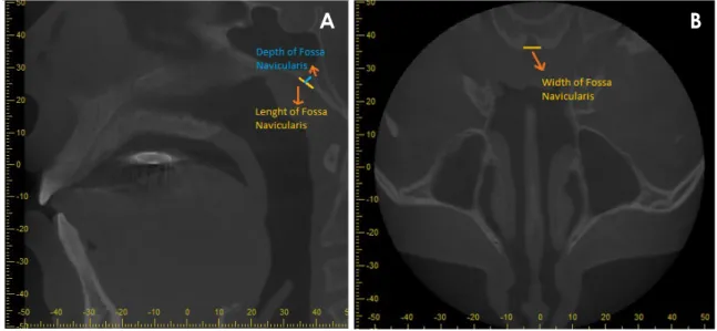
Axial and sagittal images at different levels were visual- ized on the monitor. Fossa navicularis was detected in the sagittal plane on the anterior side of the clivus. The depth and length of the fossa navicularis were measured from the deepest part in the sagittal plane(Fig. 1A). The width of the fossa navicularis was then measured on the axial plane in the same cross-section as the software’s router(Fig. 1B).
All measurements were repeated twice, with 3 weeks be- tween each measurement by the same observer.
Statistical analysis was performed using IBM SPSS® version 21.0(IBM Corp., Armonk, NY, USA). The descrip- tive statistical values were given. The normality of the data distribution was assessed by the Kolmogorov-Smirnov test. The measurements were statistically compared by sex using the Mann-Whitney U test. Correlations between age and measurements were evaluated using the Spearman cor- relation test. Intraobserver agreement was evaluated using the intraclass correlation coefficient, with results between 0.86 and 0.91. Statistical significance was set at P<0.05.
Results
Fossa navicularis was identified in 46(27.5%) patients.
Among these patients, 22 were female(22.9% of all female patients), whereas 24 were male(33.8% of all male pa- tients). The mean age of patients with fossa navicularis was 31.8±16.4years(range, 18-80years).
The mean length, depth, and width of the fossa navicu- laris in the 46 patients in whom it was present were 8.55 mm, 2.22mm, and 5.37mm, respectively(Table 1). Sex was not significantly related with the depth, length, and width of the fossa navicularis(P>0.05). A significant pos- itive correlation was found between age and length of the fossa navicularis(P=0.005; rho=0.408).
Discussion
In the present study, the prevalence and morphomet- ric properties of fossa navicularis within the clivus were evaluated in a Turkish population using CBCT. Our study
A B
Fig. 1. A. Measurements of fossa navicularis depth and length on a sagittal image. B. Measurements of fossa navicularis width on an axial image.
- 211 -
Guldane Magat
showed that the prevalence of fossa navicularis was 27.5%.
The mean length, width, and depth of the fossa navicularis in the 46 patients with fossa navicularis were 8.55mm, 5.37 mm, and 2.22mm, respectively.
Fossa navicularis is very important because it is close to various anatomical structures, such as the nasopharynx and sphenoid sinus. Various pathological lesions, such as local or metastatic tumors, adenoid retention cyst, adenoid hypertrophy, Rathke pouch cyst, and dermoid teratoma of the posterior nasopharyngeal wall, may mimic its anatomy or vice versa.6-8 For this reason, it is important to know this anatomical variation in detail.
In some studies, it was stated that fossa navicularis was the route through which an infection of the oropharynx spread to the skull. Surgical obliteration of this defect re- sulted in complete recovery.9-11 Therefore, understanding the anatomy of embryology and bone variations may help to diagnose conditions affecting this region.3
In the literature, fossa navicularis has generally been an- alyzed in dry skulls or CT images.12-14 There are a limited number of studies using CBCT.1,3 Therefore, we preferred to use CBCT images in this study. The prevalence of fos- sa navicularis was found to be between 0.9% and 5.3% in studies using dry skull. In the study of Cankal et al.,13 it was found to be 3% in CT images. Ersan3 and Bayrak et al.1 also evaluated fossa navicularis on CBCT images in the same population. Ersan3 reported the prevalence of fossa navicularis to be 6.6% in 732 patients. Bayrak et al.1 stud- ied 1,059 patients, and found the prevalence to be 7.6%. In this study, the prevalence of fossa navicularis was 27.5%.
This result is markedly higher than other studies in the lit- erature. This discrepancy might be due to different method- ologies, ethnic differences, and different sample sizes. In a recent study conducted by Ersan,5 it was reported that the prevalence of fossa navicularis was 28.8% in cleft palate patients, similar to our result.
In this study, the mean length, width, and depth of the fossa navicularis in 46 patients were 8.55mm, 5.37mm, and 2.22mm, respectively. In the literature, it has been re- ported that the length varies from 7 to 13mm, the width
from 6 to 8mm, and the depth from 2 to 5mm.15 Bayrak et al.1 reported a length of 7.15mm, width of 5.23mm, and depth of 2.76mm in CBCT measurements of 59 patients with fossa navicularis. In addition, they also studied CT scans and found that the length, width, and depth of the fossa navicularis in 22 patients were 4.12mm, 4.08mm, and 4.17mm, respectively.
Ersan3 reported no difference in the length of the fossa navicularis between age groups, while Bayrak et al.1 stated that there was a statistically significant difference between age groups in terms of the length of the fossa navicu- laris. In this study, a significant positive correlation was found between age and the length of the fossa navicularis (P=0.005; rho=0.408). There was an increase in the size of the fossa navicularis as age progressed.
In the literature, it has been reported that infections are carried through caudocranial structures via the fossa na- vicularis.10,11,16 Therefore, it is important to know this an- atomical variation in detail. Insufficient studies have been conducted on this area in the field of dentomaxillofacial radiology. Therefore, it is necessary to increase the number of studies of such anatomical variations, in particular by conducting studies with larger samples and in more ethni- cally diverse populations.
In conclusion, the prevalence of fossa navicularis in this study was higher than reported in the literature. However, it was still an uncommon variation. The anatomical structures of the fossa navicularis can be studied effectively on CBCT images. Because CBCT findings include areas at the base of the skull, which are not intended to be displayed, den- tomaxillofacial radiologists have an obligation to identify, examine, and report anatomical variations of the skull base and to prevent unnecessary requests for further imaging by dental practitioners. The findings of this study may be use- ful for radiologists, anatomists, and surgeons interested in the base of the skull.
References
1. Bayrak S, Göller Bulut D, Orhan K. Prevalence of anatomical Table 1. The mean values of fossa navicularis measurements according to sex
Sex Number Length Depth Width
Range(mean±SD) P value* Range(mean±SD) P value* Range(mean±SD) P value*
Female 22 4.93-15.00(9.32±2.59)
0.095 0.74-4.65(2.32±0.94)
0.391
2.45-9.87(5.21±1.60)
0.538
Male 24 2.41-15.94(7.85±3.56) 0.92-4.65(2.13±1.04) 1.44-10.44(5.52±2.43)
Total 46 2.41-15.94(8.55±3.19) 0.74-4.65(2.22±0.98) 1.44-10.44(5.37±2.06)
*Mann-Whitney U test
Evaluation of morphometric features of fossa navicularis using cone-beam computed tomography in a Turkish subpopulation
- 212 - variants in the clivus: fossa navicularis magna, canalis basilaris medianus, and craniopharyngeal canal. Surg Radiol Anat 2019;
41: 477-83.
2. Syed AZ, Mupparapu M. Fossa navicularis magna detection on cone-beam computed tomography. Imaging Sci Dent 2016; 46:
47-51.
3. Ersan N. Prevalence and morphometric features of fossa na- vicularis on cone beam computed tomography in Turkish popu- lation. Folia Morphol(Warsz)(in press).
4. Ahmad M, Jenny J, Downie M. Application of cone beam com- puted tomography in oral and maxillofacial surgery. Aust Dent J 2012; 57 Suppl 1: 82-94.
5. Ersan N. Prevalence of fossa navicularis among cleft palate pa- tients detected by cone beam computed tomography. Yeditepe J Dent 2017; 13: 21-3.
6. Kizilkilic O, Yalcin O, Yildirim T, Sener L, Parmaksiz G, Erdo- gan B. Hypothalamic hamartoma associated with a craniopha- ryngeal canal. AJNR Am J Neuroradiol 2005; 26: 65-7.
7. Pinilla-Arias D, Hinojosa J, Esparza J, Muñoz A. Recurrent meningitis and persistence of craniopharyngeal canal: case re- port. Neurocirugia(Astur) 2009; 20: 50-3.
8. Abele TA, Salzman KL, Harnsberger HR, Glastonbury CM.
Craniopharyngeal canal and its spectrum of pathology. AJNR Am J Neuroradiol 2014; 35: 772-7.
9. Currarino G. Canalis basilaris medianus and related defects of the basiocciput. AJNR Am J Neuroradiol 1988; 9: 208-11.
10. Prabhu SP, Zinkus T, Cheng AG, Rahbar R. Clival osteomyelitis resulting from spread of infection through the fossa navicularis magna in a child. Pediatr Radiol 2009; 39: 995-8.
11. Segal N, Atamne E, Shelef I, Zamir S, Landau D. Intracrani- al infection caused by spreading through the fossa naviclaris magna - a case report and review of the literature. Int J Pediatr Otorhinolaryngol 2013; 77: 1919-21.
12. Ray B, Kalthur SG, Kumar B, Bhat MR, D’souza AS, Gulati HS, et al. Morphological variations in the basioccipital region of the South Indian skull. Nepal J Med Sci 2014; 3: 124-8.
13. Cankal F, Ugur HC, Tekdemir I, Elhan A, Karahan T, Sevim A. Fossa navicularis: anatomic variation at the skull base. Clin Anat 2004; 17: 118-22.
14. Alalade AF, Briganti G, McKenzie JL, Gandhi M, Amato D, Panizza BJ, et al. Fossa navicularis in a pediatric patient: ana- tomical skull base variant with clinical implications. J Neuro- surg Pediatr 2018; 22: 523-7.
15. Beltramello A, Puppini G, El-Dalati G, Girelli M, Cerini R, Sbarbati A, et al. Fossa navicularis magna. AJNR Am J Neu- roradiol 1998; 19: 1796-8.
16. Akyel NG, Alımlı AG, Demirkan TH, Sivri M. Persistent cra- niopharyngeal canal, bilateral microphthalmia with coloboma- tous cysts, ectopic adenohypophysis with Rathke cleft cyst, and ectopic neurohypophysis: case report and review of the litera- ture. Childs Nerv Syst 2018; 34: 1407-10.